
Can left or right bundle branch spontaneously depolarize?
"Purkinje fibers" AND "automaticity"
- Overdrive suppression: The SA node depolarizes faster than the bundle branches' intrinsic rate. Each propagated impulse resets them before their own phase 4 depolarization reaches threshold.
- Intrinsic rate hierarchy: The usual figures are SA node 60 to 100/min, AV junction 40 to 60/min, and His-Purkinje/ventricular tissue about 20 to 40/min. These are standard teaching values, not taken from the retrieved passage.
- Escape rhythm: If the SA node and AV node fail, or there is complete AV block, a ventricular escape rhythm arises from the His-Purkinje system. It is slow, with a wide QRS if it originates below the bifurcation.
- Enhanced or abnormal automaticity: Ischemia, catecholamines, hypokalemia, and digoxin toxicity can raise Purkinje automaticity. This can cause accelerated idioventricular rhythm, ventricular parasystole, or PVCs. Braunwald's Heart Disease lists "depolarization-induced automaticity in Purkinje myocytes" as a mechanism of accelerated ventricular rhythms.
- Triggered activity: Delayed afterdepolarizations in Purkinje cells can also fire ectopic beats.
What is junctional escape rhythm
junctional escape rhythm ECG
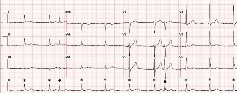
A 12-lead electrocardiogram (ECG) demonstrating severe sinus bradycardia with a heart rate of approximately 60 beats per minute. The rhythm exhibits atrial quadrigeminy, characterized by a repeating pattern where every fourth complex is a premature atrial complex (APC), marked by arrows in the rhythm strip (lead II). These APCs are followed by a compensatory pause and are coupled at an interval of approximately 550 ms. Several QRS complexes, marked with asterisks (*), lack preceding visible P-waves, suggesting a competing junctional escape rhythm or junctional beats emerging due to the underlying sinus bradycardia. The QRS complexes maintain a narrow morphology, and the ST segments and T-waves appear normal without signs of acute ischemia or electrolyte-induced peaking. This ECG illustrates complex rhythm disturbances including escape-capture sequences and ectopy in the clinical context of chronic kidney disease and electrolyte fluctuations.
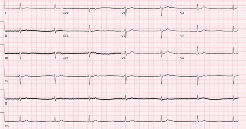
This diagnostic image is a 12-lead electrocardiogram (ECG) demonstrating junctional bradycardia. The tracing shows a slow, regular ventricular rate with narrow QRS complexes (duration within normal limits, <120 ms). A key diagnostic feature is the absence of clear, consistent sinus P waves preceding each QRS complex, suggesting an escape rhythm originating from the atrioventricular junction. In some leads, such as II and aVF, P waves are either absent or dissociated, consistent with the clinical context of a progressive bradyarrhythmia. The precordial leads (V1-V6) show preserved R-wave progression. There is no evidence of acute ST-segment elevation or significant T-wave inversions. This ECG illustrates a junctional escape rhythm, which may occur in the setting of sinus node dysfunction, high-grade AV block, or metabolic disturbances. The educational focus is on identifying non-sinus bradycardias and distinguishing junctional rhythms by their narrow QRS morphology and lack of preceding P waves.
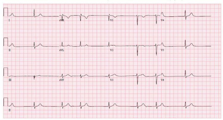
A 12-lead electrocardiogram (ECG) with a rhythm strip (Lead II) at the bottom, demonstrating a complex arrhythmia characterized by 'escape-capture bigeminy'. The tracing shows a slow junctional rhythm competing with sinus activity. Key features include an irregular rhythm with alternating morphology: narrow QRS complexes (captured beats) follow P-waves with a prolonged PR interval, while wider or differently contoured complexes represent junctional escape beats. This pattern results in a repetitive bigeminal sequence. The baseline rate is bradycardic with evidence of a slow ventricular response. Distinct P-waves are visible in several leads but show variable relationship to the QRS complexes, indicating a degree of AV dyssynchrony. T-waves are generally upright and morphology is otherwise stable with a QTc interval of approximately 410 msec. This ECG illustrates electrophysiological changes often associated with metabolic derangements or drug toxicities, such as lithium toxicity, affecting the cardiac conduction system.
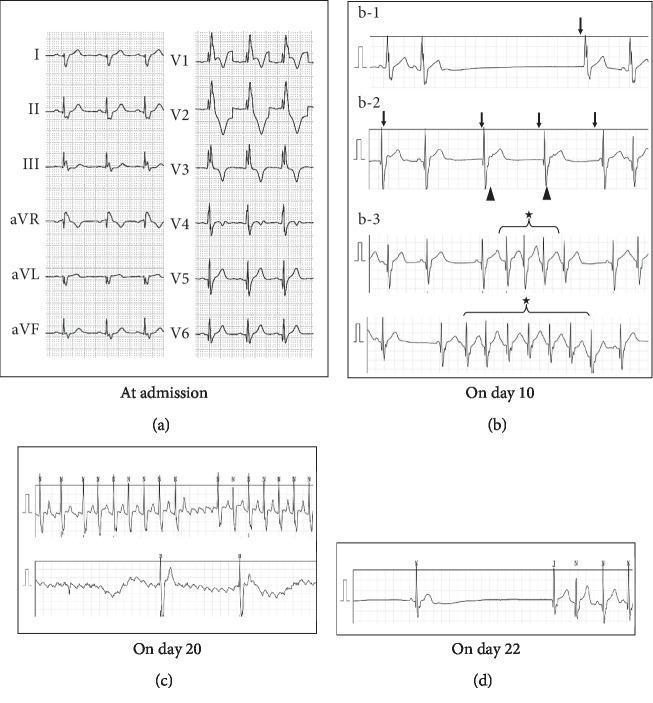
A composite clinical diagnostic image showing four sets of electrocardiogram (ECG) strips (a-d) tracking a patient's cardiac rhythm progression. (a) A standard 12-lead admission ECG demonstrating a regular sinus rhythm with a complete right bundle branch block (RBBB), characterized by rsR' patterns in V1-V2 and wide S waves in leads I and V6. (b) Multiple rhythm strips from day 10 showing sinus arrest (b-1), junctional escape rhythms with retrograde P waves indicated by arrowheads (b-2), and short bursts of non-sustained atrial tachycardia marked with stars (b-3). (c) Rhythm strips from day 20 demonstrating atrial flutter with a characteristic sawtooth baseline and variable atrioventricular (AV) conduction ratios ranging from 2:1 to 4:1. (d) A rhythm strip from day 22 showing a significant sinus arrest lasting approximately 5.7 seconds, followed by a slow junctional escape beat. This visual sequence is representative of sick sinus syndrome or complex cardiac conduction disturbances, illustrating the transition between bradyarrhythmias and tachyarrhythmias (tachy-brady syndrome).
"junctional escape rhythm"
- The SA node normally overdrives the AV junction. If the sinus rate drops below the junction's own rate, or sinus impulses stop reaching the ventricles, the junctional pacemaker fires.
- A single junctional beat is an escape beat. Several in a row make a junctional rhythm.
- In third-degree AV block at the AV node level, a junctional escape pacemaker takes over - Tintinalli's Emergency Medicine.
- Rate: 40 to 60 beats/min - Tintinalli's Emergency Medicine.
- QRS: Narrow, because the impulse starts above the bifurcation of the bundle of His. The ventricles are activated normally through the bundle branches.
- P waves: Absent, or retrograde. Retrograde P waves are inverted in the inferior leads and can fall just before, during, or just after the QRS. Tintinalli notes that in most cases junctional escape beats do not conduct retrograde into the atria, so a QRS appears without a P wave.
- Rhythm: Regular.
- Sinus bradycardia or sinus arrest, including sick sinus syndrome
- AV block (second-degree or third-degree)
- Drugs: digoxin, beta-blockers, calcium channel blockers, amiodarone
- Increased vagal tone, inferior MI, hyperkalemia, hypothyroidism
- Post-cardiac surgery
- Accelerated junctional rhythm: Rate is about 60 to 100/min. It can occur with medication toxicity (particularly digitalis), acute rheumatic fever, or inferior ischemia - Tintinalli's Emergency Medicine.
- Junctional tachycardia: Rate is above 100/min.
- If it is asymptomatic, observe and treat the cause (stop the offending drug, correct electrolytes).
- If it is symptomatic, treat the underlying cause. Atropine can raise the SA node rate and improve AV nodal conduction - Tintinalli's Emergency Medicine. If that fails, use transcutaneous or transvenous pacing, and consider a permanent pacemaker for irreversible sinus node or AV node disease.
- A narrow escape rhythm suggests the block is above the His bundle and is usually more stable. A slow, wide QRS escape suggests a lower, less reliable pacemaker - Textbook of Family Medicine.

Explain
| Pathway A | Pathway B | |
|---|---|---|
| Conduction speed | Slow | Fast |
| Recovery (refractory period) | Quick, so it is ready to fire again soon | Slow, so it stays refractory longer |
-
Sinus rhythm. The impulse goes down both pathways and meets in the common distal pathway. The two wavefronts cancel each other out. Both limbs are then refractory, shown in red, and nothing circulates.
-
Premature impulse. An early beat, such as a PAC or PVC, arrives before pathway B has recovered. B is still refractory (red), so the impulse is blocked in B. A has recovered quickly, so the impulse conducts down A. This is unidirectional block: B blocks the forward direction, but it may still conduct backwards.
-
Re-entry. The impulse travels slowly down A. By the time it reaches the common distal pathway, B has recovered. The impulse enters B from below and travels retrogradely up it. It then reaches the top of A, which has also recovered, and goes down A again, so a closed loop forms.
-
Tachycardia. The impulse keeps circling. Each circuit also sends a wavefront out to the rest of the heart, so the heart is driven at a fast rate. This continues until the circuit is interrupted, either by a change in conduction or refractoriness (a drug, vagal manoeuvre, or adenosine) or by electrical depolarisation (cardioversion).
- Two pathways joined at both ends, forming a loop.
- Unidirectional block in one limb. Braunwald notes that a drug that depresses conduction can turn unidirectional block into bidirectional block, which terminates re-entry.
- Slow conduction in the other limb, so the tissue ahead has time to recover. Fuster and Hurst's The Heart says the circuit length must exceed the wavelength (conduction velocity × refractory period).
- AVNRT (AV nodal slow and fast pathways)
- AVRT in WPW syndrome (AV node plus accessory pathway)
- Atrial flutter (a large circuit around an anatomical obstacle)
- Ventricular tachycardia around a post-MI scar