I now have all the comprehensive information needed. Let me compile the full detailed answer.
Organs of the Respiratory Tract
Upper Respiratory Tract
- Nose (external nose + nasal cavity)
- Paranasal sinuses
- Pharynx (nasopharynx, oropharynx, laryngopharynx)
- Larynx
Lower Respiratory Tract
- Trachea
- Right and left main (primary) bronchi
- Lobar (secondary) bronchi
- Segmental (tertiary) bronchi
- Bronchioles (including terminal and respiratory bronchioles)
- Alveolar ducts and alveoli
- Lungs (right and left)
THE LUNGS - Detailed Anatomy
a) Size and Shape
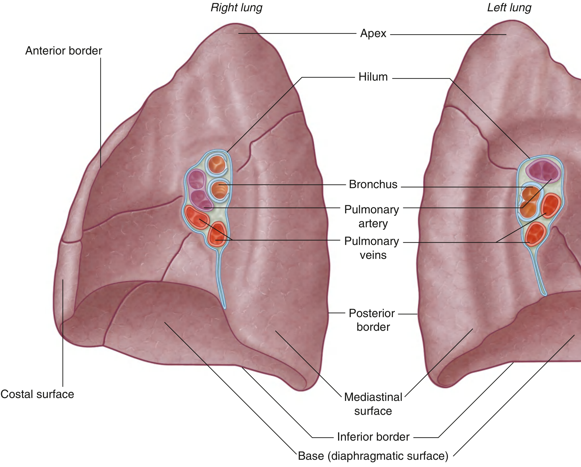
The lungs are a pair of light, spongy, cone-shaped organs situated in the thoracic cavity, one on each side of the mediastinum. Each lung is enclosed in a pleural sac.
Right Lung:
- Larger and heavier than the left (approximately 625 g vs 565 g in the adult male)
- Divided into three lobes - superior, middle, and inferior - by two fissures: the oblique fissure and the horizontal fissure
- Shorter and broader than the left because the right dome of the diaphragm is higher and the liver lies beneath it
Left Lung:
- Smaller than the right
- Divided into two lobes - superior and inferior - by the oblique fissure alone
- The oblique fissure of the left is slightly more oblique than that of the right
- Has a cardiac notch on its anterior border due to the heart's projection into the left pleural cavity
- Bears a tongue-like extension called the lingula at the lower part of the superior lobe, which projects over the cardiac notch
General shape: Each lung has:
- An apex - rounded superior tip that projects 2.5 cm above the clavicle into the root of the neck
- A base (diaphragmatic surface) - concave, resting on the diaphragm
- Three surfaces: costal, medial (mediastinal), and diaphragmatic
- Three borders: anterior, posterior, and inferior
b) Surface Anatomy
Each lung has the following surfaces and borders:
Surfaces:
| Surface | Description |
|---|
| Costal surface | Large, convex; lies against the ribs and intercostal muscles |
| Medial (mediastinal) surface | Concave; faces the mediastinum; contains the hilum |
| Diaphragmatic surface (base) | Concave; rests on the dome of the diaphragm |
Borders:
- Anterior border - thin, sharp; overlaps the pericardium. On the left side it carries the cardiac notch and lingula.
- Posterior border - broad, rounded; lies in the paravertebral gutter alongside the vertebral column (T2-T10)
- Inferior border - separates the base from the costal and mediastinal surfaces; sharp anteriorly, rounded posteriorly
Fissures - Surface Projections:
- Oblique fissure (both lungs): From the spine of T4 posteriorly → crosses the 5th interspace laterally → follows the contour of rib VI anteriorly. It ends near the 6th costal cartilage anteriorly.
- Horizontal fissure (right lung only): Runs horizontally from the oblique fissure (where it crosses rib V) anteriorly along the 4th intercostal space to the sternal border.
Apex: Projects approximately 2.5 cm above the medial third of the clavicle into the root of the neck.
Base: At full expiration reaches the 6th rib in the midclavicular line, 8th rib in the midaxillary line, and 10th rib posteriorly.
c) Blood Supply
The lungs receive a dual blood supply: pulmonary (functional) and bronchial (nutritional).
Pulmonary Arteries (Functional)
- The pulmonary trunk arises from the right ventricle and bifurcates into the right and left pulmonary arteries at the level of T4/T5, just below the carina.
- Right pulmonary artery - longer than the left; passes horizontally across the mediastinum, anterior to the right main bronchus; enters the root of the lung and gives off a branch to the superior lobe, then divides to supply the middle and inferior lobes.
- Left pulmonary artery - shorter; lies anterior to the descending aorta and posterior to the superior pulmonary vein; passes through the root and hilum and branches within the lung.
- Both arteries carry deoxygenated blood to the lungs for gas exchange.
Bronchial Arteries (Nutritional)
The bronchial arteries supply the bronchial walls, pulmonary connective tissue, and visceral pleura with oxygenated blood:
- Right side: A single right bronchial artery normally arises from the 3rd posterior intercostal artery (occasionally from the upper left bronchial artery).
- Left side: Two left bronchial arteries arise directly from the anterior surface of the thoracic aorta - the superior at vertebral level T5, and the inferior just below the left main bronchus.
- They run on the posterior surfaces of the bronchi and ramify throughout the lung tissue.
d) Venous and Lymphatic Drainage
Venous Drainage
Pulmonary veins (functional drainage - oxygenated blood):
- On each side, a superior pulmonary vein and an inferior pulmonary vein begin at the hilum and carry oxygenated blood directly to the left atrium.
- The veins are positioned inferiorly and anteriorly at the hilum.
Bronchial veins (nutritional drainage):
- Drain into:
- The pulmonary veins or the left atrium (for the more distal part)
- The azygos vein on the right, or the superior intercostal vein or hemiazygos vein on the left (for the proximal part at the hilum)
Lymphatic Drainage
The lymphatics of the lung run in two plexuses:
- Superficial (subpleural) plexus - beneath the visceral pleura
- Deep plexus - runs along the bronchi and pulmonary vessels
Both drain into tracheobronchial lymph nodes around the lobar and main bronchi and alongside the trachea. These form a chain extending from within the lung through the hilum and root into the posterior mediastinum.
Efferent vessels from these nodes pass superiorly along the trachea to unite with vessels from parasternal nodes and brachiocephalic nodes to form the right and left bronchomediastinal trunks, which drain directly into deep veins at the base of the neck or into the right lymphatic trunk / thoracic duct.
Clinical note: Lymph nodes are identified by numbered stations important in staging lung cancer.
e) Nerve Supply
The lungs are innervated via the anterior and posterior pulmonary plexuses, which lie anteriorly and posteriorly to the tracheal bifurcation and main bronchi. The posterior plexus is much larger than the anterior.
Branches from these plexuses are distributed along the airways and blood vessels. The fibers originate from:
| Component | Source | Action |
|---|
| Parasympathetic (vagal) | Vagus nerve (CN X) | Bronchoconstriction; increases secretions |
| Sympathetic | Sympathetic trunks (T1-T5) | Bronchodilation; reduces secretions |
| Visceral afferents | Both systems | Pain (pleura), stretch/cough reflexes (bronchi) |
- Vagal efferents constrict bronchioles
- Sympathetic efferents dilate bronchioles
- The visceral pleura (lung surface) is insensitive to pain; the parietal pleura is supplied by somatic nerves and is pain-sensitive
- In the mediastinum, the vagus nerves pass posterior to the roots of the lungs and the phrenic nerves pass anterior to them
f) Relations
Hilum
Within the root and hilum of each lung are:
- Pulmonary artery (superior)
- Two pulmonary veins (inferior and anterior)
- Main bronchus (posterior)
- Bronchial vessels, nerves, and lymphatics
Right Lung Relations (Medial Surface)
| Structure | Relation |
|---|
| Heart (right atrium and right ventricle) | Anteromedial impression |
| Inferior vena cava | Inferior medial |
| Superior vena cava | Anterior medial |
| Azygos vein | Arches over root superiorly |
| Esophagus | Posterior medial |
| Right subclavian artery and vein | Arch over the superior lobe (apex) |
| Trachea | Superior medial |
Left Lung Relations (Medial Surface)
| Structure | Relation |
|---|
| Heart (left ventricle, left atrium) | Produces the cardiac notch |
| Aortic arch | Superior medial |
| Thoracic (descending) aorta | Posterior medial |
| Esophagus | Posterior medial |
| Left subclavian artery and vein | Arch over the superior lobe (apex) |
Costal Surface (both lungs)
- Related to the ribs, intercostal muscles, and costal cartilages
- Anteriorly related to the sternum and internal thoracic vessels
Base (Diaphragmatic Surface)
- Right lung base: related to the liver via the right dome of the diaphragm
- Left lung base: related to the stomach and spleen via the left dome
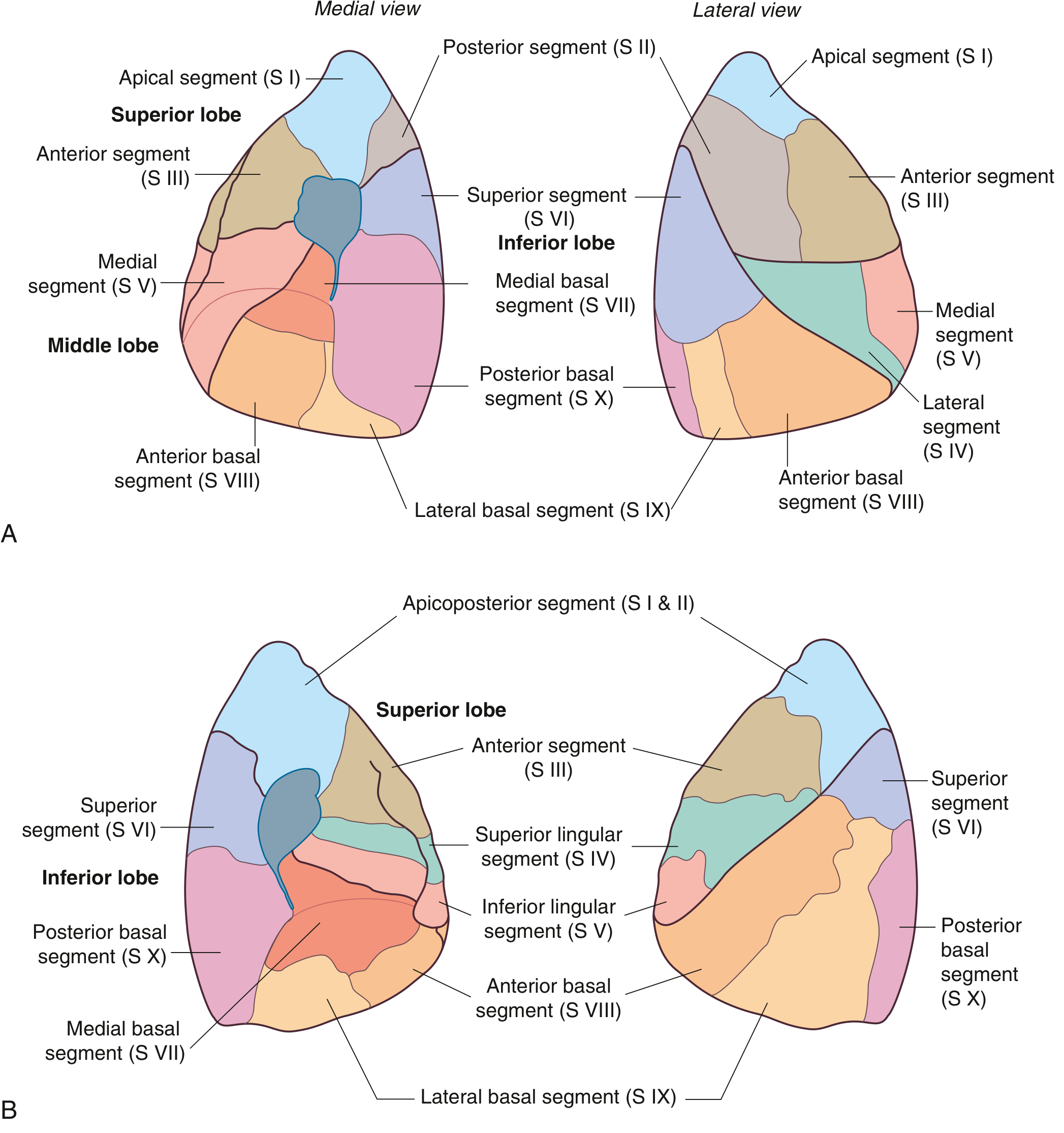
g) Bronchopulmonary Segments
A bronchopulmonary segment is defined as the area of lung supplied by a segmental (tertiary) bronchus and its accompanying branch of the pulmonary artery. Each segment is:
- Shaped like an irregular cone, with the apex at the origin of the segmental bronchus and the base projected onto the surface of the lung
- The smallest functionally independent region of the lung
- The smallest area of lung that can be surgically resected without affecting adjacent regions
- Separated from adjacent segments by intersegmental connective tissue septa
- Pulmonary vein tributaries tend to pass intersegmentally (between segments)
There are 10 bronchopulmonary segments in each lung (some fuse in the left lung).
Right Lung (10 Segments)
| Lobe | Segment No. | Segment Name |
|---|
| Superior lobe | S I | Apical |
| S II | Posterior |
| S III | Anterior |
| Middle lobe | S IV | Lateral |
| S V | Medial |
| Inferior lobe | S VI | Superior (apical) |
| S VII | Medial basal |
| S VIII | Anterior basal |
| S IX | Lateral basal |
| S X | Posterior basal |
Left Lung (8-10 Segments)
In the left lung, segments I and II typically fuse into a single apicoposterior segment, and segment VII (medial basal) may be absent or fused, giving 8-9 functional segments. The lingula corresponds to segments IV and V.
| Lobe | Segment No. | Segment Name |
|---|
| Superior lobe | S I + II | Apicoposterior |
| S III | Anterior |
| S IV | Superior lingular |
| S V | Inferior lingular |
| Inferior lobe | S VI | Superior (apical) |
| S VII | Medial basal (may be absent) |
| S VIII | Anterior basal |
| S IX | Lateral basal |
| S X | Posterior basal |
Clinical Significance of Bronchopulmonary Segments
- Aspiration pneumonia preferentially affects the superior segment of the right lower lobe (S VI) when supine, because it is the most dependent segment
- Foreign body inhalation more commonly affects the right side (especially right lower lobe) due to the wider, more vertical right main bronchus
- Surgeons can perform segmentectomy to remove individual diseased segments while preserving surrounding lung tissue
- Bronchoscopy is guided by knowledge of segment anatomy to drain abscesses or remove foreign bodies
Sources: Gray's Anatomy for Students (9780323934237); Bailey and Love's Short Practice of Surgery 28th Ed. (9780367548117)