
How to do heparin 4000 iu
heparin 4000 IU subcutaneous injection technique administration nursing
subcutaneous heparin injection technique abdomen site
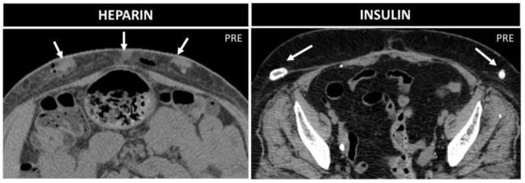
This figure presents a side-by-side comparison of axial non-enhanced CT images of the abdominal wall, illustrating complications from different subcutaneous injection types. The left panel (labeled 'HEPARIN') demonstrates multiple subcutaneous collections characterized by parenchymatous density, consistent with localized hematomas or bleeding resulting from repeated heparin injections. Within these soft-tissue nodules, small punctate air bubbles are visible. The right panel (labeled 'INSULIN') illustrates insulin-induced lipohypertrophy. These lesions appear as focal, heterogeneous subcutaneous masses that prominently feature dense peripheral calcifications (white arrows). The comparison highlights the different imaging signatures of injection-related sequelae: hemorrhagic collections and air pockets associated with anticoagulants versus calcified fatty proliferation (lipodystrophy) associated with chronic insulin therapy. Both images provide a cross-sectional view of the anterior abdominal wall at the level of the pelvis and mid-abdomen, emphasizing the clinical importance of recognizing benign injection-related findings that may mimic more serious abdominal wall pathology.
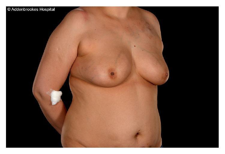
Clinical photograph of a 37-year-old female patient's torso, illustrating preoperative findings before a right mastectomy and DIEP (Deep Inferior Epigastric Perforator) flap reconstruction. The image focuses on the abdomen and chest. Extensive ecchymosis (bruising) is visible across the bilateral lower abdomen, characterized by irregular patches of purple and blue discoloration. These lesions are consistent with subcutaneous heparin or enoxaparin injection sites, commonly used for perioperative thromboprophylaxis. The upper torso shows the right breast area prepared for surgical intervention, with visible skin changes and subtle scarring in the infraclavicular region. A medical dressing is present on the right antecubital fossa, likely covering an intravenous access site. This photograph serves as an educational example of the local dermatological effects of anticoagulation therapy in patients undergoing complex reconstructive surgery.
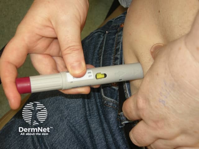
This image depicts a clinical photography of a subcutaneous injection being administered with a disposable pen-type injector. The device features a dose dial and a plunger, with a yellow indicator visible near the advance mechanism. A clinician or patient stabilizes the skin with the non-dominant hand by pinching subcutaneous tissue, while the dominant hand positions and depresses the plunger to deliver medication into the adipose layer. The needle enters the skin at a near perpendicular angle (approximately 90 degrees) to deposit the drug into subcutaneous fat for slower absorption. The abdomen is a common site for injections, but other practical regions include the outer thigh or upper arm; site rotation is advised to minimize lipohypertrophy and ensure consistent pharmacokinetics. After needle insertion and dose delivery, the device is withdrawn and the skin is gently blotted to reduce bleeding. The procedure emphasizes correct technique, avoiding intramuscular injection, excessive tissue compression, or needle bending. This image is a useful educational resource for medical trainees, diabetic patients, and healthcare professionals to practice safe subcutaneous administration, review needle depth and angle, and reinforce dose accuracy and device function. The DermNet watermark indicates dermatology education and skin-focused clinical documentation.
Heparin 4000 IU - Administration Guide
What is it and why 4000 IU?
"For prophylaxis, once-daily SC doses of 4000-5000 units are often used, whereas doses of 2500-3000 units are given when the drug is administered twice daily."
Step-by-Step Administration
1. Preparation
- Wash hands thoroughly with soap and water
- Gather: pre-filled syringe (or vial + syringe), alcohol swab, gauze, sharps container
- Check the drug label: name, dose (4000 IU), expiry, appearance (clear, colorless - discard if discolored or particulate)
- Do not expel the air bubble in pre-filled syringes - it helps ensure the full dose is delivered and seals the track afterward
2. Select and Prepare the Injection Site
| Site | Notes |
|---|---|
| Anterolateral abdominal wall (preferred) | 5 cm away from the umbilicus on either side; best absorption |
| Outer thigh | Alternative if abdomen not available |
| Upper outer arm | Less preferred |
- Avoid areas that are bruised, tender, scarred, or have previous injection sites
- Rotate sites with each injection to prevent hematomas and lipohypertrophy
- Clean skin with alcohol swab and allow to dry completely before injecting
3. Injection Technique
- Hold the pre-filled syringe like a dart in your dominant hand
- With the non-dominant hand, gently pinch a fold of subcutaneous fat (1-2 inches) - this lifts the SC layer away from muscle
- Insert the needle at 90 degrees (for adequate SC fat) - or 45 degrees if the patient is very thin
- Do NOT aspirate before injecting - aspiration is not needed for SC heparin and increases bruising risk
- Inject the solution slowly and steadily
- Hold the needle in place for 5-10 seconds after injecting
- Withdraw the needle at the same angle it was inserted
- Release the skin fold after withdrawing
4. After the Injection
- Apply gentle pressure with dry gauze for a few seconds
- Do NOT rub the site - rubbing causes bruising by spreading heparin into surrounding tissue
- Activate needle safety guard (if present) immediately after withdrawal
- Dispose in a sharps container
Key Special Points for Heparin SC
| Point | Reason |
|---|---|
| Do NOT rub the site | Prevents bruising/hematoma |
| Do NOT aspirate | Not required for SC; increases bruising |
| Keep the air bubble in pre-filled syringes | Seals the needle track, ensures full dose delivery |
| Rotate sites every injection | Prevents tissue damage and unpredictable absorption |
| Avoid 2 cm around umbilicus | Less SC fat, more vascular, higher bruising risk |
| Pinch skin throughout injection | Ensures SC (not IM) placement |
Monitoring and Side Effects
- Monitoring: Prophylactic doses of UFH at 4000-5000 IU do not typically require aPTT monitoring. LMWH at prophylactic doses does not need monitoring in most patients; anti-Xa levels (target 0.2-0.5 units/mL) can be checked in renal impairment or obesity.
- Side effects to watch for:
- Local hematoma or bruising at injection site (most common)
- Bleeding (major complication)
- Heparin-Induced Thrombocytopenia (HIT) - monitor platelets if on heparin >4 days
- Osteoporosis with prolonged use (less common with LMWH)
Important Notes
- If the dose is for LMWH (e.g., enoxaparin 4000 IU = 40 mg), the same technique applies
- If for unfractionated heparin, it may also be given IV for therapeutic anticoagulation, but the 4000 IU SC dose is specifically prophylactic
- Always verify the specific order: drug name, dose, route, frequency
- In patients with renal impairment (CrCl <30 mL/min), monitor anti-Xa levels as LMWH can accumulate
Clinical Reminder: This is general guidance. Always follow your institution's specific protocol and confirm the prescriber's order before administering any anticoagulant.