
Kortkoff sounds physiology
Korotkoff sounds are the sounds heard during blood pressure measurement using a sphygmomanometer. They arise from turbulent blood flow as the cuff is deflated. There are 5 phases, and systolic BP is recorded at Phase 1, diastolic BP at Phase 5 (or Phase 4 in some cases).
Searching sources...
Reading File
Searching Images
Korotkoff sounds blood pressure measurement phases diagram
Finding Sources
Finding Sources
I have excellent sources. Let me read the most authoritative ones - Guyton & Hall (physiology), Braunwald's Heart Disease, and Roberts & Hedges.
Reading File
Reading File
Reading File
Let me fetch the Korotkoff sounds diagram from Roberts & Hedges and the Guyton BP cuff image:
Loading Image
Loading Image
I now have excellent content and diagrams. Here is the full answer:
Korotkoff Sounds - Physiology
Historical Note
Korotkoff sounds are named after Nikolai Korotkoff, a Russian physician who first described them in 1905. They are the sounds heard through a stethoscope placed over the brachial artery during indirect blood pressure measurement using a sphygmomanometer.
- Guyton and Hall Textbook of Medical Physiology, p. 191
Mechanism of Production
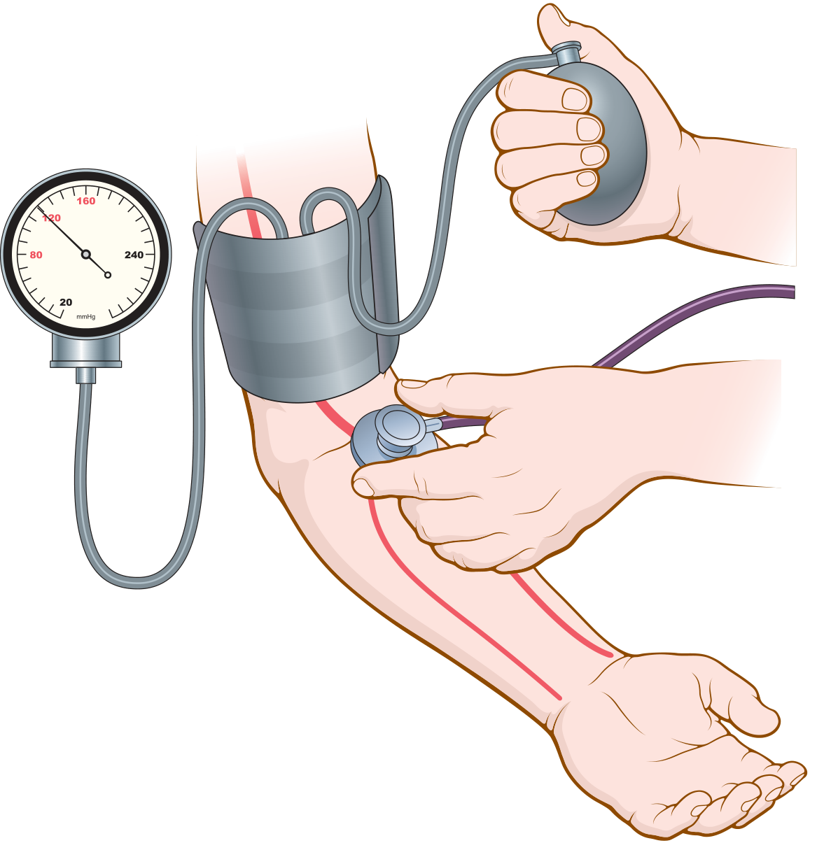
Korotkoff sounds arise from two simultaneous phenomena:
- Turbulent blood flow - When the cuff pressure is between systolic and diastolic BP, the cuff partially occludes the brachial artery. Blood jets through this narrowed segment at high velocity, generating turbulence distal to the cuff.
- Vessel wall vibration - The turbulent jet causes the arterial wall to oscillate, and these vibrations are transmitted to the stethoscope as sound.
Under normal conditions, laminar blood flow in wide-open arteries produces no turbulence, so no sounds are heard. It is the partial occlusion that creates the conditions for sound generation.
"Korotkoff sounds are believed to be caused mainly by blood jetting through the partly occluded vessel and by vibrations of the vessel wall. The jet causes turbulence in the vessel beyond the cuff, and this turbulence sets up the vibrations heard through the stethoscope."
- Guyton and Hall, p. 191
The Five Phases
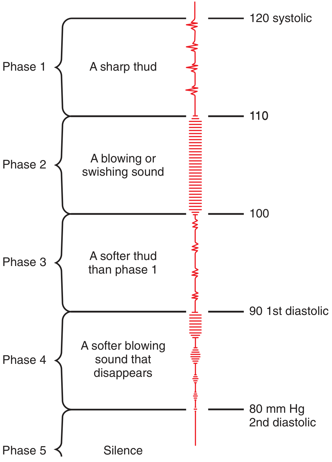
| Phase | Sound Character | Physiological Basis | Clinical Significance |
|---|---|---|---|
| I | Faint, clear, sharp tapping/thud that gradually increases | Cuff pressure just falls below systolic; blood first jets through with each heartbeat | = Systolic BP |
| II | Blowing or swishing murmur | Blood flows through partially compressed artery more freely; turbulence broadens | Auscultatory gap can occur here |
| III | Louder, crisper thud (softer than Phase I per some sources) | Artery opens more widely; flow increases | No clinical endpoint |
| IV | Sudden muffling - soft, blowing quality | Artery no longer closes fully in diastole; turbulence begins to diminish | = Diastolic BP in children & pregnancy (1st diastolic point ~90 mmHg) |
| V | Complete silence | Artery remains open throughout the cardiac cycle; laminar flow restored, no more turbulence | = Diastolic BP in adults (2nd diastolic point ~80 mmHg) |
- Roberts and Hedges' Clinical Procedures in Emergency Medicine, p. 41
Which Phase Marks Diastolic BP?
- Phase V (disappearance) is used for diastolic BP in most adults - this is the standard clinical endpoint.
- Phase IV (muffling) is used for diastolic BP in:
- Children
- Pregnant women
- Patients with aortic regurgitation - in AR, Korotkoff sounds can persist all the way to zero mmHg even though intra-arterial diastolic pressure rarely falls below 30 mmHg. Phase IV (muffling) correlates better with true diastolic pressure in these patients.
- States of high cardiac output (hyperdynamic circulation)
"In certain patients, the Korotkoff sounds do not disappear... SBP should be noted with the appearance of Korotkoff sounds (phase I) and DBP with the disappearance of sounds (phase V)."
- Washington Manual of Medical Therapeutics
"Korotkoff sounds often persist to zero even though the intra-arterial pressure rarely falls below 30 mm Hg. The point of change in Korotkoff sounds (i.e., the muffling of these sounds in phase IV) correlates with the diastolic pressure [in aortic regurgitation]."
- Braunwald's Heart Disease, 15th Ed.
Auscultatory Gap
- An auscultatory gap is a silent interval that may appear between Phase I and Phase II (or within Phase II) where sounds disappear and then reappear.
- It occurs most commonly in elderly hypertensive patients and those with atherosclerosis (stiff vessel walls alter sound transmission).
- If unrecognized, it can lead to false low readings for systolic BP.
Practical Technique Points
-
Stethoscope placed over the brachial artery at the antecubital fossa
-
Use the bell of the stethoscope (low-frequency sounds) with minimal pressure
-
Cuff deflated at 2-3 mmHg/second - too fast misses the transition points
-
Arm should be at the level of the right atrium - if the arm hangs dependently, readings are 9-14 mmHg higher
-
The cuff bladder should be placed over the artery, ~2.5 cm above the antecubital fossa
-
Roberts and Hedges' Clinical Procedures in Emergency Medicine, pp. 40-41
Limitations
-
Stiff arteries (atherosclerosis) can alter or dampen Korotkoff sounds, causing unreliable readings.
-
Obesity - thick arm tissue attenuates sounds.
-
Arrhythmias - irregular heartbeat makes phase identification inconsistent.
-
Very low cardiac output states (shock) - sounds may be absent or very faint.
-
In such cases, direct intra-arterial monitoring or Doppler methods are preferred.
-
Pfenninger and Fowler's Procedures for Primary Care, 3rd Ed.
This is a shared conversation. Sign in to Orris to start your own chat.