Labor analgesia for md anaesthesia exam . With possible table viva questions

Reading File
Finding Sources
Finding Sources
Finding Sources
Reading File
Reading File
Reading File
Reading File
Reading File
Reading File
Reading File
Finding Sources
Reading File
Reading File
Reading File
Finding Sources
Reading File
Finding Sources
Searching Images

labor pain pathways spinal levels dermatomal epidural analgesia

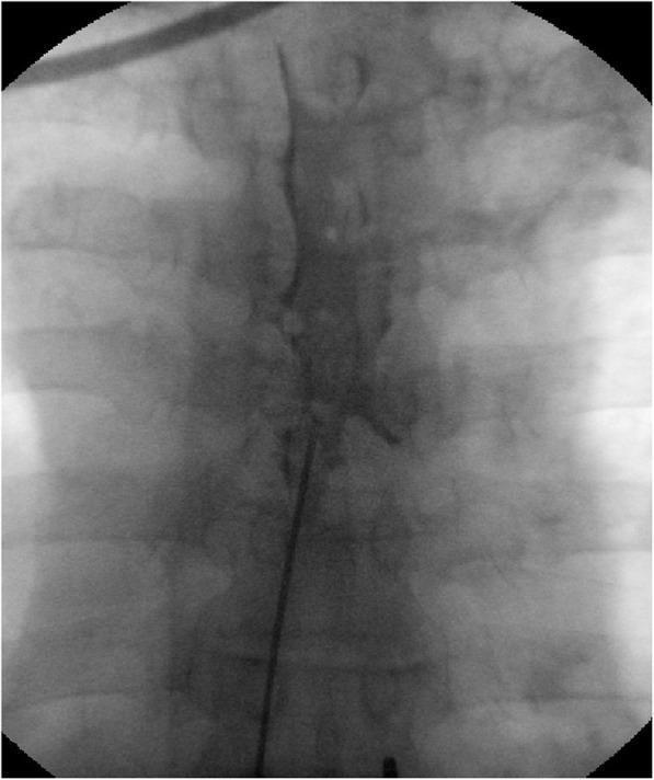
This diagnostic fluoroscopic image in an anteroposterior view demonstrates the confirmation of an epidural catheter placement for a continuous epidural block. The image shows a radiopaque epidural catheter positioned within the spinal canal, having entered via an interlaminar approach. A radiopaque contrast agent has been injected through the catheter, revealing a characteristic longitudinal and patchy distribution pattern within the epidural space. This contrast enhancement outlines the vertebral bodies and highlights the extent of the epidural spread across several vertebral levels. The visualization serves to verify the catheter's tip position and ensure adequate drug distribution for pain management, such as for herpes zoster-related neuralgia. Anatomical landmarks including the pedicles, vertebral bodies, and ribs are visible as less dense structures providing spatial context for the procedure. This clinical imaging is essential for ensuring correct dermatomal coverage during interventional pain procedures.

This diagnostic fluoroscopic image in an anteroposterior view demonstrates the confirmation of an epidural catheter placement for a continuous epidural block. The image shows a radiopaque epidural catheter positioned within the spinal canal, having entered via an interlaminar approach. A radiopaque contrast agent has been injected through the catheter, revealing a characteristic longitudinal and patchy distribution pattern within the epidural space. This contrast enhancement outlines the vertebral bodies and highlights the extent of the epidural spread across several vertebral levels. The visualization serves to verify the catheter's tip position and ensure adequate drug distribution for pain management, such as for herpes zoster-related neuralgia. Anatomical landmarks including the pedicles, vertebral bodies, and ribs are visible as less dense structures providing spatial context for the procedure. This clinical imaging is essential for ensuring correct dermatomal coverage during interventional pain procedures.

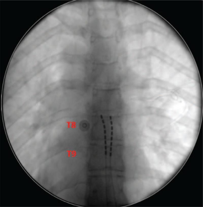
This diagnostic fluoroscopic image demonstrates the surgical placement of spinal cord stimulation (SCS) hardware. Two octopolar linear leads are visible as radiopaque, segmented lines positioned within the posterior epidural space, parallel to the midline of the thoracic vertebral column. The electrode contacts on each lead are clearly visible as discrete dark markings. The leads are centered at the level of the T8 and T9 vertebrae, which are annotated with red text labels. Anatomical landmarks include the thoracic vertebrae and the bilateral rib cage, providing a reference for the cephalocaudal positioning of the neurostimulation hardware. The image illustrates a common interventional pain management procedure designed for neural targeting to address chronic pain in the lower back and lower extremities through the modulation of dorsal column signals at specific dermatomal levels.

This diagnostic fluoroscopic image demonstrates the surgical placement of spinal cord stimulation (SCS) hardware. Two octopolar linear leads are visible as radiopaque, segmented lines positioned within the posterior epidural space, parallel to the midline of the thoracic vertebral column. The electrode contacts on each lead are clearly visible as discrete dark markings. The leads are centered at the level of the T8 and T9 vertebrae, which are annotated with red text labels. Anatomical landmarks include the thoracic vertebrae and the bilateral rib cage, providing a reference for the cephalocaudal positioning of the neurostimulation hardware. The image illustrates a common interventional pain management procedure designed for neural targeting to address chronic pain in the lower back and lower extremities through the modulation of dorsal column signals at specific dermatomal levels.

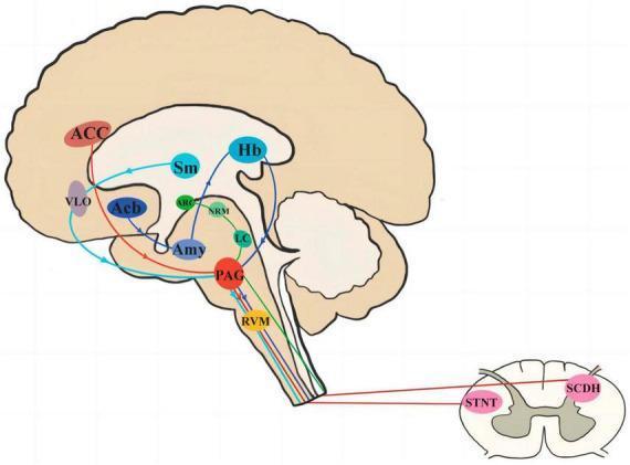
This medical diagram illustrates the pathophysiology and neural pathways of the periaqueductal gray (PAG) central to pain signal transduction and endogenous analgesia. Set against a sagittal brain outline and a cross-sectional spinal cord illustration, the diagram identifies four primary circuits. 1. The brainstem descending inhibitory system: including the anterior cingulate cortex (ACC), PAG, rostral ventral medulla (RVM), and spinal cord dorsal horn (SCDH)/spinal tract nucleus of the trigeminal nerve (STNT). 2. The midbrain limbic circuit: involving the nucleus accumbens (Acb), amygdala (Amy), and habenular nucleus (Hb). 3. The arcuate nucleus (ARC), nucleus raphe medius (NRM), and locus coeruleus (LC) descending pathway. 4. The thalamic loop: connecting the nucleus submedius (Sm) and ventrolateral orbital cortex (VLO) to the PAG. Color-coded directional arrows represent specific neurotransmission routes from higher cortical and subcortical regions through the midbrain and brainstem to the spinal cord. This infographic is designed for medical education regarding neuromodulation and pain management mechanisms.

This medical diagram illustrates the pathophysiology and neural pathways of the periaqueductal gray (PAG) central to pain signal transduction and endogenous analgesia. Set against a sagittal brain outline and a cross-sectional spinal cord illustration, the diagram identifies four primary circuits. 1. The brainstem descending inhibitory system: including the anterior cingulate cortex (ACC), PAG, rostral ventral medulla (RVM), and spinal cord dorsal horn (SCDH)/spinal tract nucleus of the trigeminal nerve (STNT). 2. The midbrain limbic circuit: involving the nucleus accumbens (Acb), amygdala (Amy), and habenular nucleus (Hb). 3. The arcuate nucleus (ARC), nucleus raphe medius (NRM), and locus coeruleus (LC) descending pathway. 4. The thalamic loop: connecting the nucleus submedius (Sm) and ventrolateral orbital cortex (VLO) to the PAG. Color-coded directional arrows represent specific neurotransmission routes from higher cortical and subcortical regions through the midbrain and brainstem to the spinal cord. This infographic is designed for medical education regarding neuromodulation and pain management mechanisms.

I now have all the information needed. Let me write a comprehensive MD exam-focused guide on labor analgesia.

Labor Analgesia - MD Anaesthesia Exam Guide

Sources: Miller's Anesthesia 10e, Barash's Clinical Anesthesia 9e, Morgan & Mikhail's Clinical Anesthesiology 7e

1. PAIN PATHWAYS IN LABOR

First Stage of Labor (Cervical dilation, uterine contractions)

  • Pain is visceral in nature
  • Carried by type C afferent fibers accompanying sympathetic nerves
  • Enter spinal cord at T10 - L1
  • Referred to lower abdomen, lumbar region, and thighs

Second Stage of Labor (Fetal descent, perineal distension)

  • Pain becomes somatic in addition to visceral
  • Perineal/vaginal vault distension carried by pudendal nerve (S2-S4)
  • Late first stage and second stage require coverage up to T10-S4

Key Viva Point:

Q: What dermatomal levels must be blocked for complete labor analgesia? A: First stage - T10 to L1; Second stage - T10 to S4 (pudendal component added)

2. METHODS OF LABOR ANALGESIA

Classification Table (High-yield for viva)

CategoryMethod
Non-pharmacologicLamaze, massage, TENS, hydrotherapy, acupuncture, hypnosis, doula support
SystemicOpioids (meperidine, fentanyl, morphine, remifentanil), ketamine, N2O
NeuraxialEpidural, spinal (single-shot), CSE, DPE
Regional blocksParacervical block, pudendal block, lumbar sympathetic block
InhalationEntonox (50% N2O + 50% O2)

3. NON-PHARMACOLOGIC METHODS

  • Lamaze method: breathing techniques + focused attention to reduce cortical pain perception
  • TENS: transcutaneous electrical nerve stimulation - mild efficacy
  • Continuous labor support (Doula): associated with shorter labor, fewer operative deliveries, less analgesic use (high-quality RCT data)
  • Hydrotherapy: Cochrane review - reduces pain in active labor, improves satisfaction
  • Acupuncture: may minimally reduce pain vs. sham; acupressure - no significant benefit
  • Massage: reduces pain in first stage only, not second/third stage
  • Evidence base for most non-pharmacologic techniques is limited by poor study design

4. SYSTEMIC OPIOIDS

Meperidine (Pethidine)

  • Historically most used; now rarely used due to side effects
  • IV: up to 50 mg; IM: 50-100 mg
  • Half-life: mother 2.5-3 h; normeperidine (active metabolite) 13-23 h
  • Normeperidine half-life 3x longer in fetus/neonate - risk of neurotoxicity, accumulation
  • Causes lower Apgar scores and prolonged neonatal respiratory depression

Morphine

  • Rarely used for labor pain
  • Active metabolite: morphine-6-glucuronide (long neonatal half-life)
  • Used IM by obstetricians for rest/sedation - onset 10-20 min, duration 4-6 h

Fentanyl

  • IV/PCIA (patient-controlled IV analgesia); short-acting, titratable
  • Remifentanil PCIA - gaining popularity; ultra-short acting (T½ ~3 min)
    • Dose: 0.1-0.4 mcg/kg per bolus, lockout 2 min
    • Requires continuous SpO2 monitoring (risk of maternal apnea)
  • All opioids cross the placenta; cause dose-related neonatal respiratory depression and decreased FHR variability

Ketamine

  • Low-dose IV (0.25-0.5 mg/kg): good adjunct for imminent delivery or when neuraxial contraindicated
  • Preserves airway reflexes; causes uterine stimulation (useful)
  • Avoid >1 mg/kg (neonatal depression, maternal dysphoria)

5. INHALATION ANALGESIA

Entonox (50% N2O + 50% O2)

  • Widely used in UK/Europe; self-administered via face mask/mouthpiece
  • Must inhale 30-60 seconds before peak of contraction
  • Advantages: rapid onset/offset, patient-controlled, no fetal depression at analgesic doses
  • Disadvantages: incomplete analgesia (~50% of patients), nausea, dizziness, occupational exposure concerns

6. NEURAXIAL ANALGESIA - THE GOLD STANDARD

6a. Epidural Analgesia

  • Most reliable and effective method for labor pain (large meta-analysis)
  • Level: L2-L3 or L3-L4 interspace (below conus medullaris)
  • Initial bolus: 10 mL of 0.0625-0.125% bupivacaine + fentanyl 50-100 mcg or sufentanil 10-20 mcg
  • Maintenance options:
    • Continuous infusion: 0.0625-0.125% bupivacaine + fentanyl 1-5 mcg/mL at 10 mL/h
    • PCEA (Patient-Controlled Epidural Analgesia): 5 mL bolus, 5-10 min lockout, 0-12 mL/h basal rate
    • PIEB (Programmed Intermittent Epidural Bolus): automated boluses > continuous infusion; better spread, less motor block, less breakthrough pain

6b. Spinal (Intrathecal) Analgesia - Single Shot

  • Fentanyl 15-25 mcg OR sufentanil 2-5 mcg ± bupivacaine 1.25-2.5 mg (plain)
  • Appropriate for multiparous patients with imminent delivery or when epidural catheter not in situ
  • Disadvantage: fixed duration; new anesthetic needed if cesarean required

6c. Combined Spinal-Epidural (CSE)

  • "Needle-through-needle" technique: epidural space identified → 25-27G pencil-point spinal needle through epidural needle → CSF confirmed → intrathecal drug injected → spinal needle withdrawn → epidural catheter inserted
  • Advantages:
    • Rapid onset (fastest of all neuraxial techniques)
    • Excellent early analgesia
    • Flexible duration via epidural catheter
    • Lower catheter failure rate vs. epidural alone
    • Less need for epidural top-up boluses
  • Disadvantages:
    • Cannot assess catheter function until spinal wears off
    • Higher incidence of pruritus
    • Higher incidence of fetal bradycardia (vs. epidural alone)
    • Failed catheter recognition may be delayed

6d. Dural Puncture Epidural (DPE) - Emerging Technique

  • Epidural space identified → spinal needle (25-26G) punctures dura but no drug injected intrathecally
  • Drug given epidurally; dural hole facilitates migration into intrathecal space
  • Advantages over standard epidural: faster onset (~2 min less), better sacral spread, fewer asymmetric blocks, fewer top-up requirements
  • No increase in PDPH vs. epidural; less pruritus and fetal bradycardia than CSE
  • 27G needle for DPE has no benefit (single study)

7. NEURAXIAL DRUGS - TABLE (HIGH-YIELD VIVA)

Spinal Opioid Doses

DrugIntrathecal DoseEpidural Dose
Morphine0.1-0.5 mg5 mg
Meperidine10-15 mg50-100 mg
Fentanyl10-25 mcg50-150 mcg
Sufentanil3-10 mcg10-20 mcg

Local Anesthetics

  • Bupivacaine 0.0625-0.125%: long duration, popular; cardiotoxic at higher doses
  • Ropivacaine 0.1-0.2%: preferred by many due to less cardiotoxicity; equipotent motor block to bupivacaine at equianalgesic doses
  • Both at ultra-dilute concentrations (0.0625%) allow "walking epidural" (mobile epidural) - no motor blockade

Adjuvants

AdjuvantMechanismNotes
Epinephrineα1 vasoconstriction → delays LA uptake; α2 → additional analgesiaUse 1:400,000-1:800,000; higher doses risk uterine artery vasoconstriction
Clonidineα2-agonistFDA warning - not recommended in obstetrics (hemodynamic instability)
DexmedetomidineHighly selective α2-agonistNot FDA approved for neuraxial use; studied as adjunct
Neostigmine (epidural)↑ Acetylcholine → spinal muscarinic/nicotinicReduces LA dose without N&V (intrathecal neostigmine causes unacceptable N&V)

8. ALTERNATIVE REGIONAL TECHNIQUES

Paracervical Block

  • Blocks uterine/cervical afferents during first stage only
  • 5-10 mL dilute LA injected submucosally into left and right lateral vaginal fornices
  • Fallen out of favor due to association with fetal bradycardia (fetal distress)

Pudendal Nerve Block

  • Pudendal nerve = S2-S4; supplies vaginal vault, perineum, rectum
  • Blocked transvaginally where it loops around ischial spine
  • 10 mL LA deposited behind each sacrospinous ligament
  • Used for: outlet forceps delivery, episiotomy repair
  • Does NOT block uterine contractions (first stage pain)

Lumbar Sympathetic Block

  • Paravertebral block at L2-L3 level
  • Alternative when neuraxial is contraindicated
  • Technically difficult; rarely used

9. IMPACT OF NEURAXIAL ANALGESIA ON LABOR PROGRESS

Key Points (Extremely Common Viva Topic)

Q: Does epidural analgesia increase cesarean delivery rate?
  • No. 2018 Cochrane review (33 studies, 10,350 women): epidural vs. non-epidural analgesia - no difference in cesarean delivery rate
  • Observational studies falsely showed association (confounding: women with dysfunctional labor/more pain are more likely to request epidural AND have higher CS rates)
  • Early epidural does NOT increase CS rate (multiple RCTs confirm)
Q: Does epidural prolong labor?
  • First stage: No significant prolongation (meta-analyses)
  • Second stage: Modest prolongation of ~15 minutes (meta-analyses/RCTs)
    • Due to: dense motor block impairing coordinated pushing; possible malposition of vertex
    • Modern dilute solutions minimize this
Q: Does timing of epidural placement matter?
  • Early neuraxial analgesia (even in latent phase, <4 cm) does NOT increase CS rate
  • Earlier placement may actually improve patient experience and benefit
Q: What is "laboring down"?
  • Technique where with epidural in situ, onset of second stage is allowed to progress passively (using uterine contractions to lower fetal station) before active pushing
  • Useful in patients with significant cardiac or vascular conditions (avoids Valsalva hemodynamic effects)
Q: ACOG definition of prolonged second stage with epidural?
  • Traditionally: >3 h in nulliparas with epidural; >2 h without
  • 2014 ACOG/SMFM consensus: no absolute maximum defined; longer durations acceptable individually

10. CONTRAINDICATIONS TO NEURAXIAL ANALGESIA

AbsoluteRelative
Patient refusalCoagulopathy (borderline)
Infection at siteThrombocytopenia (<70,000-80,000/mm³)
Uncorrected hypovolemia/hemorrhagic shockPrior back surgery
True LA allergySepticemia
Raised intracranial pressure (with posterior fossa lesion)Neurological disease
Low-molecular-weight heparin within 12 h (prophylactic) / 24 h (therapeutic)

11. COMPLICATIONS OF NEURAXIAL ANALGESIA

ComplicationDetails
HypotensionMost common; treat with IV fluids + vasopressor (ephedrine or phenylephrine); left uterine displacement
PDPH (Post-dural puncture headache)Postural, bilateral, frontal/occipital; incidence ~1% with epidural; treat with bed rest, hydration, caffeine, or blood patch
Total spinalInadvertent intrathecal injection of epidural dose; → apnea, hypotension, unconsciousness; manage ABCs
Local anesthetic systemic toxicity (LAST)Slow administration of dilute solutions; fractionate dose in 5 mL increments; treat with 20% intralipid
PruritusCommon with intrathecal/epidural opioids (especially morphine); treat with naloxone or ondansetron
Fetal bradycardiaEspecially with CSE (rapid decrease in catecholamines); manage with intrauterine resuscitation
Neurologic injuryRare; epidural hematoma, abscess, nerve damage
FeverEpidural-associated fever, especially in nulliparas with long labor

12. SPECIAL CLINICAL SCENARIOS (VIVA GOLD)

Q: Neuraxial analgesia in a parturient with severe pre-eclampsia?
  • Epidural preferred - blunts catecholamine surges during pain/contractions, maintains more stable BP
  • Check platelet count before placement
  • Avoid IM/IV opioids if possible (respiratory risk with MgSO4)
Q: Neuraxial analgesia in a parturient with cardiac disease (e.g., aortic stenosis)?
  • Pure intrathecal opioid technique is preferred (no sympathectomy, no hypotension)
  • Avoid large bolus neuraxial doses that cause precipitous sympathectomy
  • Low-concentration epidural with careful titration is second option
Q: When is general anesthesia needed for labor/vaginal delivery?
  • Uterine relaxation needed (e.g., breech extraction, delivery of second twin)
  • Absolute contraindication to neuraxial
  • Emergency where neuraxial cannot be placed in time
  • Rarely indicated
Q: Contraindications to epidural in patient on anticoagulants?
  • LMWH prophylactic dose: wait 12 h after last dose
  • LMWH therapeutic dose: wait 24 h after last dose
  • Unfractionated heparin: check PTT; acceptable after 4-6 h of last dose
  • Warfarin: check INR <1.5
Q: Why does CSE cause more fetal bradycardia?
  • Rapid and profound analgesia → sudden decrease in circulating catecholamines (especially epinephrine) → loss of β2-mediated uterine relaxation → uterine tetany → reduced uteroplacental flow → fetal bradycardia
  • Treat with: left lateral position, IV fluids, terbutaline (tocolytic), supplemental O2

13. PCEA vs. PIEB vs. CONTINUOUS INFUSION - TABLE

ParameterContinuous InfusionPCEAPIEB
Drug useModerateLessLeast
Motor blockModerateLessLeast
Breakthrough painModerateLessLeast
Patient satisfactionModerateBetterBest
Uniform spreadPoorModerateBest
Current preferenceOlder standardCommonPreferred at many centers

14. TEST DOSE FOR EPIDURAL

Standard Test Dose: 3 mL of 1.5% lidocaine + 1:200,000 epinephrine (15 mcg)
If injected intravascularlyTachycardia ≥20 bpm within 60 s (epinephrine response)
If injected intrathecallyDense motor + sensory block within 3-5 min (spinal LA dose)
  • Inject between contractions to avoid false positive tachycardia from pain
  • Lidocaine preferred over bupivacaine for test dose (less cardiotoxicity if intravascular)

15. QUICK VIVA Q&A BANK

QuestionAnswer
Gold standard for labor analgesiaNeuraxial (epidural)
Nerve fibers carrying first stage painC-fibers (visceral afferents)
Spinal levels - first stageT10-L1
Spinal levels - second stageT10-S4
Preferred LA for labor epiduralBupivacaine 0.0625-0.125% or Ropivacaine 0.1-0.2%
Why ropivacaine preferred over bupivacaineLess cardiotoxicity
Most common side effect of neuraxial analgesiaHypotension
Fentanyl dose intrathecal for labor10-25 mcg
Sufentanil dose intrathecal for labor3-10 mcg
CSE advantage over standard epiduralFaster onset + catheter flexibility
DPE advantage over standard epiduralBetter sacral spread, less asymmetric block, faster onset
Why epidural doesn't increase CS rateRCT/Cochrane evidence (confounders in old observational data)
Management of total spinalABCs: intubate, vasopressors (ephedrine/phenylephrine), L uterine displacement
PDPH treatmentConservative → epidural blood patch (gold standard)
Entonox ratio50% N2O + 50% O2
When to use intrathecal opioid aloneCardiac disease patients who can't tolerate sympathectomy
Paracervical block side effectFetal bradycardia (fetal distress)
Pudendal nerve block levelS2-S4, at ischial spine
Mobile/walking epidural0.0625% bupivacaine - no motor block
Meperidine dangerous metaboliteNormeperidine (T½ 13-23 h, neurotoxic, longer in neonate)
Most popular current epidural maintenance modePIEB (Programmed Intermittent Epidural Bolus)
Remifentanil PCIA advantageUltra-short T½ (~3 min), rapid titration
Epinephrine concentration in labor epidural1:400,000 to 1:800,000
Why is clonidine FDA-warned in obstetricsRisk of hemodynamic instability (hypotension, bradycardia)
Management of fetal bradycardia post-CSELLD, IV fluids, O2, terbutaline, consider delivery if persistent

16. MATERNAL BENEFITS OF EPIDURAL ANALGESIA (Beyond Pain Relief)

  1. Blunts sympathetic surges during contractions → prevents tachycardia, hypertension, and decreased uterine blood flow
  2. Reduces catecholamine secretion → may convert dysfunctional labor to normal
  3. Prevents maternal hyperventilation → avoids leftward shift of oxyhemoglobin curve → preserves fetal oxygenation
  4. Protects perineum → controlled fetal expulsion allows stretching rather than tearing
  5. Allows "laboring down" in cardiac patients
  6. Ready conversion to surgical anesthesia if CS needed (epidural catheter already in place)

Sources: Miller's Anesthesia 10e (pp. 8840-8860), Barash's Clinical Anesthesia 9e (pp. 3486-3494), Morgan & Mikhail's Clinical Anesthesiology 7e (pp. 1611-1618)

Discuss the perioperative management principles of patients with suspected morbidly adherent placenta . Placenta acreta spectrum . For md anaesthesia exam with possible viva questions .

Finding Sources
Finding Sources
Finding Sources
Reading File
Reading File
Reading File
Finding Sources
Reading File
Finding Sources
Finding Sources
Reading File
Finding Sources
Finding Sources
Finding Sources
Reading File
Searching Images

placenta accreta percreta histology ultrasound uterus

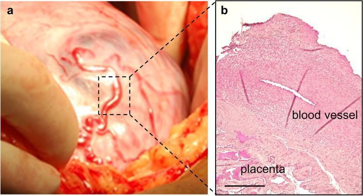
This composite image illustrates the clinical and histological features of Placenta Accreta Spectrum (PAS) and associated neoangiogenesis. Panel A is an intraoperative clinical photograph showing the anterior wall of a gravid uterus during a cesarean section. It demonstrates prominent, tortuous, newly formed epiplacental vessels characteristic of placenta percreta (FIGO grade 3a). A dashed box indicates the surgical sampling site for pathological analysis. Panel B is a corresponding histological diagnostic image showing a sagittal section of the sampled blood vessel and underlying placental tissue, stained with Weigert-Van Gieson (WVH). The histology reveals a thick muscular vessel wall; notably, the black-violet staining typically associated with internal and external elastic laminae is absent, suggesting abnormal vascular remodeling or degradation by trophoblast cells. Other visible components include yellow-stained cytoplasm/muscle cells and red-stained collagen fibers. The image serves as an educational resource for identifying surgical signs of invasive placentation and understanding the microscopic structural abnormalities of maternal-fetal neoangiogenesis.

This composite image illustrates the clinical and histological features of Placenta Accreta Spectrum (PAS) and associated neoangiogenesis. Panel A is an intraoperative clinical photograph showing the anterior wall of a gravid uterus during a cesarean section. It demonstrates prominent, tortuous, newly formed epiplacental vessels characteristic of placenta percreta (FIGO grade 3a). A dashed box indicates the surgical sampling site for pathological analysis. Panel B is a corresponding histological diagnostic image showing a sagittal section of the sampled blood vessel and underlying placental tissue, stained with Weigert-Van Gieson (WVH). The histology reveals a thick muscular vessel wall; notably, the black-violet staining typically associated with internal and external elastic laminae is absent, suggesting abnormal vascular remodeling or degradation by trophoblast cells. Other visible components include yellow-stained cytoplasm/muscle cells and red-stained collagen fibers. The image serves as an educational resource for identifying surgical signs of invasive placentation and understanding the microscopic structural abnormalities of maternal-fetal neoangiogenesis.

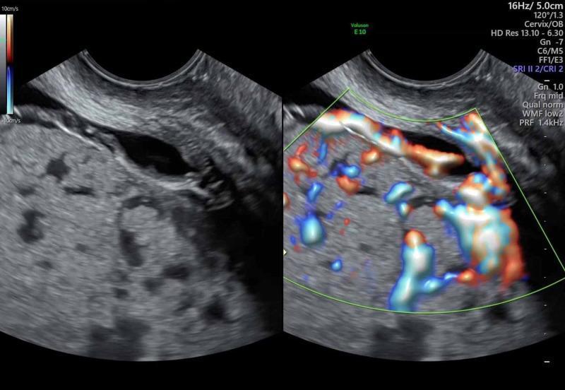
A side-by-side diagnostic ultrasound image illustrating signs of placenta percreta in a patient with placenta previa. The left panel shows a gray-scale transabdominal ultrasound of the pelvic region, revealing a heterogeneous placenta with multiple hypoechoic spaces (placental lacunae) and an indistinct retroplacental clear zone. The interface between the uterus and the posterior wall of the urinary bladder appears irregular and disrupted. The right panel displays a color Doppler ultrasound mapping significant neovascularization. It shows hypervascularity with engorged, tortuous vessels exhibiting turbulent flow (mixed red and blue signals) extending along the junction of the placenta and the uterine wall, with apparent protrusion into the bladder wall. These features are highly suggestive of placenta accreta spectrum (PAS), specifically placenta percreta, demonstrating abnormal placental invasion beyond the serosa. The image is intended for advanced obstetric and radiologic education regarding high-risk pregnancy complications.

A side-by-side diagnostic ultrasound image illustrating signs of placenta percreta in a patient with placenta previa. The left panel shows a gray-scale transabdominal ultrasound of the pelvic region, revealing a heterogeneous placenta with multiple hypoechoic spaces (placental lacunae) and an indistinct retroplacental clear zone. The interface between the uterus and the posterior wall of the urinary bladder appears irregular and disrupted. The right panel displays a color Doppler ultrasound mapping significant neovascularization. It shows hypervascularity with engorged, tortuous vessels exhibiting turbulent flow (mixed red and blue signals) extending along the junction of the placenta and the uterine wall, with apparent protrusion into the bladder wall. These features are highly suggestive of placenta accreta spectrum (PAS), specifically placenta percreta, demonstrating abnormal placental invasion beyond the serosa. The image is intended for advanced obstetric and radiologic education regarding high-risk pregnancy complications.

I now have comprehensive content from the textbooks. Let me compile the complete exam-focused guide.

Placenta Accreta Spectrum (PAS) / Morbidly Adherent Placenta

Perioperative Management - MD Anaesthesia Exam Guide

Sources: Creasy & Resnik's Maternal-Fetal Medicine, Barash's Clinical Anesthesia 9e, Miller's Anesthesia 10e

1. DEFINITION AND CLASSIFICATION

Morbidly Adherent Placenta (MAP) = umbrella term encompassing the entire PAS
Abnormal trophoblastic invasion beyond the Nitabuch fibrinoid layer (the normal boundary between placenta and decidua basalis):
TermDepth of InvasionDescription
Placenta accretaMyometrium (superficial)Trophoblast directly attached to myometrium; no intervening decidua; most common (~75-78%)
Placenta incretaInto myometriumInvasion into the myometrial muscle (~17%)
Placenta percretaThrough serosa/beyondInvasion through uterine serosa; may involve bladder, bowel, pelvic vessels; most dangerous (~5-7%)
Placenta percreta - intraoperative tortuous epiplacental vessels and histology
Intraoperative view of placenta percreta showing tortuous epiplacental neovascularization, with corresponding histology demonstrating abnormal vascular remodeling
Ultrasound features of placenta percreta - lacunae and Doppler hypervascularity
Color Doppler ultrasound showing placental lacunae, absent retroplacental clear zone, and turbulent hypervascularity at the utero-bladder interface - hallmarks of PAS

2. RISK FACTORS

Primary risk factors:
  • Placenta previa + prior cesarean delivery = the most important combination
  • Risk of accreta with previa:
    • 0 prior CS: ~3-5%
    • 1 prior CS: ~11-25%
    • 2 prior CS: ~35-47%
    • ≥3 prior CS: >60%
Other risk factors:
  • Prior uterine surgery (myomectomy, endometrial ablation, curettage, pelvic radiation)
  • In vitro fertilization (IVF)
  • Short interpregnancy interval
  • Advanced maternal age
  • Smoking and multiparity
Viva Q: What is the most important risk factor for PAS? A: The combination of placenta previa + prior cesarean delivery. Risk exceeds 60% with previa and ≥3 cesareans.

3. DIAGNOSIS

Ultrasound (First-line, sensitivity ~80-90%; expert-blinded studies show ~55%)

  • Loss of normal hypoechoic retroplacental boundary between placenta and bladder
  • Intraplacental sonolucent spaces (lacunae) - "Swiss cheese" appearance
  • Color Doppler: turbulent flow in lacunar spaces, bridging vessels into placental tissue
  • Obliteration of well-delineated bladder wall (in percreta)
  • Can be diagnosed as early as first trimester, especially with cesarean scar ectopic

MRI

  • Used when ultrasound is inconclusive
  • No risk from MRI without gadolinium (studies including first trimester show no adverse outcomes)
  • Better soft tissue resolution for posterior placentas and determining depth of invasion
Viva Q: Why might ultrasound miss PAS? A: Considerable variation among experienced clinicians; features of accreta can be present in normal placentas; posterior placentas are particularly difficult to image. Sensitivity only ~55% in blinded expert studies.

4. ANTEPARTUM PLANNING - THE MULTIDISCIPLINARY TEAM (MDT)

This is the cornerstone of management. Outcomes are markedly better when PAS is diagnosed before delivery and managed by a multidisciplinary team at a referral center ("center of excellence").

MDT Conference - Schedule before 34 weeks' gestation

Attendees must include:
  • Obstetric Anaesthesiologist
  • Maternal-Fetal Medicine (MFM) specialist
  • Obstetric/Gynaecologic surgeon (Gyn oncology, pelvic surgery)
  • Urologist (bladder involvement in percreta)
  • Vascular surgeon / General surgeon
  • Interventional Radiologist
  • Blood bank / Haematologist
  • Neonatologist
  • Dedicated Nursing team (ICU/OR)

Conference Agenda:

  • Timing and location of delivery
  • Personnel required
  • Blood bank strategy (massive transfusion protocol, cell salvage)
  • Anesthesia plan (neuraxial vs. GA, conversion strategy)
  • Other medical issues (fetal lung maturity steroids, anticoagulation)
  • Patient counseling (including risk of hysterectomy, loss of fertility)
Anesthesiologist's specific antepartum role:
  • Meet patient at an antepartum visit
  • Discuss anesthesia plan and answer questions
  • Counsel specifically about: possible conversion from neuraxial to GA if major hemorrhage occurs
  • Discuss consent for invasive monitoring, possible blood transfusion, ICU admission

5. TIMING OF DELIVERY

  • Planned delivery at 34-35 weeks' gestation (after corticosteroids for fetal lung maturity)
  • This represents the optimal balance between:
    • Avoiding catastrophic emergency hemorrhage (risk increases with advancing gestation)
    • Neonatal morbidity from prematurity
  • Delivery is performed without amniocentesis for fetal lung maturity confirmation
  • Emergency delivery involves more blood loss than elective planned surgery
Viva Q: Why deliver at 34-35 weeks and not later? A: To avoid uncontrolled emergency surgery from spontaneous labor or hemorrhage; elective surgery reduces blood loss, transfusion requirements, and allows optimal team assembly. Maternal morbidity is significantly reduced with planned delivery.

6. PREOPERATIVE PREPARATION

6a. Investigations

  • Full blood count, coagulation profile (PT, aPTT, fibrinogen)
  • Group and crossmatch - minimum 4-6 units pRBC crossmatched (often more)
  • Renal and liver function
  • Clotting factors
  • Baseline arterial blood gas (if time permits)

6b. Blood Preparation

  • Massive Transfusion Protocol (MTP) activated and on standby
    • Typical ratio: pRBCs : FFP : Platelets = 1:1:1 (damage control resuscitation principle)
  • Pre-warmed blood products available in the room
  • Cryoprecipitate available (for fibrinogen replacement)
  • Tranexamic acid (TXA) - 1g IV at induction, repeat at 3 h or if ongoing bleeding
  • Cross-matched pRBCs in OR before knife-to-skin

6c. Intraoperative Cell Salvage (IOBS)

  • Should be set up and ready for all PAS cases (high-level recommendation)
  • Modern cell savers with leukodepletion filters safely remove amniotic fluid debris, fetal cells, immune mediators
  • Eliminates historical concern about amniotic fluid embolism (AFE) with cell salvage
  • Fetal red cells in salvaged blood may potentiate alloimmunization, but this risk already exists during delivery
  • Only absolute contraindications: microbial contamination of field or malignancy (tumor rupture)
  • Particularly important for Jehovah's Witness patients (kept in continuous circuit = often acceptable)
Viva Q: Is cell salvage safe in obstetric surgery? A: Yes, with leukodepletion filters. Reviews of cell salvage in obstetrics found no serious maternal complications. It is specifically recommended for planned cesarean hysterectomy and PAS cases. Leukodepletion filters remove amniotic fluid components and inflammatory mediators.

6d. Vascular Access and Monitoring (Setup before induction)

  • 2 large-bore IV cannulae (14-16G)
  • Arterial line (radial or femoral) - essential for:
    • Beat-to-beat BP monitoring during rapid hemorrhage
    • Frequent arterial blood gas sampling
    • Coagulation monitoring (TEG/ROTEM guided therapy)
  • Central venous catheter (CVC) or large-bore introducer sheath
    • For CVP monitoring, vasopressor/inotrope infusion, rapid transfusion
  • Consider pulmonary artery catheter or advanced cardiac output monitoring in patients with cardiac disease
  • Temperature monitoring - forced air warming, fluid warmer essential
  • Urinary catheter - to monitor UO and detect bladder involvement intraoperatively
  • Thromboelastography (TEG) or ROTEM - point-of-care coagulation monitoring to guide ratio-based transfusion and factor replacement

6e. Interventional Radiology - Prophylactic Balloon Catheters

  • Pre-operative placement of balloon-tipped catheters in the internal iliac arteries (or aorta/common iliac arteries) via the femoral arteries
  • Inflated during uterine/lower segment dissection to reduce blood flow to the operative field
  • Controversially debated: published evidence shows variable benefit; some series show no proven reduction in blood loss; embolic complications reported
  • A comprehensive literature review concluded larger RCTs are needed
  • Current status: used selectively at centers with IR expertise; timing of deployment during surgery has been inconsistent
  • Alternative: pre-operative uterine artery embolization (UAE) post-delivery in conservative management

7. ANESTHETIC MANAGEMENT

7a. Choice of Anesthesia - Key Controversy

Recommended approach: Combined Spinal-Epidural (CSE) or Epidural
  • Allows patient to be awake during fetal delivery
  • Partner can be present at birth
  • Flexible - block can be extended as long as needed
  • Epidural catheter preserved for postoperative analgesia
  • Review of 350 cases of placenta previa: neuraxial anesthesia associated with reduced blood loss and reduced transfusion requirement compared to GA
When to convert to General Anesthesia:
  • Major hemorrhage - convert early, before massive fluid resuscitation causes airway oedema
  • Hemodynamic instability not responsive to vasopressors
  • If neuraxial block is inadequate for prolonged surgery
  • Emergency presentation with no time for neuraxial
  • Patient refusal
Critical Viva Q: Should GA or regional anesthesia be used for PAS? A: No single technique is mandated. CSE is preferred at many centers - allows awake delivery, partner presence, and flexibility. Neuraxial anesthesia has been associated with less blood loss. GA is used when: major hemorrhage occurs, neuraxial is inadequate, or patient refuses. If neuraxial is planned, CSE or epidural (not single-shot spinal) must be used so the block duration can be extended. Convert to GA early if hemorrhage occurs, before airway oedema from massive fluids develops.

7b. General Anesthesia - RSI for PAS

Indications for primary GA in PAS:
  • Emergency surgery
  • Neuraxial contraindicated (coagulopathy, patient refusal, severe hemorrhage)
  • Anticipated need for prolonged complex pelvic surgery from the start
RSI Protocol:
  • Preoxygenation (3-5 min or 4 vital capacity breaths of 100% O2)
  • Rapid Sequence Induction (RSI) - cricoid pressure + RSI drugs
    • Propofol/Thiopentone + Succinylcholine 1.5 mg/kg (or rocuronium 1.2 mg/kg if sugammadex available)
  • Video laryngoscopy preferred (difficult airway anticipated due to pregnancy + potential airway oedema if hemorrhage)
  • Maintenance: volatile agent (desflurane/sevoflurane) + opioids + muscle relaxant
  • Avoid nitrous oxide if cell salvage in use (fire hazard concerns) or if bowel distension is a concern

7c. Intraoperative Drug Management

DrugRoleNote
OxytocinUterotonic after cord clamping20-50 U/L IV infusion; avoid IV boluses (hypotension)
MethylergonovineUterotonic0.2 mg IM; avoid in hypertension, preeclampsia, cardiac disease
Carboprost (PGF2α)Uterotonic0.25 mg IM; avoid in asthma, pulmonary HTN
MisoprostolUterotonic200-800 mcg rectal/vaginal/buccal; minimal side effects
Tranexamic acid (TXA)Antifibrinolytic1g IV at induction; repeat 3h or with ongoing hemorrhage
VasopressorsBP supportPhenylephrine or noradrenaline infusion for neuraxial-induced hypotension or haemorrhagic shock
Calcium chlorideIonized hypocalcaemia from massive transfusion1g IV with each 4 units pRBC

7d. Surgical Strategy

Standard recommended surgical approach:
  1. Fundal/classical uterine incision avoiding the placenta (not low transverse)
  2. Deliver fetus through uterine incision
  3. Clamp and cut the cord immediately
  4. Leave the placenta in situ - do NOT attempt manual removal (causes catastrophic hemorrhage)
  5. Proceed directly to total abdominal hysterectomy (TAH)
  6. This requires complex technique - wide resection of lower uterine segment and parametrium, possibly bladder resection (if percreta)
Viva Q: Why is the incision made fundally in PAS? A: To avoid cutting through the abnormally vascularized lower segment/placenta and causing uncontrollable hemorrhage before the fetus is delivered. A classical or fundal incision avoids the placenta entirely.
Viva Q: Why is the placenta left in situ and not manually removed? A: Attempting manual removal of a morbidly adherent placenta tears the placental tissue from the myometrium, causing massive, uncontrollable hemorrhage. The standard of care is to leave it in place and proceed to hysterectomy with the placenta still attached.

8. CONSERVATIVE MANAGEMENT (Non-hysterectomy Options)

Indications (carefully selected cases only):

  • Strong desire to preserve fertility
  • Suspicion of small focal accreta
  • Fundal location (post-myomectomy or classical CS)
  • Posterior implantation

Method:

  • Deliver via fundal hysterotomy
  • Leave placenta entirely in situ
  • Close uterus
  • Post-delivery uterine artery embolization (UAE)
  • Follow-up with serial US/MRI for placental resorption

Risks of Conservative Management:

  • 18/167 (11%) required primary hysterectomy for intraoperative bleeding in one series
  • Further 18/167 required delayed hysterectomy
  • Severe morbidity in 10/167 (~6%)
  • One death from methotrexate complications
Viva Q: Is methotrexate used for conservative management of PAS? A: No. Methotrexate is unequivocally not advised - it has not been shown to be efficacious and has caused serious complications including one death. It should not be given.

9. HEMORRHAGE MANAGEMENT - INTRAOPERATIVE

Massive Transfusion Protocol (MTP) Principles

Trigger: Clinical hemorrhage + hemodynamic instability OR anticipated blood loss >10% blood volume
Damage Control Resuscitation:
  • pRBC : FFP : Platelets = 1:1:1 ratio
  • Avoid large-volume crystalloid (worsens coagulopathy, oedema)
  • Target: Hb >8 g/dL, Platelets >50,000, PT/INR <1.5, fibrinogen >2 g/L
Point-of-care guidance (TEG/ROTEM):
  • Guides targeted factor replacement
  • Identifies specific coagulopathy pattern: dilutional, fibrinolytic, or DIC
Fibrinogen replacement:
  • Cryoprecipitate (10 units) → raises fibrinogen by ~1 g/L
  • Fibrinogen concentrate (4g) → more rapidly available
Recombinant Factor VIIa (rFVIIa):
  • Last resort for life-threatening uncontrolled hemorrhage
  • Dose: 90 mcg/kg IV
  • Works only in acidosis-corrected, normothermic, normocalcaemic patient (the "lethal triad" must be addressed first)

The "Lethal Triad" to Prevent:

ComponentTreatment
Hypothermia (<35°C)Forced air warming, warmed IV fluids, warm OR environment
Acidosis (pH <7.2)Adequate resuscitation, vasopressors, bicarbonate if severe
CoagulopathyMTP with 1:1:1 ratio, TXA, cryoprecipitate, TEG-guided

10. POSTOPERATIVE CARE

  • ICU admission is routine after cesarean hysterectomy for PAS
  • Continue invasive monitoring (arterial line, CVC) for 24-48 h
  • Monitor for:
    • Ongoing hemorrhage (drain output, peritoneal collections)
    • DIC - serial coagulation testing
    • Acute kidney injury from hypoperfusion (especially if bladder involved in percreta)
    • Ileus, bowel injury (if percreta)
    • Wound complications
  • Thromboprophylaxis:
    • High VTE risk post-caesarean hysterectomy
    • LMWH when hemostasis assured (typically 12-24 h post-op)
    • TED stockings / intermittent pneumatic compression from day 1
  • Analgesia:
    • Epidural catheter (if neuraxial used) for 24-48 h postoperative analgesia
    • If GA used: multimodal - paracetamol + NSAIDs (when hemostasis secure) + opioid PCA
  • Psychological support:
    • Loss of uterus (if hysterectomy performed) is a major psychological event
    • Debrief with patient and partner; involve counseling services

11. SPECIAL SITUATIONS

Jehovah's Witness with PAS

  • Extensive counseling - with family AND patient alone
  • Determine exactly which products are acceptable (spectrum ranges from no blood products to some derivatives)
  • Cell salvage in continuous circuit (blood never leaves the body circuit) - often acceptable
  • Acute normovolaemic haemodilution (ANH): pre-op collection, re-infusion from continuous circuit
  • Erythropoietin + IV iron pre-operatively to optimize preoperative Hb
  • Antifibrinolytics (TXA) + surgical hemostatic measures essential
  • Document refusal and capacity clearly; seek legal advice if life-threatening

Undiagnosed PAS (Intraoperative Surprise)

  • Surgeon attempts placental removal → hemorrhage
  • Immediately: call for senior anesthesiologist and obstetric surgeon, activate MTP
  • Place large-bore IV access if not already done
  • Arterial line ASAP
  • Convert to GA if neuraxial in place
  • Consider bimanual uterine compression, balloon tamponade
  • Proceed to hysterectomy if hemorrhage uncontrollable
  • Outcomes significantly worse than planned surgery

12. VIVA QUESTION BANK

QuestionAnswer
Define placenta accreta, increta, percretaAccreta: trophoblast invades to myometrium (no Nitabuch layer). Increta: into myometrium. Percreta: through serosa (and may involve bladder/bowel/vessels)
Nitabuch layerFibrinoid layer at the decidua-trophoblast junction; absent in PAS
Most common type of PASPlacenta accreta (~75-78%)
Most important risk factor for PASPlacenta previa + prior cesarean delivery (>60% risk with ≥3 CS + previa)
When should MDT conference occur?Before 34 weeks' gestation
Who should be in the MDT?MFM, Anaesthesiologist, Gyn oncologist, Urologist, Vascular surgeon, IR, Blood bank, Neonatologist
Optimal timing of planned delivery34-35 weeks (after corticosteroids); without amniocentesis
Why deliver electively and early?Planned surgery has less blood loss and better outcomes vs emergency surgery
Why is early diagnosis important?Reduces blood loss, transfusion requirements; allows planned MDT delivery
Preferred anesthetic techniqueCSE or epidural (neuraxial) - reduces blood loss, allows awake delivery; convert to GA if major hemorrhage
When to convert to GA urgently?Major hemorrhage - EARLY, before airway oedema from fluid resuscitation
Why fundal uterine incision?Avoids cutting through the highly vascular lower segment/placenta
Why leave placenta in situ?Manual removal causes catastrophic, uncontrollable hemorrhage
What is the standard surgical treatment?Cesarean delivery + total abdominal hysterectomy (with placenta in situ)
Role of balloon catheter IR?Prophylactic internal iliac balloon occlusion during surgery; controversial evidence - no proven consistent benefit, embolic complications reported
Is cell salvage safe in obstetrics?Yes, with leukodepletion filters; specifically recommended for planned cesarean hysterectomy
Contraindications to cell salvageMicrobial contamination, malignancy with tumor rupture
Lethal triad in massive hemorrhageHypothermia + Acidosis + Coagulopathy
MTP ratiopRBC : FFP : Platelets = 1:1:1
Role of TXAAntifibrinolytic; 1g IV at induction, repeat at 3h or with ongoing bleeding
Fibrinogen trigger for replacement<2 g/L; replace with cryoprecipitate or fibrinogen concentrate
Role of rFVIIaLast resort for uncontrolled hemorrhage; correct lethal triad first
Is methotrexate used for conservative management?No - unequivocally not advised; no proven benefit, causes serious morbidity/mortality
Conservative management candidatesSelected: desire fertility, small focal accreta, fundal location, posterior placenta
VTE risk post-CS hysterectomyVery high; LMWH + stockings + IPC from day 1, resume when hemostasis secure
Why does percreta involve bladder?Invasion through uterine serosa into adjacent structures; bladder lies immediately anterior to lower uterine segment
Why is posterior PAS harder to manage?Less accessible surgically; harder to visualize on ultrasound
Calcium in massive transfusion1g CaCl2 IV per 4 units pRBC; citrate in blood products chelates ionized calcium
Blood loss in PAS surgeryAverages 3-5 liters in planned; can be >10 L in emergency/undiagnosed
ICU post-CS hysterectomyRoutine - for monitoring, analgesia, hemodynamic support

13. SUMMARY CHECKLIST FOR ANAESTHETIST

Pre-op:
  • MDT conference attended, plan documented
  • Patient counseled (anesthesia options, risks, consent for transfusion, ICU)
  • Blood products crossmatched and available in OR
  • MTP activated/on standby
  • Cell salvage machine set up with leukodepletion filter
  • Arterial line, 2x large-bore IV, CVC plan
  • TEG/ROTEM available
  • Video laryngoscope checked and ready (difficult airway kit)
  • Fluid/blood warmers ready, OR temperature raised
  • Vasopressor infusions drawn up
Intra-op:
  • Regional first (CSE/epidural) if planned and stable
  • Arterial line before induction (all cases)
  • Convert to GA early if hemorrhage (before airway oedema)
  • TXA given at induction
  • TEG-guided transfusion
  • Monitor for lethal triad; correct actively
  • Calcium replacement with blood products
Post-op:
  • ICU admission
  • Ongoing coagulation/hemorrhage monitoring
  • VTE prophylaxis when hemostasis secure
  • Psychological debrief and support

Sources: Creasy & Resnik's Maternal-Fetal Medicine (pp. 1021-1022), Barash's Clinical Anesthesia 9e (pp. 1733-1734), Barash Transfusion chapter (p. 1334)
This is a shared conversation. Sign in to Orris to start your own chat.