
Hemithorax meaning in chest x ray
chest radiograph opacified hemithorax
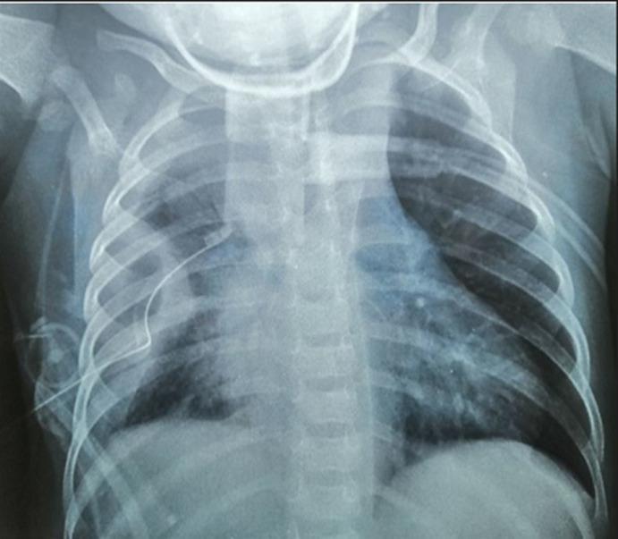
Anteroposterior (AP) pediatric chest radiograph demonstrating complex pleural and mediastinal pathology. The right hemithorax shows a large, opacified region consistent with a septated or loculated pleurisy (pleural empyema). Visible within the right pleural space is a radiopaque thoracic drain (chest tube), appropriately positioned for fluid evacuation. The mediastinum is significantly widened, indicating mediastinal enlargement or possible mediastinitis, causing a mass effect with displacement of the heart and mediastinal structures toward the left hemithorax. The underlying lung parenchyma on the right shows areas of increased density, suggesting associated pulmonary condensation or pneumonia. This diagnostic image illustrates the radiological appearance of complicated pleural effusion and mediastinal involvement in a pediatric patient, likely following clinical intervention for empyema.
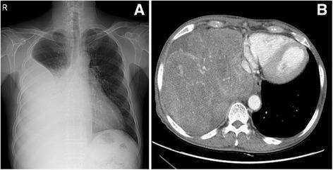
This figure presents two diagnostic imaging modalities of the chest in a patient with Doege-Potter syndrome, demonstrating a massive intrathoracic tumor. Image (A) is a posteroanterior chest radiograph showing a large, dense, opacified mass shadow occupying the middle and lower zones of the right hemithorax, causing obscuration of the right cardiac border and hemidiaphragm. Image (B) is an axial contrast-enhanced computed tomography (CT) scan at the level of the lower lobes, revealing a large, lobulated, heterogeneously enhancing mass in the right hemithorax. The mass occupies a significant portion of the right thoracic cavity, displacing the mediastinal structures toward the left. The heterogeneity on CT suggests areas of variable vascularity, internal necrosis, or fibrous tissue within the tumor. These findings are consistent with a solitary fibrous tumor (SFT) of the pleura, a mesenchymal neoplasm that can grow to a massive size and occasionally present with paraneoplastic hypoglycemia.
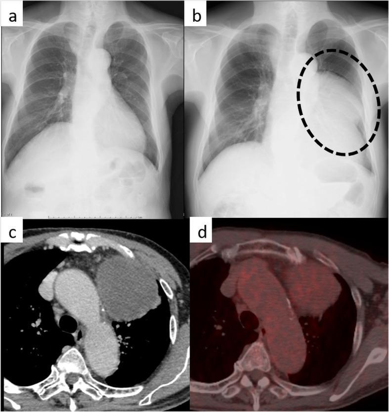
This composite figure presents multi-modal clinical imaging of a large mediastinal mass. Panel (a) is a baseline posterior-anterior (PA) chest radiograph showing a normal mediastinal contour. Panel (b) is a follow-up chest radiograph from one year later, revealing a giant, well-demarcated opacified mass (indicated by a black dotted circle) occupying the left mid-to-lower hemithorax and obliterating the left cardiac border. Panel (c) provides a chest-enhanced axial computed tomography (CT) scan, demonstrating a well-circumscribed, approximately 11-cm homogeneous mass in the left anterior mediastinum. The mass displays a CT density of approximately 40 Hounsfield units, indicating a solid soft-tissue composition without visible fat components. Panel (d) shows a fused fluorodeoxyglucose-positron emission tomography (FDG-PET) scan, illustrating mild-to-moderate radiopharmaceutical uptake throughout the tumor volume (SUVmax of 3.3). Collectively, these images illustrate the rapid progression and diagnostic workup of an anterior mediastinal tumor, later pathologically identified as a well-differentiated liposarcoma (sclerosing variant).
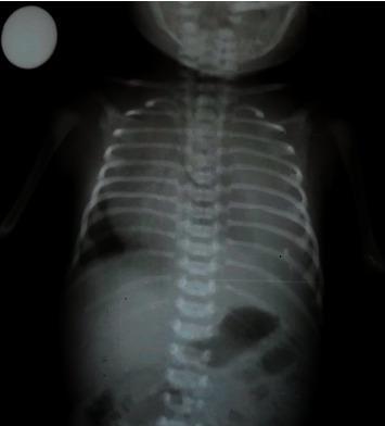
This anteroposterior (AP) chest radiograph of a neonate demonstrates a massive opacification of the left hemithorax. The dense, homogenous opacity obscures the left lung parenchyma, the cardiac border, and the left hemidiaphragm, consistent with a large-volume pleural effusion. There is evidence of significant mass effect, as indicated by a rightward mediastinal shift of the heart and trachea away from the opacified side. In contrast, the right hemithorax shows clear lung fields with normal vascular markings and a sharp diaphragmatic contour. The skeletal structures, including the infant's ribs and spine, are visible, showing normal ossification for the developmental age. Below the diaphragm, a normal neonatal bowel gas pattern is present in the upper abdomen. A radiopaque circular marker is noted in the upper left corner of the film. This clinical finding is characteristic of congenital chylothorax in a newborn presenting with respiratory distress.
"opacified hemithorax" OR "hemithorax" radiograph
- One lung, plus its pleural space, the hemidiaphragm and the chest wall on that side.
- The mediastinum sits between the two hemithoraces. The trachea and heart can be shifted toward or away from a given hemithorax.
- Opacified (or "white-out") hemithorax: the whole side looks white. Grainger & Allison's Diagnostic Radiology says whole-lung collapse gives complete opacification of the affected hemithorax. The usual cause in adults is an obstructing tumour in a main bronchus or a mucous plug.
- Large pleural effusion: an effusion over about 300 mL causes blunting of the costophrenic angle. If it is large enough, the whole hemithorax can be obscured (Fischer's Mastery of Surgery, 8th ed). Rosen's Emergency Medicine notes that on a supine film a large effusion may show only a diffuse haziness of the hemithorax.
- Hyperexpanded or hyperlucent hemithorax: this side looks larger and darker. Examples are congenital lobar emphysema and a large pneumothorax (Schwartz's Principles of Surgery, 11th ed).
- Small or contracted hemithorax: loss of volume from collapse, fibrosis, pneumonectomy or pleural thickening.
| Mediastinal position | Likely cause |
|---|---|
| Shifted toward the opaque side | Lung collapse (e.g. obstructing lesion, mucous plug), or pneumonectomy |
| Shifted away from the opaque side | Large pleural effusion or a large mass |
| Midline, not shifted | Consolidation, or a mix of effusion and collapse |