
Explain this topic and make me understand and memorise it in one hour
cd /tmp/workspace/attachments/3994cc3e-5910-4eb6-80d8-bf94d4b9a0b2/ && python3 -c " import pdfplumber with pdfplumber.open('Shock but modified .pdf') as pdf: print(f'Total pages: {len(pdf.pages)}') for i, page in enumerate(pdf.pages): text = page.extract_text() print(f'--- PAGE {i+1} ---') print(text[:3000] if text else '[No text]') print() "
python3 -c " from pypdf import PdfReader reader = PdfReader('/tmp/workspace/attachments/3994cc3e-5910-4eb6-80d8-bf94d4b9a0b2/Shock but modified .pdf') print(f'Pages: {len(reader.pages)}') # Check for images on first page page = reader.pages[0] print('Page keys:', page.keys()) if '/Resources' in page: res = page['/Resources'] print('Resources:', res.keys() if hasattr(res, 'keys') else res) "
python3 -c " from pypdf import PdfReader import base64, json reader = PdfReader('/tmp/workspace/attachments/3994cc3e-5910-4eb6-80d8-bf94d4b9a0b2/Shock but modified .pdf') page = reader.pages[0] xobj = page['/Resources']['/XObject'] print('XObjects on page 1:', list(xobj.keys())) for k in list(xobj.keys())[:3]: obj = xobj[k] print(f' {k}: subtype={obj.get(\"/Subtype\")}, keys={list(obj.keys())[:8]}') "
python3 << 'EOF' from pypdf import PdfReader from PIL import Image import io, os reader = PdfReader('/tmp/workspace/attachments/3994cc3e-5910-4eb6-80d8-bf94d4b9a0b2/Shock but modified .pdf') os.makedirs('/tmp/workspace/shock_pages', exist_ok=True) for pg_num, page in enumerate(reader.pages): xobj = page['/Resources'].get('/XObject', {}) for k in xobj: obj = xobj[k] if obj.get('/Subtype') == '/Image': data = obj.get_data() filter_type = obj.get('/Filter', '') width = obj['/Width'] height = obj['/Height'] colorspace = obj.get('/ColorSpace', '') bits = obj.get('/BitsPerComponent', 8) try: img = Image.open(io.BytesIO(data)) except: # Try raw mode if '/DeviceRGB' in str(colorspace): mode = 'RGB' elif '/DeviceGray' in str(colorspace): mode = 'L' else: mode = 'RGB' try: img = Image.frombytes(mode, (width, height), data) except Exception as e: print(f'Page {pg_num+1}: Could not decode - {e}') continue out_path = f'/tmp/workspace/shock_pages/page_{pg_num+1:02d}.jpg' img.save(out_path, 'JPEG', quality=85) print(f'Saved page {pg_num+1}: {width}x{height} -> {out_path}') break EOF
python3 -c "import pytesseract; print('pytesseract available')" 2>&1 || echo "pytesseract not available"
pip install --user pytesseract 2>&1 | tail -3; python3 -c "import pytesseract; print(pytesseract.get_tesseract_version())" 2>&1
🧠 SHOCK - Complete 1-Hour Study Guide
⏱️ MINUTE 0-10 | THE BIG PICTURE — What Is Shock?
Core Definition
Shock = Inadequate tissue perfusion → Cellular hypoxia → Cell death (if untreated)
"A state of systemic tissue hypoperfusion resulting from reduced cardiac output and/or reduced effective circulating blood volume."
Why does it happen? Three mechanisms:
- Pump fails → Cardiogenic shock
- Tank is empty → Hypovolemic shock
- Pipes too wide / broken → Distributive shock (Septic, Neurogenic, Anaphylactic)
⏱️ MINUTE 10-25 | TYPES OF SHOCK
Master Mnemonic: "CHANDA"
- Cardiogenic
- Hypovolemic
- Anaphylactic
- Neurogenic
- Distributive (septic)
- Additional: Obstructive (sometimes classified separately)
1. Cardiogenic Shock
- Tamponade (cardiac)
- Arrhythmia
- Pulmonary embolism (outflow obstruction)
- EMI (Myocardial Infarction - intrinsic pump failure)
- ↓ CO, ↓ BP
- ↑ SVR (body tries to compensate)
- ↑ PCWP (back-pressure, pulmonary congestion)
- Skin: Cold, clammy, pale
2. Hypovolemic Shock
- Hemorrhage (trauma, GI bleed, ruptured ectopic)
- Burns (massive plasma loss)
- Diarrhea/vomiting
- Third-spacing (peritonitis, pancreatitis)
- ↓ CO, ↓ BP, ↓ PCWP
- ↑ SVR (compensatory vasoconstriction)
- Skin: Cold, clammy
3. Septic Shock (Most Complex - Learn This Deeply!)
- Sepsis = Life-threatening organ dysfunction from dysregulated host response to infection
- Septic shock = Sepsis + profound circulatory + metabolic abnormalities with greater mortality
- Gram-positive bacteria (most common - e.g., Staph, Strep)
- Gram-negative bacteria
- Fungi
Microbe (Gram+/Gram-/Fungi)
        ↓
Activate Innate Immune Cells (via TLRs, G-protein receptors, C-type lectins)
        ↓
Release of Cytokines: TNF, IL-1, IL-6, IL-12, IFN-γ
        ↓
┌──────────────────────────────────────────┐
│ 1. Endothelial activation → ↑ permeability, edema
│ 2. Vasodilation → Hypotension (↓ SVR)
│ 3. ↑ NO → Systemic vasodilation
│ 4. DIC (procoagulant state: ↑ TF, ↓ thrombomodulin, ↓ protein C)
│ 5. Metabolic: Hyperglycemia, lactic acidosis, insulin resistance
│ 6. Adrenal insufficiency (cortisol failure)
└──────────────────────────────────────────┘
        ↓
Multi-Organ Dysfunction Syndrome (MODS)
- Early ("warm shock"): ↑ CO, ↓ SVR, skin WARM & flushed
- Late ("cold shock"): ↓ CO, ↓ BP, skin cold, clammy
4. Neurogenic Shock
- No compensatory tachycardia (because sympathetics are lost)
- Bradycardia + hypotension = red flag (vs other shocks which cause tachycardia)
- Skin: Warm, dry (not cold/clammy)
5. Anaphylactic Shock
- History of allergen exposure
- Urticaria, bronchospasm, angioedema
- Treatment: Epinephrine (first-line always!)
Quick Comparison Table
| Feature | Cardiogenic | Hypovolemic | Septic (early) | Neurogenic |
|---|---|---|---|---|
| CO | ↓↓ | ↓ | ↑ | ↓ |
| SVR | ↑↑ | ↑ | ↓↓ | ↓↓ |
| HR | ↑ | ↑↑ | ↑ | ↓ or normal |
| Skin | Cold, clammy | Cold, clammy | Warm, flushed | Warm, dry |
| PCWP | ↑ | ↓ | ↓ | ↓ |
⏱️ MINUTE 25-38 | STAGES OF SHOCK
Stage 1 - NON-PROGRESSIVE (Compensated)
- Baroreceptor reflexes detect ↓ BP
- Catecholamines (adrenaline, noradrenaline) released
- ADH (vasopressin) released → fluid retention
- RAAS activated: Renin → Angiotensin II → Aldosterone → Na+ and water retention
- Sympathetic stimulation → Tachycardia + vasoconstriction
- Tachycardia
- Peripheral vasoconstriction (cold, pale skin)
- Blood shunted to VITAL ORGANS (heart + brain are protected)
- Oliguria (kidney conserving fluid)
Stage 2 - PROGRESSIVE (Decompensated)
- Persistent oxygen deficit → Anaerobic glycolysis → Lactic acid accumulates
- Lactic acidosis → Arterioles dilate (pH blunts vasomotor response)
- Blood pools in microcirculation
- Endothelial injury → DIC begins
- Vital organs start to fail
Stage 3 - IRREVERSIBLE
- Lysosomal enzymes leak → Further cell destruction
- Myocardial contractility worsens severely
- Gut ischemia → Bacterial translocation → Bacteremia can be superimposed
- Renal failure (ATN)
- Death inevitable despite treatment
⏱️ MINUTE 38-48 | ATLS CLASSES OF HEMORRHAGIC SHOCK
| Parameter | Class I | Class II | Class III | Class IV |
|---|---|---|---|---|
| Blood loss (%) | 0-15% | 15-30% | 30-40% | >40% |
| Volume (adult 70kg) | <750 mL | 750-1500 mL | 1500-2000 mL | >2000 mL |
| HR (bpm) | <100 | >100 | >120 | >140 |
| BP | Normal | Normal | Decreased | Decreased |
| Pulse pressure | Normal | Decreased | Decreased | Decreased |
| RR (/min) | 14-20 | 20-30 | 30-40 | >35 |
| Urine (mL/hr) | >30 | 20-30 | 5-15 | Negligible |
| CNS | Slightly anxious | Mildly anxious | Anxious/confused | Confused/lethargic |
| Fluid | Crystalloid | Crystalloid | Crystalloid + Blood | Crystalloid + Blood |
Memory Trick: "Rule of 15s and 30s"
- Class I ends at 15% loss
- Class II = 15-30%
- Class III = 30-40%
- Class IV = >40%
- HR jumps to >100 at Class II, >120 at III, >140 at IV
- BP only drops at Class III (before that, the body compensates!)
Key Clinical Pearl:
BP is the LAST thing to fall! Pulse pressure narrows first (↑ diastolic from vasoconstriction, then ↓ systolic). Don't wait for hypotension to diagnose shock!
⏱️ MINUTE 48-55 | ORGAN EFFECTS IN SHOCK (Morphology)
- Brain (hypoxic encephalopathy)
- R - Rarely affected in pure hypovolemic shock, but in sepsis...
- Adrenals (lipid depletion; Waterhouse-Friderichsen syndrome in DIC/meningococcemia)
- Intestine (ischemia → bacterial translocation)
- Nephron/Kidney (Acute Tubular Necrosis - most common cause of death in progressive shock)
- Kidney (oliguria → anuria → ATN)
- Adrenals and Gut ischemia
| Organ | Lesion in Shock |
|---|---|
| Brain | Hypoxic encephalopathy, watershed infarcts |
| Heart | Subendocardial hemorrhage/necrosis |
| Kidney | ATN (tubular ischemia) → oliguria → anuria |
| Adrenals | Cortical cell lipid depletion (stress response) |
| GI tract | Ischemic colitis, stress ulcers, bacterial translocation |
| Lungs | Resistant in hypovolemic shock; BUT in sepsis → DAD ("Shock Lung" = ARDS) |
| Liver | Centrilobular necrosis (zone 3) |
Fibrin Thrombi
- Form in any tissue but most visible in kidney glomeruli
- Sign of DIC complicating shock
⏱️ MINUTE 55-60 | CLINICAL RECOGNITION + MANAGEMENT OVERVIEW
Clinical Features by Type:
- Hypovolemic/Cardiogenic: Hypotension, weak rapid pulse, tachypnea, cool clammy cyanotic skin
- Septic (early): Fever, warm flushed skin, tachycardia, hypotension
- Neurogenic: Bradycardia + hypotension (paradox!), warm dry skin
Management Principles
- Stop the bleeding (source control)
- Large-bore IV access x2
- Crystalloids initially (Lactated Ringer's preferred over NS)
- Blood products for Class III/IV (1:1:1 ratio: pRBC:FFP:Platelets in massive transfusion)
- Avoid sodium bicarbonate for acidosis (treat the cause, not the pH)
- Goal: Base deficit correction + lactate clearance
- Blood cultures FIRST, then antibiotics within 1 hour
- IV fluid resuscitation (30 mL/kg crystalloid)
- Vasopressors if no response: Norepinephrine (first-line)
- Stress-dose steroids (hydrocortisone 200mg/day) if vasopressor-refractory
- Monitor lactate (goal: clearance >10% in 2 hours)
- Epinephrine IM 0.3-0.5 mg (thigh) - ALWAYS first-line
- Antihistamines + steroids (adjuncts, not first-line)
- IV fluids + airway management
- Spinal immobilisation
- Vasopressors (phenylephrine or norepinephrine)
- Bradycardia → Atropine
🧠 MASTER MNEMONICS SUMMARY
| Concept | Mnemonic |
|---|---|
| Types of shock | CHANDA (Cardiogenic, Hypovolemic, Anaphylactic, Neurogenic, Distributive/Septic, Additional) |
| Cardiogenic causes | TAPE (Tamponade, Arrhythmia, PE, EMI) |
| Stages of shock | NPI (Non-progressive → Progressive → Irreversible) |
| Organs in shock | Brain-Heart-Kidney-Adrenal-GI-Lung |
| Septic shock features | V-EDMA (Vasodilation, Edema, DIC, Metabolic, Adrenal failure) |
| Sepsis organisms | G+, G-, Fungi (in order of frequency) |
⚡ RAPID REVISION - HIGH-YIELD FACTS (Read This Last)
- Shock = tissue hypoperfusion = cellular hypoxia
- Septic shock has WARM SKIN early (all others = cold, clammy)
- Neurogenic shock = bradycardia (others = tachycardia) - no sympathetics!
- BP falls only at Class III haemorrhage - don't wait for it!
- Lactic acidosis = marker of anaerobic metabolism = progressive shock
- Kidney (ATN) = most clinically significant organ failure in shock
- Lungs = "shock lung" = ARDS (only in septic/traumatic shock, NOT pure hypovolemic)
- DIC seen in up to 50% of septic shock patients
- Adrenal failure in DIC → Waterhouse-Friderichsen syndrome
- First-line vasopressor in septic shock = Norepinephrine
- First-line in anaphylaxis = Epinephrine (IM, not IV)
- Septic shock mortality remains 20-40% despite treatment
- In irreversible shock: lysosomal enzyme leak + gut bacterial translocation = death spiral
- Blood is shunted to heart + brain in compensated shock (skin sacrificed first)
- Pulse pressure narrows BEFORE systolic BP falls - earliest BP change in hemorrhage
📅 1-HOUR SCHEDULE
| Time | Activity |
|---|---|
| 0-10 min | Read Big Picture + Types overview |
| 10-25 min | Study each type deeply with the comparison table |
| 25-38 min | Learn 3 Stages of shock (NPI) |
| 38-48 min | Memorise ATLS table - drill all 4 classes |
| 48-55 min | Organ effects table |
| 55-60 min | Rapid revision of all 15 high-yield facts |
- Minutes 0-10: Core definition and the 3 fundamental mechanisms of shock
- Minutes 10-25: All 5 types (Cardiogenic, Hypovolemic, Septic, Neurogenic, Anaphylactic) with hemodynamic profiles, a comparison table, and the mnemonic CHANDA
- Minutes 25-38: The 3 stages of shock (Non-progressive → Progressive → Irreversible) with the exact pathophysiology at each stage
- Minutes 38-48: The full ATLS 4-class hemorrhagic shock table with every parameter - this is the most commonly tested content in surgery exams
- Minutes 48-55: Organ-specific morphological changes (ATN, shock lung/ARDS, DIC, adrenal failure, Waterhouse-Friderichsen)
- Minutes 55-60: Management principles + 15 high-yield rapid-revision facts
So now explain the topics
SHOCK - Full Explanation (Topic by Topic)
🔴 TOPIC 1: WHAT IS SHOCK?
Shock = Inadequate tissue perfusion → Cells starve of oxygen → Cells die
"A state of systemic tissue hypoperfusion resulting from reduced cardiac output and/or reduced effective circulating blood volume."
Why does shock kill you?
- They switch from aerobic respiration (efficient, normal) to anaerobic glycolysis (inefficient, produces lactic acid)
- Lactic acid builds up → Metabolic acidosis
- Acidosis damages cell membranes, enzymes stop working
- Lysosomes burst → Release digestive enzymes inside the cell → Cell destroys itself
- Organ after organ fails → Death
🔴 TOPIC 2: TYPES OF SHOCK
- The pump (heart)
- The tank (blood volume)
- The pipes (blood vessels)
TYPE 1 - CARDIOGENIC SHOCK ("Pump Failure")
- T - Tamponade (fluid around heart squeezes it, can't fill properly)
- A - Arrhythmia (heart beating too fast/irregularly - no time to fill)
- P - Pulmonary Embolism (clot blocks outflow from right heart)
- E - MI / Extensive myocardial damage (muscle is dead, can't contract)
- CO drops → Blood pressure falls
- Body senses danger → Releases catecholamines (adrenaline) → Vasoconstriction
- Skin becomes cold, pale, clammy (blood is being shunted away from skin to vital organs)
- Pulse is weak and fast
- Because the pump is failing, blood backs up → Pulmonary congestion, breathlessness
| Parameter | Value |
|---|---|
| Cardiac Output | ↓↓ |
| Blood Pressure | ↓ |
| SVR (resistance) | ↑↑ (body compensating) |
| PCWP (lung pressure) | ↑ (blood backing up) |
TYPE 2 - HYPOVOLEMIC SHOCK ("Empty Tank")
- Hemorrhage - trauma, GI bleed, ruptured ectopic pregnancy, aortic aneurysm
- Burns - you lose massive amounts of plasma through burned skin
- Diarrhea/Vomiting - fluid leaves the gut
- Third-spacing - fluid goes to wrong compartments (pancreatitis, peritonitis, bowel obstruction) - it's still in the body but not in circulation
- Less blood → Less filling of heart chambers → Reduced stroke volume → ↓ CO → ↓ BP
- Body compensates with tachycardia + vasoconstriction
- Kidneys activate RAAS → Try to retain salt and water
- ADH is released → Kidneys retain water
- PCWP is LOW (tank is empty, no back-pressure into lungs)
- No pulmonary congestion
TYPE 3 - SEPTIC SHOCK ("Broken Pipes + Poison in the Blood")
Sepsis = Life-threatening organ dysfunction caused by a dysregulated host response to infection
Septic shock = Sepsis + persistent hypotension despite fluid resuscitation + need for vasopressors + lactate >2 mmol/L
THE PATHOGENESIS - Step by Step:
- Gram-positive bacteria (most common) - release exotoxins, have cell wall components
- Gram-negative bacteria - release LPS (lipopolysaccharide / endotoxin) from their outer membrane
- Fungi - release cell wall components (glucans)
- TLRs (Toll-Like Receptors) - like alarm bells that recognise "foreign patterns" (PAMPs = Pathogen-Associated Molecular Patterns)
- G-protein coupled receptors - detect bacterial peptides
- C-type lectin receptors (Dectins) - detect fungal wall components
- Macrophages, neutrophils, dendritic cells activate
- Release: TNF, IL-1, IL-6, IL-12, IFN-γ and many others
- These are the molecules that cause ALL the downstream damage
- Cytokines → Endothelium produces massive amounts of NO (Nitric Oxide)
- NO is a potent vasodilator
- Blood vessels dilate everywhere → Blood pressure drops → Tissue hypoperfusion
- This is why early septic shock = WARM, flushed skin (unlike all other shocks!) - blood is still flowing to skin, just at low pressure
- Cytokines loosen tight junctions between endothelial cells
- Fluid leaks out of vessels into tissues → Massive edema
- Proteins leak out → Oncotic pressure drops → More edema
- Lung edema → ARDS ("Shock Lung")
- Proinflammatory cytokines:
- ↑ Tissue Factor expression (clotting ON switch)
- ↓ Thrombomodulin (clotting OFF switch is broken)
- ↓ Protein C (another anti-clot mechanism lost)
- ↑ PAI-1 (prevents clot breakdown)
- Result: Tiny clots form in capillaries throughout the body
- Eventually, all clotting factors and platelets are consumed
- Patient ends up BLEEDING paradoxically (coagulation factors exhausted)
- Organ infarcts from microthrombi + bleeding from factor depletion = lethal combination
- Insulin resistance + hyperglycemia (cytokines block GLUT-4)
- ↑ Gluconeogenesis (TNF, IL-1, glucocorticoids, glucagon all drive it)
- ↑ Lactate (anaerobic metabolism) → Lactic acidosis
- Adrenal insufficiency - cortisol production fails OR frank adrenal necrosis from DIC (Waterhouse-Friderichsen syndrome)
| Phase | CO | SVR | Skin | Temperature |
|---|---|---|---|---|
| Early ("warm shock") | ↑ (hyperdynamic) | ↓↓ | Warm, flushed | Fever |
| Late ("cold shock") | ↓ | ↓ | Cold, mottled | Hypothermia |
TYPE 4 - NEUROGENIC SHOCK ("Cut Nerve Wires to the Pipes")
- All other shocks → Tachycardia (sympathetic compensates)
- Neurogenic shock → Bradycardia (the very system that would cause tachycardia is destroyed)
- Hypotension + Bradycardia = neurogenic shock (called "warm shock" too - but caused by spinal injury)
TYPE 5 - ANAPHYLACTIC SHOCK ("Allergic Pipe Explosion")
- Prior sensitisation to allergen → IgE antibodies produced and coated on mast cells
- Re-exposure to allergen → IgE cross-links → Mast cell degranulation
- Releases: Histamine, leukotrienes, prostaglandins
- Histamine → Vasodilation + ↑ capillary permeability → Hypotension
- Bronchospasm (wheeze, dyspnoea)
- Urticaria/Angioedema (skin rash, swelling)
- Hypotension (cardiovascular collapse)
- Adrenaline IM 0.3-0.5mg into the outer thigh
- It reverses bronchospasm AND vasoconstriction simultaneously
- Antihistamines and steroids are adjuncts only
🔴 TOPIC 3: STAGES OF SHOCK
STAGE 1: NON-PROGRESSIVE (Compensated) Shock
- Baroreceptors in aortic arch and carotid sinus detect ↓ BP → Send signal to brain
- Sympathetic nervous system activates:
- Heart rate ↑ (tachycardia)
- Force of contraction ↑
- Peripheral vasoconstriction (skin, gut, skeletal muscle)
- Blood shunted to brain and heart (these vessels are NOT sensitive to sympathetic vasoconstriction)
- Adrenal medulla releases catecholamines (adrenaline, noradrenaline) → Amplifies above
- ADH (vasopressin) released from posterior pituitary → Kidneys retain water
- RAAS activated:
- ↓ Renal perfusion → Juxtaglomerular cells release Renin
- Renin → converts Angiotensinogen → Angiotensin I
- ACE converts Angiotensin I → Angiotensin II (powerful vasoconstrictor)
- Angiotensin II → stimulates Aldosterone from adrenal cortex
- Aldosterone → Kidney retains Na+ and water → Urine output drops
- Tachycardia ✓
- Cold, pale skin ✓
- Mild anxiety ✓
- Urine output slightly reduced ✓
- Blood pressure may still be NORMAL (body is compensating!)
STAGE 2: PROGRESSIVE (Decompensated) Shock
- Persistent ↓ O₂ delivery → Cells switch to anaerobic glycolysis → Lactic acid produced
- Lactic acidosis (pH falls) → Arterioles dilate (pH blunts smooth muscle vasomotor response)
- Blood pools in capillaries → Venous return drops → CO drops further → Vicious cycle
- Endothelial cells become ischemic → Tight junctions break down → Edema worsens
- DIC begins (especially in septic shock) - microthrombi block capillaries
- Vital organs begin to fail:
- Kidneys → Oliguria
- Brain → Confusion
- Lungs → ↑ work of breathing
↓O₂ → Anaerobic glycolysis → Lactic acid
    → ↓pH → Arteriole dilation
    → Blood pooling → ↓CO → ↓O₂ (back to start, worse each time)
STAGE 3: IRREVERSIBLE Shock
- Lysosomes rupture inside cells → Release digestive enzymes → Cell auto-digests
- Myocardium fails severely (not just from poor perfusion - cardiomyocytes are dying)
- Gut barrier breaks down:
- Intestinal mucosal cells die
- Gut bacteria translocate across the wall into the bloodstream
- Bacterial sepsis is now superimposed on top of whatever caused the shock in the first place
- Renal failure (ATN - acute tubular necrosis) → Anuria
- Multi-Organ Dysfunction Syndrome (MODS) → Death
🔴 TOPIC 4: ATLS HEMORRHAGIC SHOCK CLASSES (The 4-Class System)
CLASS I - Minor Hemorrhage
- Blood loss: 0-15% (up to ~750 mL - think donating blood)
- HR: <100 | BP: Normal | RR: 14-20/min
- Urine: >30 mL/hr
- CNS: Slightly anxious (you'd be too if you're bleeding!)
- Treatment: Oral fluids + crystalloids. Body compensates easily.
CLASS II - Moderate Hemorrhage
- Blood loss: 15-30% (750-1500 mL - significant, like a bad trauma)
- HR: >100 | BP: Still normal (body still compensating!) | Pulse pressure: Narrowed
- RR: 20-30/min | Urine: 20-30 mL/hr
- CNS: Mildly anxious
- Key point: Pulse pressure narrows because diastolic rises (vasoconstriction) even as systolic is maintained - earliest BP change!
- Treatment: Crystalloid IV fluids
CLASS III - Severe Hemorrhage
- Blood loss: 30-40% (1500-2000 mL)
- HR: >120 | BP: Now FALLS | RR: 30-40/min
- Urine: 5-15 mL/hr (kidneys shutting down)
- CNS: Anxious or confused
- This is the crossover point - compensation is failing, BP drops here
- Treatment: Crystalloid + Blood transfusion required
CLASS IV - Life-threatening Hemorrhage
- Blood loss: >40% (>2000 mL)
- HR: >140 | BP: Markedly decreased | RR: >35/min
- Urine: Negligible (kidney shut down)
- CNS: Confused/lethargic (brain hypoxia)
- Immediately life-threatening. Minutes matter.
- Treatment: Massive transfusion protocol (1:1:1 - pRBC:FFP:Platelets)
The KEY exam insight:
Blood pressure is the LAST parameter to fall (only drops at Class III+). Tachycardia and narrowed pulse pressure come first. If you wait for hypotension, you've already lost Class II.
🔴 TOPIC 5: ORGAN EFFECTS (What Shock Does to Organs)
🧠 Brain
- Hypoxic encephalopathy - neurons die first (most sensitive to hypoxia)
- Watershed infarcts - areas between two arterial territories (most vulnerable when pressure drops)
- Clinical: Confusion → Lethargy → Coma → Death
❤️ Heart
- Subendocardial ischemia/necrosis (inner lining of heart gets least blood)
- Worsens CO further → Vicious cycle
🫘 Kidneys (Most clinically important organ in shock)
- Acute Tubular Necrosis (ATN) - tubular cells are exquisitely sensitive to ischemia
- Presents as: Oliguria → Anuria
- If patient survives shock, ATN causes a phase of renal failure (managed with dialysis)
- Tubular cells can regenerate - so this is potentially reversible if patient survives!
🫁 Lungs
- In hypovolemic shock alone: Relatively resistant
- In septic shock / trauma: Diffuse Alveolar Damage (DAD) → ARDS = "Shock Lung"
- Mechanism: Inflammatory mediators damage alveolar-capillary membrane → Flooding of alveoli
- Clinical: Progressive hypoxia, bilateral infiltrates, poor O₂ despite high FiO₂
🦠 Adrenal Glands
- Cortical cell lipid depletion - adrenals are using up stored lipids to make cortisol (stress response)
- In severe DIC (especially meningococcaemia): Bilateral adrenal haemorrhage = Waterhouse-Friderichsen Syndrome → Acute adrenal insufficiency
🍽️ GI Tract
- Ischemic colitis and stress ulcers
- Mucosal barrier breaks down → Bacterial translocation (gut bacteria enter bloodstream)
- This can turn hypovolemic shock into septic shock in late stages
🔬 Fibrin Thrombi
- Form in small vessels throughout the body
- Most easily seen in kidney glomeruli on microscopy
- Sign of DIC complicating shock
🔴 TOPIC 6: MANAGEMENT OVERVIEW
UNIVERSAL PRINCIPLES OF SHOCK MANAGEMENT:
- Secure airway, give oxygen
- IV access (large bore, ideally 2 x wide-bore cannulas in antecubital fossa)
- Identify and treat the cause (this is the definitive treatment)
- Fluids (type and amount depend on shock type)
- Monitor response: HR, BP, urine output (target >0.5 mL/kg/hr), mental status, lactate clearance
For Hypovolemic (Hemorrhagic):
- Stop the bleeding first (pressure, surgery, endoscopy)
- Warm IV crystalloids (Lactated Ringer's is better than NS - less acidosis)
- Class III/IV → Massive Transfusion Protocol: pRBC + FFP + Platelets in 1:1:1 ratio
- Avoid over-resuscitation (causes abdominal compartment syndrome, dilutional coagulopathy)
- Permissive hypotension in penetrating trauma (target SBP 80-90 until surgical control)
For Septic:
- Hour-1 bundle:
- Measure lactate
- Blood cultures (before antibiotics)
- Broad-spectrum antibiotics within 1 hour of recognition
- 30 mL/kg IV crystalloid for hypotension or lactate ≥4
- Vasopressors if hypotension persists: Norepinephrine is first-line
- Steroids: Hydrocortisone 200 mg/day if vasopressor-refractory (adrenal insufficiency)
- Target: MAP ≥65 mmHg, lactate clearance
For Anaphylactic:
- Epinephrine IM 0.3-0.5 mg (anterolateral thigh) - IMMEDIATELY, no delay
- Remove allergen if possible
- Airway management (can swell shut rapidly)
- IV fluids (large volumes)
- Antihistamines (H1 + H2 blockers) and steroids - secondary measures only
For Neurogenic:
- Immobilise spine
- IV fluids (cautious - heart is fine, just pipes are dilated)
- Vasopressors: Phenylephrine (pure alpha - vasoconstrictor without increasing HR)
- If bradycardia: Atropine or temporary pacing
🧠 FINAL UNDERSTANDING CHECK - CAN YOU ANSWER THESE?
-
A patient with MI comes in cold, clammy, BP 80/60, pulse 120. What type of shock? What will PCWP be?
-
A 25-year-old stabbed in the abdomen, HR 115, BP normal, pulse pressure narrowed, anxious. What class of hemorrhage? What does "normal BP" tell you?
-
A septic patient has warm skin and HR 130 but BP 85/50. Why is the skin warm?
-
A spinal injury patient has BP 70/50 and HR 55. Why is the HR low?
-
In progressive shock, why does acidosis make things worse?
-
A shock patient survives but now has anuria 2 days later. What organ lesion explains this?
- Cardiogenic shock. PCWP will be HIGH (blood backs up into lungs).
- Class II hemorrhage. Normal BP means the body is still compensating - but tachycardia and narrow pulse pressure tell you significant blood loss has occurred. Never be reassured by normal BP alone.
- Early septic shock - massive vasodilation from NO and cytokines means blood is still perfusing the skin at low pressure. It's a distributive shock, not a flow-failure shock.
- Neurogenic shock - the sympathetic nervous system that would cause tachycardia is destroyed by the spinal injury. Paradoxical bradycardia with hypotension is the hallmark.
- Acidosis dilates arterioles (pH blunts smooth muscle tone) → Blood pools in capillaries → ↓ venous return → ↓ CO → More hypoxia → More lactic acid → Worse acidosis. It's a vicious cycle.
- Acute Tubular Necrosis (ATN) from ischemia during shock. The tubular cells need 48-72 hours to die and manifest as renal failure. This is why oliguria/anuria appears AFTER shock is treated.
Cardiogenic shock cause clinical features patho physiology management
cardiogenic shock pathophysiology causes clinical features management 2024
CARDIOGENIC SHOCK - Complete Deep Dive
🔴 DEFINITION
Cardiogenic shock (CS) = Low cardiac output state resulting in life-threatening end-organ hypoperfusion and hypoxia, despite adequate intravascular volume.
- Systolic BP <90 mmHg for ≥30 minutes (or MAP <65 mmHg requiring vasopressors)
- Cardiac Index <2.2 L/min/m² (severely reduced)
- PCWP >15 mmHg (blood backing up into lungs - this distinguishes it from hypovolemic shock)
- Evidence of tissue hypoperfusion (lactate >2 mmol/L, cold extremities, oliguria, altered consciousness)
🔴 PART 1: CAUSES
PRIMARY CAUSE: Acute MI - accounts for ~80% of all cardiogenic shock
- Anterior MI (larger territory = more muscle dead)
- Older age
- Diabetes mellitus
- Prior MI (already damaged heart)
- Multivessel coronary disease
Full Classification of Causes (Mnemonic: "PUMP CAVE")
| Category | Cause | Mechanism |
|---|---|---|
| Pump failure (intrinsic) | Acute MI (LV failure) | 40% LV muscle loss → no squeeze |
| Pump failure (intrinsic) | Acute MI (RV failure) | Right coronary occlusion → RV can't fill LV |
| Pump failure (intrinsic) | Myocarditis / Cardiomyopathy | Inflammation or chronic failure |
| Pump failure (intrinsic) | Takotsubo (stress) syndrome | Catecholamine-induced LV stunning |
| Compression (extrinsic) | Cardiac Tamponade | Fluid around heart squeezes it |
| Arrhythmia | VT / VF / Complete Heart Block | Too fast or too slow to fill |
| Valve failure | Acute Mitral Regurgitation | Papillary muscle rupture post-MI → massive regurgitation |
| Valve failure | Acute Aortic Regurgitation | LV floods backwards, can't maintain forward flow |
| Embolic obstruction | Massive Pulmonary Embolism | RV overloaded → interventricular septum shifts → LV fails |
Mechanical Complications of MI (Important for exams!)
| Complication | Timing | Mechanism | Clue |
|---|---|---|---|
| Papillary muscle rupture → Acute MR | Day 1-7 | Inferior MI → Posteromedial papillary muscle ruptures → Massive mitral regurgitation | Sudden new loud holosystolic murmur at apex |
| Ventricular Septal Rupture (VSR) | Day 1-14 (median 24h) | Necrotic septum ruptures → L-to-R shunt | Harsh pansystolic murmur at left sternal border, step-up in O₂ at RV |
| Free wall rupture | Day 1-7 | Necrotic LV wall tears → Haemopericardium → Tamponade | Sudden cardiovascular collapse, Beck's triad |
| RV infarction | Simultaneous with inferior MI | Proximal RCA occlusion → RV dies | ↑ JVP, clear lungs, hypotension (Triad) |
🔴 PART 2: PATHOPHYSIOLOGY
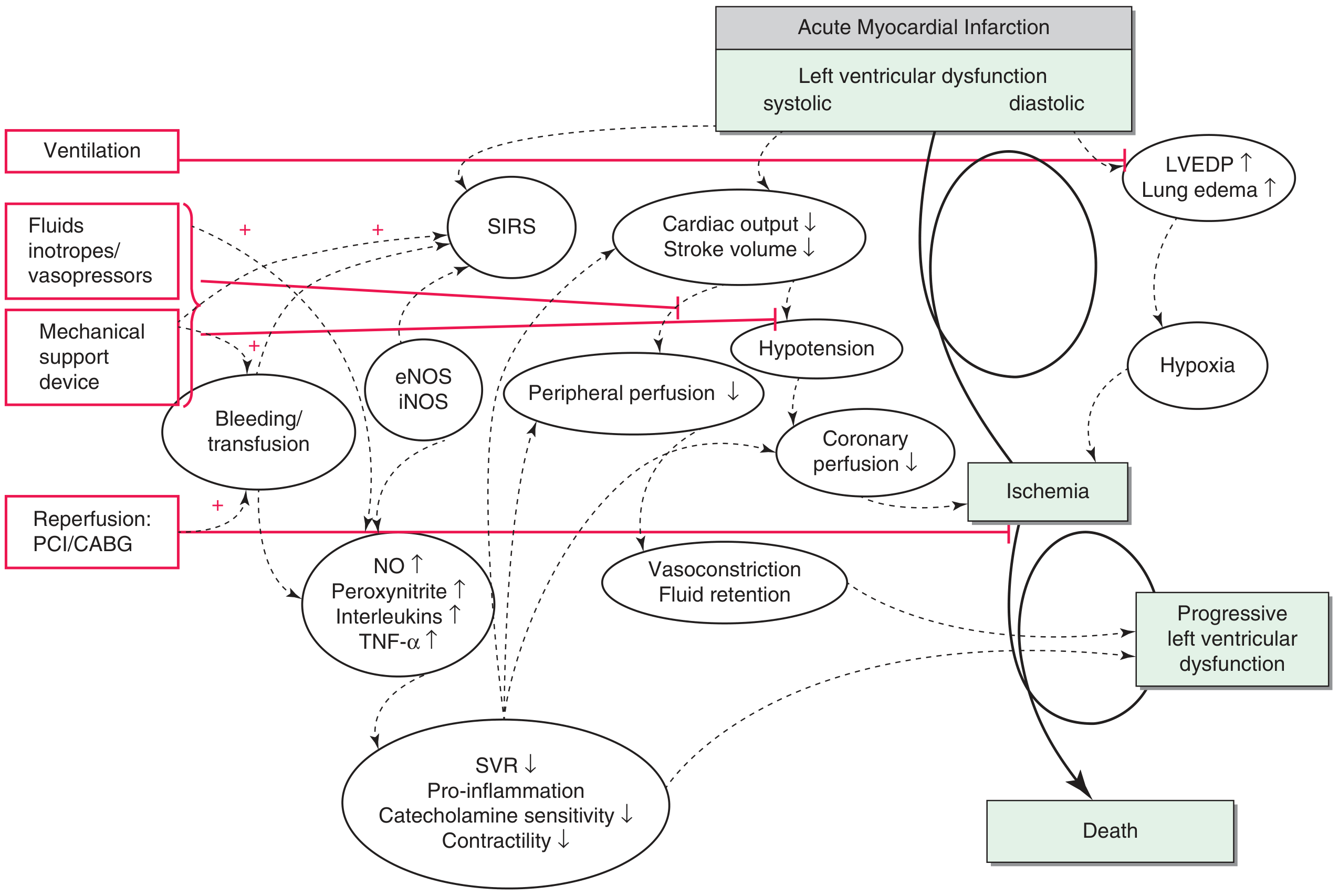
The Vicious Cycle - Step by Step:
- Coronary artery blocked → Myocardial ischemia → LV systolic and diastolic dysfunction
- Dead muscle cannot contract (systolic failure) AND cannot relax (diastolic failure)
- Stroke volume ↓ → Cardiac output ↓
- Blood pressure falls → Tissue hypoperfusion begins
- ↓ CO → ↓ Blood pressure → ↓ Coronary perfusion pressure
- The coronary arteries fill in diastole - they need adequate aortic pressure
- Lower aortic pressure = less coronary blood flow = more ischemia
- More ischemia = more muscle dies = even less CO = even less coronary perfusion
- This is the vicious spiral → Death if not broken
- LV can't empty → LV end-diastolic pressure (LVEDP) rises
- Pressure backs up into pulmonary veins → Pulmonary capillary pressure rises
- Fluid leaks into lung alveoli → Pulmonary oedema
- Pulmonary oedema → ↓ O₂ exchange → Hypoxia
- Hypoxia → more myocardial ischemia → more pump failure (vicious cycle again!)
- Baroreceptors sense ↓ BP → Activate sympathetic nervous system
- Catecholamines released → Tachycardia + Peripheral vasoconstriction
- Initially: Vasoconstriction maintains BP and shunts blood to heart and brain ✓
- Later: ↑ SVR = ↑ afterload = the failing heart has to work HARDER against more resistance = worsens pump failure ✗
- The body's own compensation becomes the enemy
- Myocardial injury (dead cells) triggers inflammatory response
- Inflammatory cytokines (TNF-α, Interleukins) released → Activate iNOS and eNOS
- iNOS/eNOS → ↑ NO production → ↑ Peroxynitrite (cardiotoxic!)
- This causes paradoxical vasodilation and directly kills more cardiomyocytes
- Catecholamine sensitivity ↓ (drugs work less well)
- Contractility ↓ further
- Anaerobic glycolysis → Lactic acidosis
- Acidosis → Reduces efficacy of catecholamines and inotropes (drugs stop working)
- Hypoxia + Acidosis + ↓ Contractility → Progressive LV dysfunction → Death
Summary Diagram (Text Version):
AMI → LV dysfunction
         ↓
    ↓ CO, ↓ BP
    /            \
Forward         Backward
failure         failure
   ↓               ↓
↓ Coronary    LVEDP↑ → Pulmonary oedema
 perfusion         ↓
   ↓           Hypoxia
More ischemia     ↓
   ↓          More ischemia
↓↓ CO ←←←←←←←←←←←←←←←←←←
   ↓
SIRS → TNF, IL → NO → Vasodilation
              → Peroxynitrite → ↓Contractility
   ↓
Lactic acidosis → Catecholamines ineffective
   ↓
DEATH (unless spiral is broken)
Haemodynamic Profile Summary:
| Parameter | Direction | Why |
|---|---|---|
| Cardiac Output (CO) | ↓↓ | Pump is failing |
| Blood Pressure | ↓ | Less blood being pumped |
| SVR | ↑↑ | Body compensates with vasoconstriction |
| PCWP / LVEDP | ↑↑ | Blood dams up behind failing LV |
| CVP (Right-sided) | ↑ (LV MI) / ↑↑ (RV MI) | Backward congestion |
| SvO₂ (mixed venous O₂) | ↓↓ | Tissues extracting more O₂ (starving) |
| Lactate | ↑↑ | Anaerobic metabolism |
| Urine output | ↓ | Renal hypoperfusion |
- Cardiogenic: PCWP HIGH (lungs are wet/congested)
- Hypovolemic: PCWP LOW (tank is empty, lungs are dry)
🔴 PART 3: CLINICAL FEATURES
"The 5 Ws of Cardiogenic Shock Presentation"
Symptoms:
- Chest pain (if AMI is the cause - may have already had it for hours)
- Severe breathlessness / dyspnoea (pulmonary oedema - lungs are flooding)
- Extreme fatigue and weakness
- Anxiety and sense of impending doom
- Altered mental state - confusion, drowsiness (cerebral hypoperfusion)
Signs (System by System):
- Pulse: Weak, thready, rapid (low stroke volume; may be slow if complete heart block)
- BP: <90 mmHg systolic (or drop of >30 mmHg from baseline)
- Narrow pulse pressure (low SBP, high DBP from vasoconstriction)
- JVP elevated (blood backing up, especially in RV failure or tamponade)
- S3 gallop on auscultation (sound of blood hitting a stiff, failing ventricle)
- S4 gallop (stiff LV - diastolic dysfunction)
- Murmurs - NEW murmur = mechanical complication! (MR, VSR)
- Tachypnoea (fast breathing, >20/min)
- Bilateral crackles (basal, spreading up as oedema worsens)
- May be in frank pulmonary oedema - pink frothy sputum, orthopnoea, sitting bolt upright, using accessory muscles
- Skin: Cold, pale, clammy, mottled, cyanotic (peripheral vasoconstriction + poor perfusion)
- Prolonged capillary refill time (>2 seconds)
- Cold extremities despite feeling feverish centrally
- Oliguria (<0.5 mL/kg/hr) progressing to anuria (renal hypoperfusion)
- Agitation, confusion, restlessness, stupor (cerebral hypoperfusion)
Classic Clinical Picture to memorise:
"A 70-year-old with known IHD presents 4 hours after onset of crushing chest pain. He is pale, sweating profusely, gasping for air, cannot lie flat. HR 120, BP 80/60, RR 28. There are bilateral crackles up to the mid-zones. JVP is elevated. S3 present. Extremities are ice cold and mottled."
SCAI Classification (Current Standard)
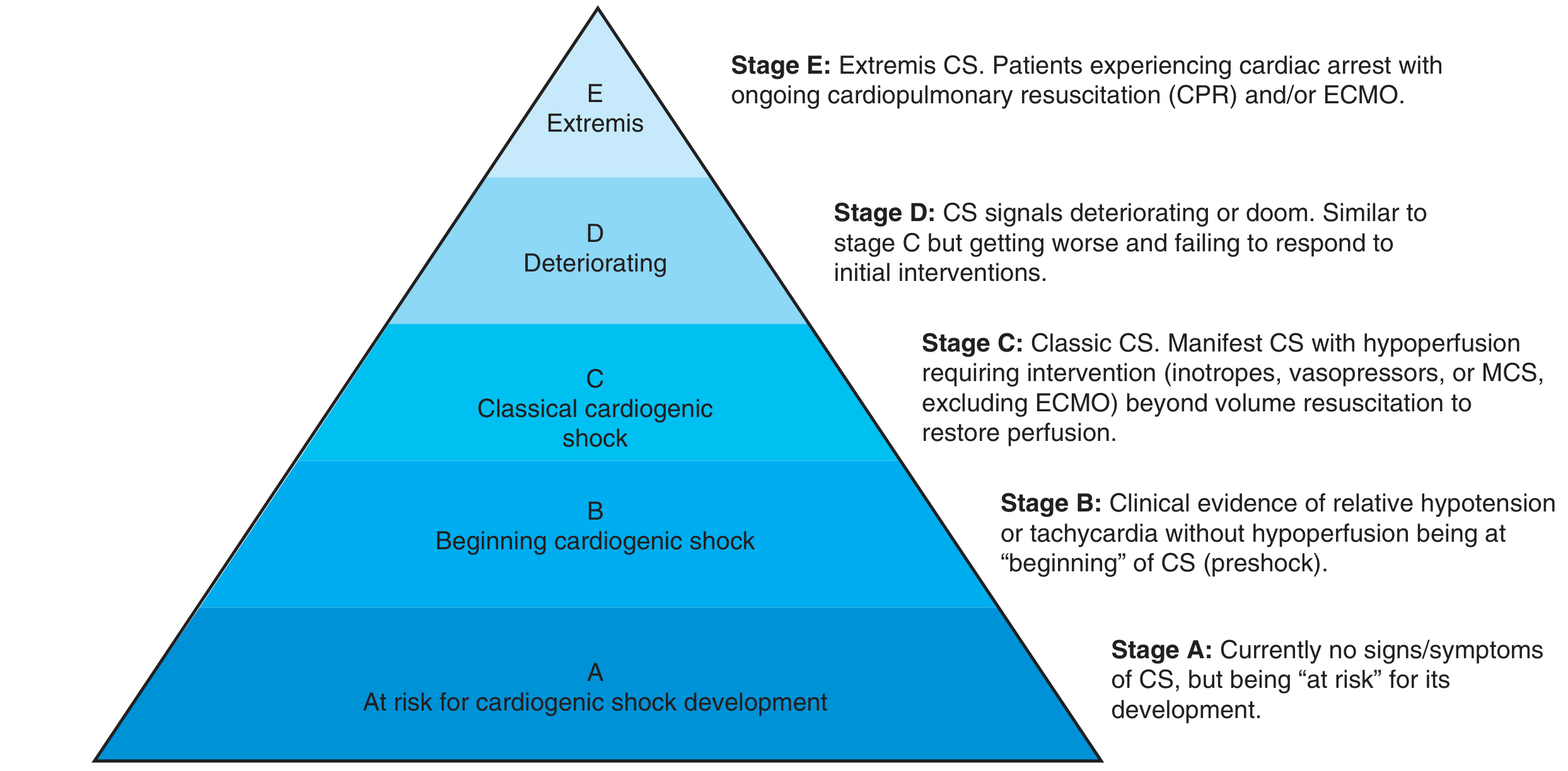
| Stage | Name | Description |
|---|---|---|
| A | At Risk | No signs of CS yet. Large MI, known HF. Watch closely. |
| B | Beginning (Preshock) | Tachycardia or relative hypotension. NO hypoperfusion yet. |
| C | Classical CS | Manifest shock: hypoperfusion requiring inotropes/MCS |
| D | Deteriorating | Failing initial treatment. Getting worse. |
| E | Extremis | Cardiac arrest, CPR ongoing, ECMO required |
🔴 PART 4: INVESTIGATIONS
ECG:
- ST-elevation (STEMI) or T-wave changes
- New LBBB
- Arrhythmias (VT, AF, Complete Heart Block)
Echocardiogram (MOST IMPORTANT investigation):
- Confirms LV dysfunction (EF severely reduced)
- Identifies mechanical complications (MR, VSR, tamponade, RV failure)
- Essential for diagnosis and guiding treatment
- Do it immediately at the bedside
Blood tests:
- Troponin ↑↑ (myocardial necrosis)
- Lactate ↑ (>2 mmol/L = tissue hypoperfusion, >5 = very poor prognosis)
- ABG: Hypoxia, metabolic acidosis, ↑ CO₂
- Creatinine ↑ (renal hypoperfusion/AKI)
- LFTs ↑ (hepatic hypoperfusion - "shock liver")
- BNP/NT-proBNP ↑↑ (heart failure marker)
- FBC, coagulation (DIC if severe)
Chest X-Ray:
- Cardiomegaly
- Bilateral pulmonary infiltrates (pulmonary oedema - "bat-wing" pattern)
- Kerley B lines (interstitial oedema)
- Upper lobe venous diversion
Invasive Monitoring (Swan-Ganz / Pulmonary Artery Catheter):
- Confirms: ↓ CO, ↑ PCWP, ↑ SVR
- Used in complex cases to guide vasopressor/inotrope titration
🔴 PART 5: MANAGEMENT
- Stabilise (buy time)
- Fix the cause (definitive treatment)
- Support failing organs
STEP 1: Immediate Stabilisation (First 30-60 minutes)
- High-flow O₂ (15L via non-rebreather mask)
- If pulmonary oedema + respiratory distress → Non-invasive ventilation (NIV/CPAP)
- CPAP reduces work of breathing AND reduces preload (helpful in oedema)
- Intubation if unable to maintain airway or respiratory failure
- Large-bore IV access x2
- Foley catheter (monitor urine output hourly)
- Continuous ECG monitoring + SpO₂ + arterial line (continuous BP)
- Cautious fluids - unlike hypovolemic shock, you do NOT aggressively push fluids
- Small bolus (250 mL) only if there are signs of hypovolaemia or RV infarction
- Too much fluid = worsens pulmonary oedema in LV failure
- Arrhythmia → Cardiovert (if VT/VF) or pace (if complete heart block)
- Cardiac tamponade → Emergency pericardiocentesis
- Tension pneumothorax → Needle decompression
STEP 2: Vasopressors and Inotropes
Vasopressors (Raise BP):
| Drug | Mechanism | When to Use |
|---|---|---|
| Norepinephrine (Noradrenaline) | α1 + β1 agonist | First-line vasopressor in CS with profound hypotension. Raises BP. |
| Vasopressin | V1 receptor | Add-on if norepinephrine inadequate |
| Dopamine | Dose-dependent (dopaminergic, β, α) | Now LESS preferred - more arrhythmias than norepinephrine |
Inotropes (Strengthen the squeeze):
| Drug | Mechanism | Use |
|---|---|---|
| Dobutamine | β1 agonist (mainly) | Positive inotrope - increases CO. Risk: causes tachycardia + arrhythmias. May also lower BP. |
| Milrinone | PDE-3 inhibitor → ↑ cAMP | Inotrope + vasodilator. Useful if β-blockers on board (doesn't need β receptor). Risk: hypotension. |
| Levosimendan | Calcium sensitiser + K-ATP channel opener | Inotrope without increasing O₂ demand. Used in acute decompensated HF. |
STEP 3: Reperfusion - THE DEFINITIVE TREATMENT for MI-related CS
Restore blood flow to the dead/dying heart muscle. Everything else is just buying time.
- Gold standard for STEMI complicated by cardiogenic shock
- Open the blocked artery with a stent → Restore flow → Stop more muscle dying → Break the vicious cycle
- Target: Door-to-balloon time <90 minutes (or <120 min if transfer required)
- Even in very sick patients, revascularisation improves survival
- Recent data: Revascularise the culprit vessel only first (not all blocked vessels at the same time - immediate multivessel PCI increases mortality)
- If anatomy not suitable for PCI (left main disease, complex multivessel disease)
- Can be done emergently but has higher operative risk in CS
- If PCI not available within 120 minutes and no contraindications
- Less effective than PCI but better than nothing
STEP 4: Mechanical Circulatory Support (MCS)
IABP (Intra-Aortic Balloon Pump):
- A balloon in the descending aorta, inflates in diastole → ↑ coronary perfusion pressure (coronaries fill in diastole!)
- Deflates rapidly in systole → Creates suction effect → ↓ afterload → Heart pumps easier
- Easy to insert percutaneously
- Note: The IABP-SHOCK II trial showed NO mortality benefit vs medical therapy alone - now used mainly as a bridge
Impella (Axial Flow Pump):
- Tiny motor pump placed across the aortic valve into the LV
- Actively pumps blood from LV into aorta → Unloads the LV
- Provides 2.5-5.0 L/min of support
- Better haemodynamic support than IABP
- Current evidence does not show clear mortality benefit over medical therapy in most patients, but remains widely used
VA-ECMO (Venoarterial Extracorporeal Membrane Oxygenation):
- Takes blood from venous system, oxygenates it outside the body, pumps it back into the arterial system
- Provides full cardiopulmonary bypass - takes over both heart and lungs
- Used in Stage E (Extremis) - cardiac arrest, refractory shock
- The ECLS-SHOCK trial (2023): VA-ECMO did NOT show mortality benefit vs medical therapy, AND had significantly more bleeding and limb ischaemia complications
- Still used selectively as a bridge to decision (bridge to transplant, bridge to VAD)
VAD (Ventricular Assist Device):
- Long-term device implanted surgically
- Used as bridge to transplant or destination therapy in refractory CS
- Decision made by multidisciplinary heart team
STEP 5: Management of Specific Causes
| Cause | Specific Management |
|---|---|
| RV Infarction CS | Fluids (target RA pressure 10-15 mmHg), avoid nitrates and diuretics (preload dependent!), catecholamines, early PCI |
| Acute MR (papillary rupture) | IABP + vasodilators as bridge → Emergency mitral valve surgery |
| VSR | IABP as bridge → Emergency surgical VSR closure (mortality >80% without surgery) |
| Cardiac Tamponade | Emergency pericardiocentesis - needle drainage |
| Arrhythmia | DC cardioversion (VT/VF) / Temporary pacing (Complete Heart Block) |
| Acute Aortic Dissection | Emergency surgery (avoid anticoagulation) |
STEP 6: Monitoring Response to Treatment
- MAP ≥65 mmHg
- Urine output >0.5 mL/kg/hr
- Lactate clearance >10-20% every 2 hours (falling lactate = organs recovering)
- SpO₂ >94%
- Mental status improving
- Extremities warming up
🔴 PROGNOSIS
- Age >73 years
- Prior stroke
- Blood glucose at admission >10.6 mmol/L (191 mg/dL)
- Creatinine >132.6 μmol/L (1.5 mg/dL) at admission
- TIMI flow <3 after PCI
- Lactate >5 mmol/L at admission
- Score 0-2 = lower risk (~24% mortality)
- Score 4-6 = very high risk (~75%+ mortality)
🧠 THE BIG PICTURE - How Everything Connects
CAUSE (AMI, Arrhythmia, Tamponade, etc.)
         ↓
    LV/RV Dysfunction
         ↓
    ↓ Cardiac Output
    /             \
Forward failure  Backward failure
↓ Coronary flow  ↑ LVEDP → Pulmonary oedema
↓ BP            ↓ O₂ exchange
↑ Ischaemia     ↑ Hypoxia
         \       /
          ↓↓ CO
          ↓
SIRS + NO ↑ → ↓ Contractility further
          ↓
    Lactic acidosis
          ↓
MANAGEMENT INTERRUPTS THE SPIRAL:
→ O₂, Ventilation (fix hypoxia)
→ Vasopressors (fix BP)
→ Inotropes (improve contractility)
→ PCI/CABG (fix the cause - most important!)
→ MCS (mechanical support when drugs fail)
⚡ HIGH-YIELD EXAM FACTS
- Most common cause = Acute MI (anterior) causing LV failure (~80% of cases)
- Classic haemodynamics: ↓ CO, ↓ BP, ↑ SVR, ↑ PCWP, ↓ SvO₂
- PCWP is HIGH - this differentiates it from hypovolemic shock (PCWP low)
- Skin: Cold, pale, clammy (NOT warm - that's septic shock)
- JVP elevated + pulmonary oedema + hypotension = cardiogenic shock until proven otherwise
- New murmur after MI = mechanical complication (MR, VSR, free wall rupture)
- RV infarction = elevated JVP + clear lungs + hypotension (no pulmonary oedema - lungs are DRY)
- In RV infarction: NEVER give nitrates or diuretics (reduce preload → catastrophic BP drop)
- Definitive treatment for MI-CS = Emergency PCI (restore coronary flow)
- First-line vasopressor = Norepinephrine
- First-line inotrope = Dobutamine
- IABP works by: Inflating in diastole (↑ coronary flow) + Deflating in systole (↓ afterload)
- ECLS-SHOCK trial (2023) = VA-ECMO showed no mortality benefit + more complications
- SCAI Stage A-E: At risk → Beginning → Classical → Deteriorating → Extremis
- Mortality = 40-60%. Still one of medicine's most lethal emergencies.