
A 60 year old man reports to the medicine OPD with complaints of extreme weakness and fatigue as well as numbness and heaviness in lower limbs for past 6 months. On taking history it was found that he is a strict vegetarian. On examination of the patient he looks pale and neurological examination showed sensory and motor loss of both the legs. Findings of the laboratory investigations are: Peripheral smear showing large sized RBC.Urine showed high levels of methyl malonic acid and homocysteine. 1) Identify the nutritional deficiency and the probable diagnosis. ii) Correlate the patient symptoms and the signs on examination with the lab reports. iii) Enumerate other lab investigations that can be ordered to reach to a definite diagnosis. iv) Analyse the lab findings and give biochemical explanation of the same. v) Plan a treatment for this patient.(2+3+2+5+3)[
Vitamin B12 (Cobalamin) Deficiency - Complete Case Analysis
i) Nutritional Deficiency & Probable Diagnosis (2 marks)
- Megaloblastic (Macrocytic) Anemia due to Vitamin B12 deficiency
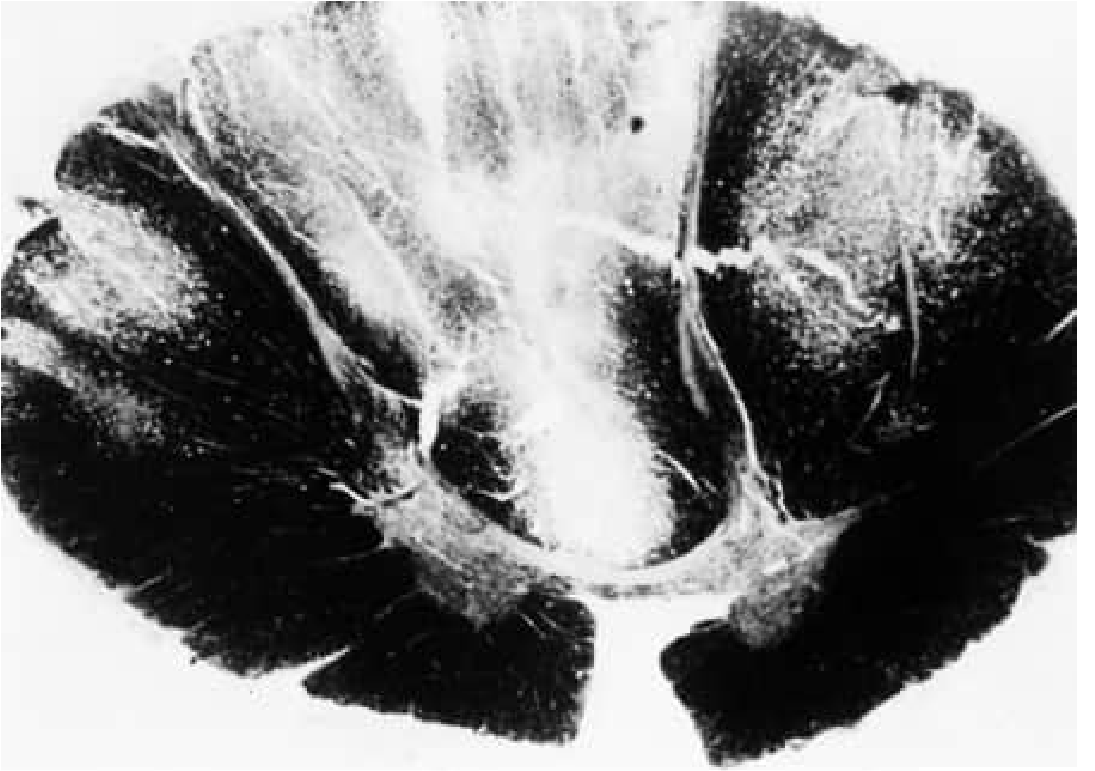
- Subacute Combined Degeneration (SCD) of the Spinal Cord - the neurological syndrome resulting from severe, prolonged B12 deficiency
ii) Correlation of Symptoms, Signs, and Lab Reports (3 marks)
| Clinical Feature | Biochemical/Pathological Explanation |
|---|---|
| Extreme weakness and fatigue | Megaloblastic anemia - large, defective RBCs with impaired oxygen-carrying capacity lead to tissue hypoxia and fatigue |
| Pallor on examination | Anemia from ineffective erythropoiesis; bone marrow produces fewer, abnormally large erythroid precursors that are destroyed before release |
| Numbness and heaviness in both lower limbs | Demyelination of the posterior and lateral columns of the spinal cord (SCD). Posterior column involvement causes loss of proprioception and vibration (numbness/heaviness); lateral column (corticospinal tract) involvement causes motor weakness |
| Sensory and motor loss of both legs (bilateral) | Symmetric demyelination of both posterior columns (sensory loss) and lateral columns (motor/pyramidal loss) of the spinal cord |
| Large RBCs on peripheral smear (macrocytosis/megalocytosis) | B12 deficiency → "methylfolate trap" → impaired thymidine synthesis → defective DNA synthesis → cells enlarge but cannot divide normally → macrocytes/megaloblasts |
| High urinary methylmalonic acid (MMA) | B12 is a cofactor for L-methylmalonyl-CoA mutase. Without B12, L-methylmalonyl-CoA cannot be converted to succinyl-CoA and accumulates, getting hydrolyzed to methylmalonic acid which spills into urine |
| High homocysteine | B12 (as methyl-cobalamin) is required by methionine synthase to convert homocysteine to methionine. Without B12, homocysteine accumulates in serum and urine |
iii) Further Lab Investigations for Definitive Diagnosis (2 marks)
- Serum Vitamin B12 (Cobalamin) level - low in deficiency (normal: 200-900 pg/mL). Note: can be falsely normal in up to 30% of cases, hence metabolite testing is also needed.
- Complete Blood Count (CBC) - macrocytic anemia (elevated MCV >100 fL), leukopenia, possible thrombocytopenia
- Peripheral blood smear - hypersegmented neutrophils (>5 lobes; more than 5% with ≥5 lobes, or any with ≥6 lobes is diagnostic), oval macrocytes
- Serum Folate and RBC Folate levels - to differentiate folate deficiency (which also causes megaloblastic anemia but NOT SCD), and to rule out combined deficiency
- Bone Marrow Examination - reveals megaloblastic changes (nuclear-cytoplasmic maturation asynchrony), giant metamyelocytes
iv) Biochemical Explanation of Lab Findings (5 marks)
A. Large RBCs (Macrocytosis / Megaloblastosis)
Methyl-THF  ---[B12 + Methionine Synthase]--->  THF
                       ↑ BLOCKED in B12 deficiency
- THF is needed to generate 5,10-methylene-THF, which is the one-carbon donor for thymidylate synthase to convert dUMP → dTMP (thymidine)
- Without adequate thymidine, DNA synthesis is impaired
- Cells continue to grow and accumulate RNA/protein (cytoplasmic maturation continues) but cannot divide
- Result: large cells with immature, open-chromatin nuclei - megaloblasts in the marrow, macrocytes/oval macrocytes in peripheral blood
- Rapidly dividing precursors (erythroid, myeloid) are most affected → anemia + leukopenia
B. Elevated Urinary Methylmalonic Acid (MMA)
Propionyl-CoA → L-Methylmalonyl-CoA → [Adenosyl-B12 + Mutase] → Succinyl-CoA (enters TCA cycle)
                                                  ↑ BLOCKED in B12 deficiency
L-Methylmalonyl-CoA accumulates → hydrolyzed to Methylmalonic Acid → excreted in urine
- MMA elevation is specific for B12 deficiency (not seen in folate deficiency)
- MMA >400 nmol/L is diagnostic of cobalamin deficiency
- This is also implicated in neurological damage: abnormal fatty acid synthesis from MMA may disrupt myelin structure
C. Elevated Homocysteine
Homocysteine + Methyl-THF  ---[Methionine Synthase + B12]--->  Methionine + THF
                                           ↑ BLOCKED
Homocysteine accumulates → elevated in serum and urine
- Elevated homocysteine is seen in both B12 AND folate deficiency
- However, elevated MMA combined with elevated homocysteine specifically points to B12 (cobalamin) deficiency
- Elevated homocysteine itself is toxic to vascular endothelium (contributes to atherosclerosis risk)
D. Neurological Damage (SCD) - Biochemical Basis
- Impaired methionine/SAM synthesis: Methionine is activated to S-adenosylmethionine (SAM), the universal methyl donor. SAM is required for methylation of myelin basic protein and phospholipids. Without SAM, myelin maintenance fails → demyelination of posterior and lateral columns
- MMA toxicity: Methylmalonic acid may substitute for malonyl-CoA in fatty acid synthesis, generating abnormal odd-chain fatty acids that disrupt myelin integrity
v) Treatment Plan (3 marks)
- Cyanocobalamin or Hydroxocobalamin 1000 μg IM or SC daily for 7 days, then
- 1000 μg weekly for 4 weeks (some regimens use every-other-day for 2 weeks, then weekly x8 weeks)
- 1000 μg IM/SC monthly (for cyanocobalamin) or every 2-3 months (for hydroxocobalamin)
- Alternatively: High-dose oral B12 1000-2000 μg/day - effective even in pernicious anemia via passive (IF-independent) diffusion (~1% absorbed by mass action); appropriate for dietary deficiency especially
- Strict vegetarians should be advised to regularly consume B12-fortified foods (fortified cereals, plant-based milks)
- If unwilling to change diet, long-term oral B12 supplementation is mandatory
- Reticulocyte count rises in 2-3 days (confirms diagnosis)
- Hemoglobin improves over 4-8 weeks
- Serum MMA and homocysteine normalize within ~2 weeks of treatment
- Neurological improvement is delayed (6-12 months) and may be incomplete - early diagnosis and treatment is therefore critical, as neurological remission inversely correlates with duration before treatment
- Robbins & Kumar Basic Pathology, pp. 396-397 (Megaloblastic anemia, Vitamin B12 deficiency)
- Bradley and Daroff's Neurology in Clinical Practice, pp. 1797-1799 (Subacute combined degeneration, lab studies, treatment)
- Goldman-Cecil Medicine, Chapter 150 (Cobalamin: pathobiology, diagnosis, treatment)
- Katzung's Basic and Clinical Pharmacology, 16e, Chapter 33 (Vitamin B12 pharmacology)