
What is cancer?
cancer cell division tumor growth hallmarks
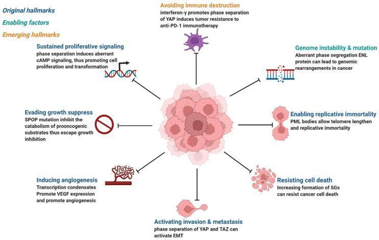
A pathophysiology diagram illustrating the 'hallmarks of cancer' in the context of phase separation and biomolecular condensates. Centrally, a pink, irregular mass represents a cluster of proliferating cancer cells. Radiating from this center are various mechanisms linked to oncogenesis and tumor maintenance, categorized as original, enabling, or emerging hallmarks. These include: (1) Sustained proliferative signaling via aberrant cAMP signaling; (2) Genome instability and mutation through ENL protein phase segregation; (3) Enabling replicative immortality via PML body-mediated telomere lengthening; (4) Resisting cell death through stress granule (SG) formation; (5) Activating invasion and metastasis via YAP/TAZ phase separation and EMT; (6) Inducing angiogenesis through VEGF-promoting transcription condensates; (7) Evading growth suppression via SPOP mutations; and (8) Avoiding immune destruction involving YAP-mediated resistance to anti-PD-1 therapy. The diagram uses distinct icons (DNA strands, blood vessels, spiky immune cells) and text annotations to connect specific phase-separation abnormalities to established cancer phenotypes, emphasizing how aberrant condensates drive pathological signaling cascades and malignant transformation.
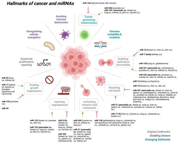
This medical infographic illustrates Hanahan and Weinberg’s hallmarks of cancer and their regulation by specific microRNAs (miRNAs). Centered around a primary tumor mass, ten hallmarks are arranged radially: Sustained proliferative signaling (DNA helix), Deregulating cellular energetics (mitochondrion), Avoiding immune destruction (leukocyte), Tumor-promoting inflammation, Genome instability & mutation (fragmented DNA), Enabling replicative immortality (dividing cells), Resisting cell death (apoptotic bodies), Activating invasion and metastasis (migrating cells), Inducing angiogenesis (vessel sprouting), and Evading growth suppressors. The diagram classifies these into original hallmarks, enabling factors, and emerging hallmarks. Specific miRNAs (oncomiRs and tumor-suppressor miRs) are linked to each hallmark with green arrows (activation) or red bars (inhibition). For example, miR-21 is shown promoting metastasis and resisting cell death across multiple cancers (pancreatic, breast, lung), while miR-155 is linked to inflammation and angiogenesis. The diagram serves as an educational summary of how non-coding RNA dysregulation drives malignant transformation and progression in various human cancers, including lung, colorectal, and prostate carcinomas.
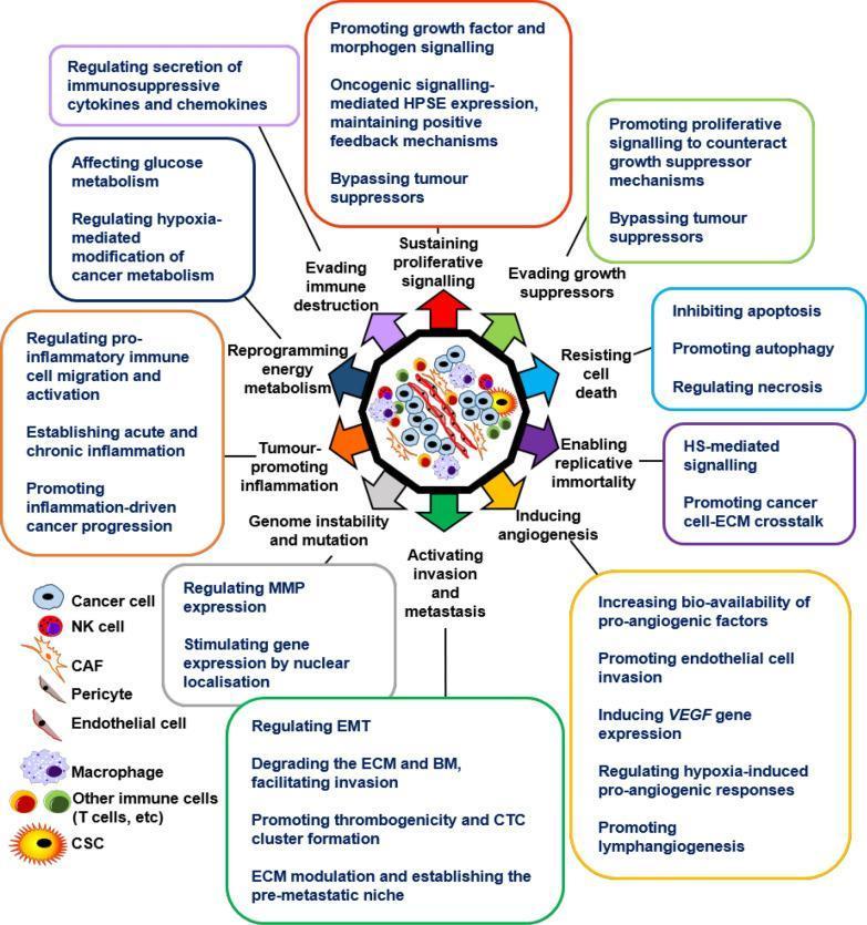
A pathophysiology diagram illustrating the regulatory role of Heparanase (HPSE) in the hallmarks of cancer within the tumor microenvironment (TME). The central graphic depicts a heterogeneous TME containing various cell types including cancer cells, cancer stem cells (CSCs), NK cells, macrophages, T cells, cancer-associated fibroblasts (CAFs), pericytes, and endothelial cells. Radiating from this center are several thematic categories of cancer hallmarks, each linked to specific HPSE-mediated mechanisms: 1) Evading immune destruction (cytokine regulation); 2) Sustaining proliferative signaling (growth factor and feedback signaling); 3) Evading growth suppressors; 4) Resisting cell death (apoptosis inhibition, autophagy); 5) Enabling replicative immortality (HS-mediated signaling); 6) Inducing angiogenesis (VEGF and pro-angiogenic factors); 7) Activating invasion and metastasis (MMP regulation, EMT, and ECM degradation); 8) Genome instability and mutation; 9) Tumor-promoting inflammation; and 10) Reprogramming energy metabolism (hypoxia modification). The diagram serves as a comprehensive educational summary of how HPSE enzymatic and non-enzymatic activities drive oncogenic progression and therapeutic resistance across oncology specialties.
What Is Cancer?
Normal Cells vs. Cancer Cells
The 8 Hallmarks of Cancer
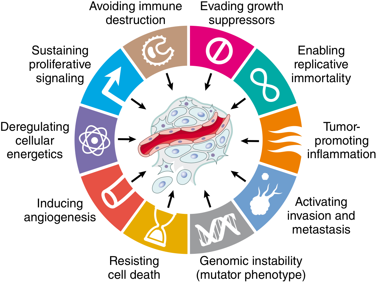
| Hallmark | What it means |
|---|---|
| Self-sufficiency in growth signals | Cancer cells produce their own growth signals or have constantly "on" receptors (via oncogenes), so they proliferate without external stimulation |
| Insensitivity to growth-inhibitory signals | They ignore stop signals normally provided by tumor suppressor genes (e.g., RB, TP53) |
| Evasion of apoptosis | Cells resist programmed death, often by overexpressing anti-apoptotic proteins like BCL-2 |
| Limitless replicative potential (immortality) | Normal cells are limited to ~50-70 divisions (Hayflick limit); cancer cells bypass this via telomerase reactivation and stem cell-like self-renewal |
| Sustained angiogenesis | Tumors secrete VEGF and other factors to grow new blood vessels, ensuring nutrient and oxygen supply |
| Ability to invade and metastasize | Cells break through the basement membrane, enter blood/lymph vessels, and colonize distant organs - responsible for most cancer deaths |
| Evasion of immune surveillance | Cancer cells downregulate surface markers or express checkpoint molecules (e.g., PD-L1) to hide from immune cells |
| Deregulated cellular metabolism (Warburg effect) | Even in the presence of oxygen, cancer cells preferentially use aerobic glycolysis - generating biosynthetic precursors for rapid growth |
- Robbins, Cotran & Kumar Pathologic Basis of Disease, Chapter 7
Genetic Basis: Oncogenes and Tumor Suppressors
-
Proto-oncogenes → Oncogenes: Proto-oncogenes are normal genes that promote cell growth (e.g., RAS, MYC, HER2). A single gain-of-function mutation converts them to oncogenes - the cellular equivalent of a stuck accelerator pedal. Oncoproteins are constitutively active and drive continuous proliferation even without external signals.
-
Tumor suppressor genes: These normally restrain cell growth (e.g., RB, TP53, BRCA1). Loss-of-function mutations in both alleles ("two-hit hypothesis") remove the brakes. TP53 ("guardian of the genome") is mutated in over 50% of all human cancers.
Types of Cancer
| Type | Origin | Examples |
|---|---|---|
| Carcinoma | Epithelial cells | Lung, breast, colon, prostate cancer |
| Sarcoma | Mesenchymal cells (connective tissue, bone, muscle) | Osteosarcoma, liposarcoma |
| Leukemia | Hematopoietic (blood-forming) cells | AML, CML |
| Lymphoma | Lymphoid cells | Hodgkin lymphoma, diffuse large B-cell lymphoma |
| Central nervous system tumors | Glial or neuronal cells | Glioblastoma, medulloblastoma |
| Germ cell tumors | Gonads/germ cells | Testicular seminoma |
What Causes Cancer? (Carcinogenic Agents)
- Chemical carcinogens - tobacco smoke, asbestos, aflatoxin (direct-acting or indirect-acting after metabolic activation)
- Radiation - ultraviolet light (skin cancer), ionizing radiation (leukemias, thyroid cancer)
- Oncogenic viruses and microbes - HPV (cervical cancer), hepatitis B/C (liver cancer), EBV (Burkitt lymphoma, nasopharyngeal carcinoma), H. pylori (gastric cancer)
- Inherited mutations - germline mutations in BRCA1/2 (breast/ovarian cancer), APC (colorectal cancer), TP53 (Li-Fraumeni syndrome)
Benign vs. Malignant Tumors
| Feature | Benign | Malignant (Cancer) |
|---|---|---|
| Growth rate | Slow | Rapid (variable) |
| Invasion | No - stays encapsulated | Yes - breaks through barriers |
| Metastasis | Never | Hallmark feature |
| Cell differentiation | Well differentiated | Poorly differentiated (anaplastic) |
| Recurrence after removal | Rare | Common |