
White stuff in baby eye along with cuff
neonatal conjunctivitis baby eye discharge swelling
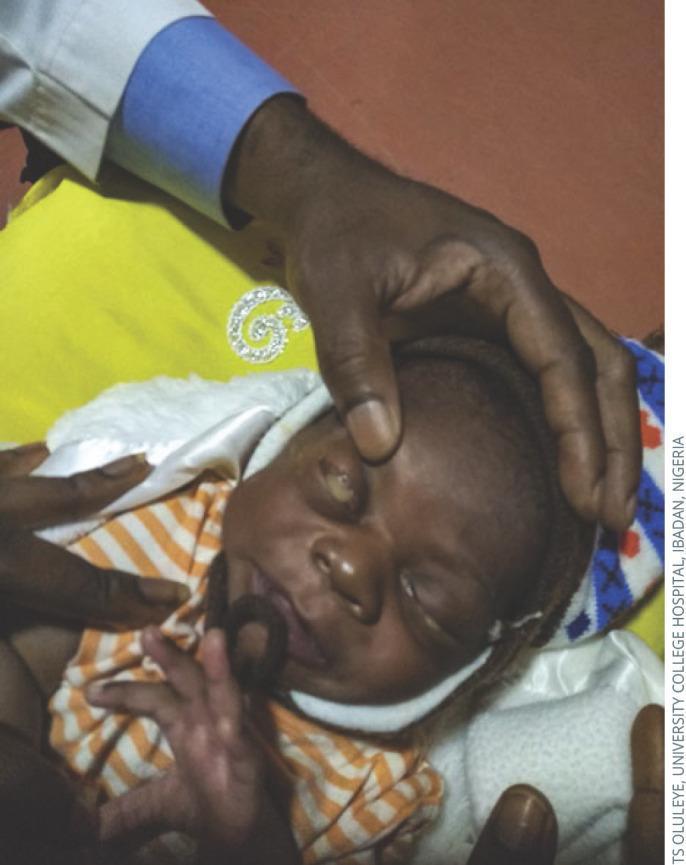
This clinical photograph shows an infant presenting with ophthalmia neonatorum, a form of neonatal conjunctivitis. The primary clinical signs include severe bilateral eyelid edema (swelling) and profuse, thick, purulent (pus-like) discharge emanating from the palpebral fissure. The discharge is thick and yellowish, characteristic of a hyperacute bacterial infection such as gonococcal conjunctivitis. An adult hand is shown gently retracting the upper eyelid to reveal the extent of the inflammation and ocular discharge. The infant is swaddled and using a pacifier, indicating a clinical examination setting. This image serves as a significant educational tool for recognizing the signs of neonatal eye infections, which require urgent diagnosis and treatment to prevent serious complications such as corneal ulceration, perforation, and permanent visual impairment. The anatomical focus is on the ocular and periorbital region, demonstrating the hallmark presentation of severe infectious conjunctivitis in the first weeks of life.
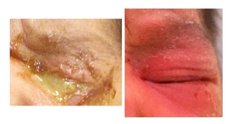
This clinical photograph set provides a side-by-side comparison of a neonatal eye before and after treatment for hyperacute bacterial conjunctivitis. The left panel shows the eye at presentation, characterized by severe blepharitis with marked eyelid edema and erythema. A profuse, thick, yellowish-green purulent discharge is seen emanating from the palpebral fissure, with associated crusting and maceration of the periocular skin. This presentation is classically associated with Neisseria species infection. The right panel demonstrates the clinical resolution following seven days of intravenous ceftriaxone therapy. There is a significant reduction in periorbital swelling and erythema, the complete absence of purulent exudate, and restoration of normal skin texture. The comparison illustrates the rapid clinical response of ophthalmia neonatorum to appropriate systemic antimicrobial therapy, emphasizing the importance of recognizing purulent neonatal conjunctivitis as a medical emergency.
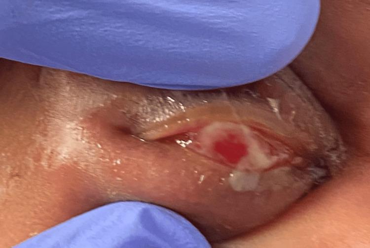
This clinical photograph displays a close-up, procedural view of the right eye of a neonate, with the upper eyelid everted by a clinician wearing blue nitrile gloves. The image demonstrates a primary clinical presentation of ophthalmia neonatorum (neonatal conjunctivitis). Significant findings include diffuse conjunctival injection (hyperemia) and a prominent accumulation of thick, yellowish mucopurulent discharge covering the palpebral and bulbar surfaces. The surrounding periorbital tissues and eyelids show marked erythema and edema. This visual illustrates the hallmark signs of infectious conjunctivitis in a newborn, highlighting the importance of assessing for N. gonorrhoeae or Chlamydia trachomatis. The photograph captures a key diagnostic step in the physical examination of a pediatric patient presenting with eye swelling and discharge, focusing on the character of the ocular secretion and the integrity of the ocular surface.
White Discharge + Eyelid Swelling (Cuff) in a Baby
What Is It?
Causes (by Timing)
| Timing | Likely Cause |
|---|---|
| Day 1-2 | Chemical (silver nitrate drops given at birth) |
| Day 2-5 | Neisseria gonorrhoeae - most dangerous, can cause blindness |
| Day 5-14 | Chlamydia trachomatis - most common overall cause |
| Any time | Staph, Strep, other bacteria; rarely Candida |
"A variety of organisms are implicated - N. gonorrhoea, C. trachomatis (commonest), staphylococcus, streptococcus, Candida spp, etc. The most serious cause is N. gonococcus as it can rapidly cause blindness." - Park's Textbook of Preventive and Social Medicine
Key Clinical Features
- White/yellow/green sticky discharge from one or both eyes
- Eyelid swelling (the "cuff") - periorbital edema and redness
- Conjunctival redness (red eye)
- In gonococcal infection: the discharge is thick, profuse, and yellowish-green (hyperacute); eyelid swelling is very marked
- In chlamydial infection: discharge tends to be more mucopurulent, onset is slower
What To Do - Treatment
By Cause (from Textbook of Family Medicine)
| Type | Treatment |
|---|---|
| Gonococcal | Ceftriaxone 125 mg IM single dose + erythromycin/tetracycline; ophthalmology referral |
| Chlamydial | Oral erythromycin base 50 mg/kg/day in 4 divided doses for 14 days; azithromycin is an alternative |
| Other bacterial | Gram-positive: erythromycin ointment; Gram-negative: gentamicin or tobramycin eye drops |
- For chlamydia, systemic (oral) treatment is preferred over topical, because it also treats nasopharyngeal co-infection (which can progress to pneumonia).
- Cultures and Gram stain of the discharge should be done to identify the exact organism.
When It Is Urgent
- Gonococcal ophthalmia neonatorum is a medical emergency - it can perforate the cornea and cause permanent blindness within days if untreated.
- Marked eyelid swelling + profuse thick yellow discharge + onset in the first 5 days = treat for gonorrhea until proven otherwise.
- Fever + swelling may also suggest dacryocystitis (infection of the tear sac), which in infants with a swelling/mass at the inner corner of the eye requires urgent ophthalmology referral and systemic antibiotics.
Prevention
- 1% silver nitrate drops OR tetracycline 1% ointment OR erythromycin 0.5% ointment applied to both eyes at birth prevents gonococcal ophthalmia neonatorum.
- Treating maternal genital infections before delivery is the primary prevention.
Sources: Textbook of Family Medicine 9e; Park's Textbook of Preventive and Social Medicine; Harrison's Principles of Internal Medicine 22E; Red Book 2021 (AAP)
Bo i meant cough
White Eye Discharge + Cough in a Baby
The Classic Diagnosis: Chlamydia trachomatis Infection
How It Presents
Eye Findings (Conjunctivitis)
- Appears a few days to several weeks after birth
- Ocular congestion, eyelid swelling, and white/mucopurulent discharge
- Lasts 1-2 weeks or longer if untreated
Respiratory Findings (Pneumonia)
- Onset typically 2 to 19 weeks after birth
- The baby is characteristically afebrile (no fever) - this is a key clue
- Staccato cough - a repetitive, machine-gun-like cough (similar to whooping cough)
- Tachypnea (fast breathing), crackles (rales) on lung exam
- Wheezing is uncommon
- Chest X-ray shows hyperinflation with bilateral infiltrates
- Nasal stuffiness and ear infection (otitis media) may also occur
"A repetitive staccato cough, tachypnea, and rales in an afebrile 1-month-old infant are characteristic." - Red Book 2021, AAP
Why This Combination?
Treatment
| Target | Drug | Dose |
|---|---|---|
| First choice | Oral erythromycin base | 50 mg/kg/day in 4 divided doses for 14 days |
| Alternative | Azithromycin | 20 mg/kg once daily for 3 days |
- Systemic (oral) treatment is required - topical eye drops alone are not enough because they do not treat the nasopharyngeal or lung infection
- The mother (and her partner) also need treatment for chlamydial genital infection
"Systemic treatment with erythromycin base has been recommended for ophthalmia neonatorum and for C. trachomatis pneumonia in infants." - Harrison's Principles of Internal Medicine 22E
Other Things to Consider
| Condition | Clues |
|---|---|
| Whooping cough (Pertussis) | Paroxysmal cough + whooping sound, can also cause eye discharge from coughing; check immunization status |
| RSV / viral bronchiolitis | Cough + wheeze, but eye discharge less typical |
| Gonococcal conjunctivitis | Eye-only, thick yellow discharge, onset day 2-5, NO pneumonia |
What To Do
- Age of the baby
- When the eye discharge started vs. when the cough started
- Whether the baby has a fever (chlamydia pneumonia is typically afebrile)
- Mother's STI screening history during pregnancy
Sources: Red Book 2021 (AAP); Medical Microbiology 9e; Sherris & Ryan's Medical Microbiology 8e; Harrison's Principles of Internal Medicine 22E
Even for c-section baby
chlamydia trachomatis neonatal conjunctivitis cesarean section transmission
chlamydia trachomatis neonatal infection cesarean delivery
PMID: 7806879
PMID: 29850935
Can a C-Section Baby Get Chlamydial Eye Discharge + Cough?
Short Answer: Yes
The Evidence
Study 1 - Bell et al., 1994 (Journal of Infection, PMID 7806879)
| Delivery Mode | Transmission Rate |
|---|---|
| Vaginal delivery | 58/125 infants (46%) culture-positive |
| C-section with ruptured membranes | 2/10 infants (20%) culture-positive |
| C-section without ruptured membranes | 1/6 infants (~17%) culture-positive |
"Infants born to infected women are at risk of C. trachomatis infection regardless of route of delivery."
Study 2 - Xu et al., 2018 (European Journal of Pediatrics, PMID 29850935)
- 78 (22.7%) were C-section babies
- 266 (77.3%) were vaginally delivered
How Does a C-Section Baby Get Infected?
| Mechanism | Explanation |
|---|---|
| Ruptured membranes before C-section | If the waters break before surgery, bacteria can ascend into the amniotic fluid and infect the baby |
| Ascending infection before rupture | Even with intact membranes, low-level ascending infection is possible |
| In utero transplacental spread | Less common but documented |
| Post-natal contact | Infected secretions from the mother's hands, breast, or caregivers |
Key Takeaway
- White eye discharge
- Staccato cough
- No fever (afebrile is the hallmark)
- Age 2-12 weeks
The baby is 2 months old, first there was fever, then cough and now eye infection, she sleeps in ac with 30°c
2-Month-Old: Fever → Cough → Eye Discharge
Most Likely Diagnosis: Adenovirus (Pharyngoconjunctival Fever)
- Fever first - the virus causes a systemic febrile illness
- Cough - respiratory tract involvement
- Eye discharge last - conjunctivitis develops as the infection spreads
"Adenovirus causes pharyngitis, which is often accompanied by conjunctivitis (pharyngoconjunctival fever)... Affected patients have mild, flu-like symptoms including nasal congestion, cough, fever, and possible conjunctivitis lasting 3-5 days." - Medical Microbiology 9e
Why the AC at 30°C Is Relevant
- Still blowing air over the baby - causing dry mucous membranes in the nose and throat, making the baby more prone to respiratory infections and worsening congestion
- Not cool enough to be the cause of illness (cold temperatures don't cause infections by themselves), but the dry moving air irritates airways in an already-sick baby
- The AC filter can also circulate dust particles and allergens
Full Differential Diagnosis for This Baby
| Diagnosis | Fits? | Reasoning |
|---|---|---|
| Adenovirus (Pharyngoconjunctival Fever) | ✅ Best fit | Fever → cough → conjunctivitis is the textbook sequence |
| RSV / Viral bronchiolitis | Possible | Common at this age, causes fever + cough, but eye discharge less typical |
| Common cold (Rhinovirus) | Possible | Can cause all three symptoms |
| Chlamydia trachomatis | Less likely now | Typically no fever, and eye symptoms usually come first, not last |
| Bacterial conjunctivitis | Possible but secondary | Thick yellow-green discharge, could be secondary to a viral illness |
| Pertussis (Whooping cough) | Needs ruling out | Very dangerous at 2 months - causes severe paroxysmal cough; check vaccination status |
⚠️ Red Flag - This Baby Needs to Be Seen TODAY
- The immune system is immature
- Infections can deteriorate very rapidly
- Fever in a baby under 3 months always requires a doctor's assessment - they may need blood tests, urine tests, and sometimes a spinal tap to rule out serious bacterial infection (meningitis, sepsis)
- Breathing very fast (more than 60 breaths/minute) or working hard to breathe
- Nostrils flaring, chest sucking in with each breath
- Lips or fingertips turning blue
- Fever over 38°C (100.4°F) - at 2 months, ANY fever needs urgent evaluation
- Baby is very limp, unusually drowsy, or cannot be woken
- Not feeding / fewer wet nappies than normal
- High-pitched or weak cry
What To Do Right Now
- Go to a doctor or paediatric emergency today - do not wait. A 2-month-old with fever + cough + eye discharge needs examination
- Do not give any medicine (not even infant paracetamol/Calpol) without a doctor's guidance at this age
- Keep the baby fed - breast milk or formula gives antibodies and prevents dehydration
- Gently clean the eye - wipe from the inner corner outward with a clean wet cotton ball, one wipe per swab
- Adjust the AC - point vents away from the baby, set to 26-28°C, avoid direct airflow
Sources: Medical Microbiology 9e; Sherris & Ryan's Medical Microbiology 8e; Goldman-Cecil Medicine
No the fever recided 4 days back after that cough remains persistent with runny nose sometimes and today the eye problem
- ~4+ days ago: Fever (now gone)
- Ongoing since: Persistent cough + runny nose
- Today (new): White eye discharge
Updated Assessment: 2-Month-Old
Full Timeline
| Day | What happened |
|---|---|
| ~5-7 days ago | Fever started |
| ~4 days ago | Fever resolved |
| Since then | Persistent cough + runny nose |
| Today | White eye discharge appeared |
Most Likely: Post-Viral Conjunctivitis (Adenovirus or similar)
"Viral conjunctivitis is classically preceded by a viral infection with upper respiratory symptoms, with sequential involvement of the eyes."
Is the White Discharge Viral or Bacterial?
| Feature | Suggests Viral | Suggests Bacterial |
|---|---|---|
| Discharge color | Watery, white, clear | Thick yellow-green pus |
| Morning eye matting | Mild | Both eyes stuck shut |
| Both eyes involved | Often starts in one, spreads | Often bilateral |
| Preceded by URTI | Yes - fits this baby | Less common pattern |
| Preauricular lymph node | May be swollen | Usually not |
What This Baby Actually Needs
✅ Reassuring signs (if all of these are present)
- Fever has been gone 4 days - good, the acute phase is over
- Baby is feeding normally
- No fast/laboured breathing
- White (not yellow-green) eye discharge
- Baby is alert and responsive
Home Care (if the baby is otherwise well)
- Eye cleaning - gently wipe from inner to outer corner with clean wet cotton, fresh cotton for each wipe, 3-4 times a day
- Saline nasal drops (not medicated) - 1-2 drops in each nostril before feeds helps with the runny nose and may actually reduce the eye congestion too, since the nose and eye drain together
- AC setting - keep at 26-28°C, never blow directly onto baby, avoid dry air
- Keep feeding well - breast milk especially helps because it contains maternal antibodies against the exact virus causing this infection
When to See a Doctor (do not delay if any of these appear)
- Discharge becomes thick yellow-green / pus-like
- Eyelids swollen and red (the "cuff" from before)
- Fever returns
- Cough gets significantly worse or baby breathes fast (over 60 breaths/minute)
- Baby seems to be in pain when eye is touched
- Eye looks red/cloudy
Bottom Line
Sources: Rosen's Emergency Medicine; Medical Microbiology 9e