
Massive transfusion protocol
"massive transfusion protocol" AND "damage control resuscitation"
massive transfusion protocol blood products ratio
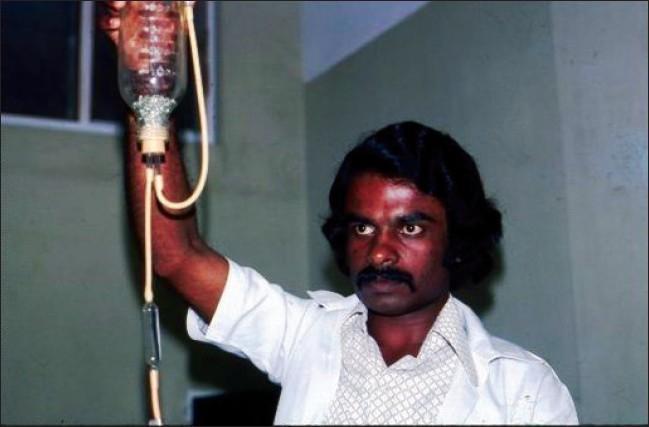
A clinical photograph of a historical blood transfusion setup from the early 1970s. The image displays a clear glass transfusion bottle held vertically by a medical professional. The interior of the bottle contains numerous small, round glass beads, which functioned as an early filtration system to trap clots or debris during the administration of blood products. Thick, translucent rubber tubing is connected to the base of the bottle, leading to an in-line vertical glass drip chamber. This chamber allowed for the visual monitoring of flow rates and the trapping of air bubbles before the blood reached the patient. The photograph illustrates the evolution of transfusion medicine and the transition from reusable glass and rubber equipment to modern, sterile, disposable plastic systems. This content is relevant for medical history, hematology, and the study of global health resource development in South Asia.

Summary : This image is a logo for a medical specialty or department related to blood transfusion. logo: TRANSFUSION MEDICINE Visible Elements : • The logo contains the text "TRANSFUSION MEDICINE" in uppercase white letters on a dark blue rectangular background. • To the left of the text, there are several stylized red blood cells depicted in a scattered arrangement, suggesting the theme of blood or hematology. • The background is white, and the overall design is horizontal. Dimensions & Placement : • The text occupies the right two-thirds of the logo, while the red blood cell graphics are on the left. • The logo is rectangular and oriented horizontally. Analysis : • The use of red blood cells visually reinforces the focus on transfusion medicine, which deals with blood products and their clinical use. • The dark blue background for the text provides strong contrast, making the department or specialty name highly legible.
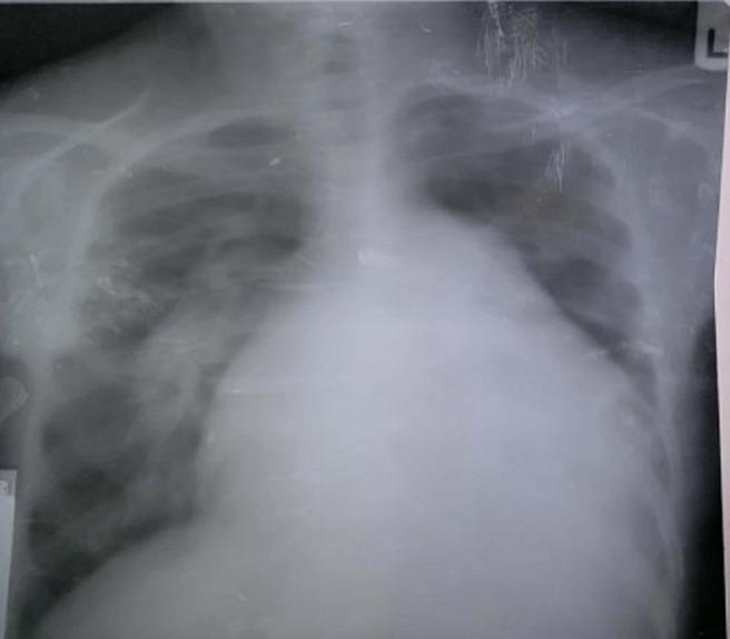
This diagnostic image is a posteroanterior (PA) view chest X-ray demonstrating significant cardiothoracic abnormalities. The most prominent feature is massive cardiomegaly, with a cardiothoracic ratio (CTR) estimated at 0.8, indicating the cardiac silhouette occupies 80% of the thoracic width. A notable bulge in the pulmonary conus is visible along the left heart border, suggestive of pulmonary hypertension or pulmonary artery dilation. The lung fields exhibit pulmonary plethora, characterized by increased vascular markings and congestion extending to the peripheral zones, typical of a left-to-right shunt (such as an atrial septal defect). The combination of these findings—cardiomegaly, a prominent pulmonary segment, and plethoric lung fields—is clinically significant for chronic volume overload and increased pulmonary blood flow, frequently seen in congenital heart disease with Eisenmenger physiology or advanced valvular disease. The image serves as an educational example of cardiac remodeling and pulmonary vascular redistribution in the context of high-flow cardiac shunts.

Summary : This image is a logo for "Transfusion Medicine," featuring stylized red blood cells and bold text. logo: TRANSFUSION MEDICINE Logo Elements : • Text: "TRANSFUSION MEDICINE" in uppercase, white font on a dark blue rectangular background. • Graphic: Multiple stylized red blood cells depicted to the left of the text, suggesting a medical or hematology theme. • Layout: The text is right-aligned with the blood cell graphics on the left, creating a balanced composition. • Colour palette: Dominant colours are dark blue (background for text) and red (blood cells), with white text for contrast. Dimensions & Placement : • Horizontal orientation. • Text box occupies the right half, blood cell graphics on the left. • No additional taglines or company names visible. Analysis : The logo visually communicates a focus on blood and transfusion medicine through the use of red blood cell imagery and clear, bold text. The colour scheme reinforces the medical theme and ensures high visibility and readability.
"massive transfusion" AND "blood product ratio"
massive transfusion protocol 2024 2025 guidelines PROPPR study damage control resuscitation
Massive Transfusion Protocol (MTP)
Definition
- Replacement of 1 complete blood volume (~10 units pRBCs) within 24 hours
- Transfusion of ≥4 units pRBCs in 30 minutes, or ≥3 units in 1 hour with ongoing hemorrhage
- Active blood loss at a rate of >150 mL/minute
- Rosen's Emergency Medicine, 10th Ed. (block 21)
- Tintinalli's Emergency Medicine (block 19)
Triggering / Activating an MTP
Scoring Tools
| Score | Threshold |
|---|---|
| ABC Score (Assessment of Blood Consumption): Penetrating mechanism + HR >120 + SBP <90 + FAST+ | ≥2 predicts need for MTP |
| Shock Index (HR ÷ SBP) | >1.0 suggests significant hemorrhage |
| Clinician gestalt | Trauma surgeon's judgment (used in PROPPR) |
Common Indications
- Traumatic hemorrhagic shock
- Ruptured aortic aneurysm
- Obstetric emergencies (placenta previa, uterine rupture, PPH)
- Major GI hemorrhage (especially with liver disease)
- Major surgery (liver transplant, complex cardiac)
Blood Product Ratios - The Core of MTP
The 1:1:1 Standard (pRBCs : FFP : Platelets)
- 1:1:1 group: achieved hemostasis faster, fewer deaths from exsanguination at 24 hours
- No statistically significant difference in overall 24-hour or 30-day mortality
- No increased complications in the 1:1:1 group despite higher plasma/platelet use
- Result: 1:1:1 is the current standard for MTP initiation
A multicenter 2025 study confirmed that most ACS TQIP trauma centers have adopted the 1:1:1 ratio aligned with damage control resuscitation principles. (PMC12346504)
MTP Pack Structure (Typical Institutional Example)
- 6 units pRBCs + 6 units FFP + 1 apheresis platelet unit (= 6 WB-derived platelets)
- Blood bank issues packs continuously until notified to stop (stabilization or death)
- Tietz Textbook of Laboratory Medicine, 7th Ed. (block 40)
Damage Control Resuscitation (DCR) Principles
- Rapid hemorrhage control - surgical/endovascular before ongoing blood loss worsens coagulopathy
- Permissive hypotension - target MAP ~50 mmHg or SBP ~80-90 mmHg until hemorrhage is controlled (except TBI patients)
- Balanced blood product transfusion - 1:1:1 ratio; avoid crystalloid resuscitation
- Prevention/correction of the "lethal triad" - hypothermia, acidosis, coagulopathy
- Minimize crystalloids - large-volume crystalloid worsens dilutional coagulopathy and outcomes
Blood Products in Detail
Red Blood Cells (pRBCs)
- Initial resuscitation: Group O uncrossmatched when type unknown
- Females of childbearing age: prefer O Rh(D)-negative
- Fully crossmatched as soon as possible; do not delay in severe hemorrhage
- Consider: once ~1 blood volume of donor products has been given, new specimens for crossmatch are needed (donor cells dilute the sample)
Fresh Frozen Plasma (FFP)
- Contains all coagulation factors
- Traditional trigger: PT/aPTT >1.5× normal
- In MTP: given empirically in 1:1 ratio with pRBCs from the start
- Type A plasma is an alternative to AB plasma (compatible with 84% of population: 40% A + 44% O) when AB supply is limited
- Liquid plasma (never-frozen, stored 2-6°C up to 5 days): faster availability, lower labile factor concentration after day 7
Platelets
- Traditional trigger: <50,000/mm³ with active bleeding; <100,000/mm³ with TBI
- In MTP: given in 1:1:1 ratio empirically
- Platelet transfusion may be less effective during rapid uncontrolled hemorrhage; surgical control first is ideal where feasible
Cryoprecipitate
- Give when fibrinogen <100 mg/dL, especially if DIC is suspected
- Note: plasma is a better choice if other factor deficiencies coexist, unless DIC causes preferential fibrinogen depletion
- CRYOSTAT-2 trial assessed early high-dose cryoprecipitate in traumatic bleeding (ongoing at time of publication)
- Dose formula:
(Desired fibrinogen - Initial fibrinogen) × Plasma volume ÷ 250 mg/unit
Tranexamic Acid (TXA)
- Anti-fibrinolytic; reduces mortality in trauma hemorrhage
- CRASH-2 evidence: give within 3 hours of injury (1g IV over 10 min, then 1g over 8 hours)
- Efficacy diminishes and may be harmful if given >3 hours post-injury
- Also used in PPH
Calcium
- Citrate in banked blood chelates ionized calcium
- Hypocalcemia impairs myocardial function and coagulation
- Recent evidence (PMID 38519359) associates higher calcium doses with improved trauma survival
- Replace empirically with IV calcium chloride or calcium gluconate during rapid transfusion
Whole Blood
- Increasingly used at select centers (especially military/trauma) as it supplies all components in physiologic ratios
- Limitation: no universally compatible type; O low-titer whole blood typically used (usually limited to 1-2 units to limit incompatible plasma load)
Coagulation Monitoring
Conventional Labs
- PT, aPTT, fibrinogen, platelet count - obtained at MTP initiation and every 30-60 minutes
- Limitation: results lag behind clinical events; cannot guide early management
Viscoelastic Testing (TEG / ROTEM)
| Test | Measures |
|---|---|
| TEG (Thromboelastography) | Clot formation, strength, and lysis in whole blood |
| ROTEM (Rotational Thromboelastometry) | Same principle, different parameter nomenclature |
- Guides targeted component therapy (plasma, platelets, cryoprecipitate, TXA)
- A 2015 systematic review found TEG/ROTEM was not clearly superior to standard tests - Rosen's EM
- A subsequent trauma trial found higher mortality in the conventional-coagulation group vs. TEG-guided group
- Most institutions use a hybrid approach: start with 1:1:1 protocol, then tailor based on lab/viscoelastic results as they return
- Henry's Clinical Diagnosis and Management by Laboratory Methods (blocks 17, 19)
Complications of Massive Transfusion
| Complication | Mechanism | Management |
|---|---|---|
| Hypothermia | Cold banked blood reduces clotting factor activity | Blood warmers, warming blankets, warmed IV fluids |
| Hypocalcemia | Citrate chelates ionized Ca²⁺ | IV calcium replacement |
| Hypomagnesemia | Dilution + citrate | Monitor and replace |
| Hyper/hypokalemia | Old stored blood releases K⁺; massive transfusion may dilute K⁺ | Frequent electrolytes |
| Metabolic acidosis | Hypoperfusion; citrate overwhelms hepatic metabolism at very high volumes | Optimize O₂ delivery; bicarbonate not routinely recommended |
| Metabolic alkalosis | Citrate → bicarbonate (when liver handles it normally) | Usually self-limiting |
| Dilutional coagulopathy | Loss/dilution of factors and platelets if balanced ratios not maintained | MTP with 1:1:1 ratio prevents this |
| TACO (Transfusion-associated circulatory overload) | Volume overload | Slow infusion rate; diuretics if tolerated |
| TRALI (Transfusion-related acute lung injury) | Leukocyte antibodies from donor | Supportive; O₂; avoid further FFP from implicated donor |
| Transfusion reactions | Hemolytic, allergic, febrile | Stop transfusion; follow reaction algorithm |
| DIC | Consumption of coagulation factors | Treat underlying cause; replace components |
Special Populations
Geriatric Trauma
Obstetric Hemorrhage
Military/Combat
Stopping the MTP
- Hemorrhage is surgically controlled
- Patient is hemodynamically stable
- Transition to goal-directed therapy based on labs
- Patient death or declaration of futility
Summary Flowchart
Hemorrhage suspected → Activate MTP → Group O uncrossmatched pRBCs immediately
         ↓
Issue MTP Pack 1: pRBCs : FFP : Platelets (1:1:1)
+ TXA within 3 hours of injury
+ Calcium supplementation
         ↓
Warm all products. Permissive hypotension (SBP ~80-90).
Minimize crystalloids.
         ↓
Serial labs / TEG/ROTEM q30-60 min → Tailor therapy
(Cryoprecipitate if fibrinogen <100; factor concentrates if available)
         ↓
Hemorrhage control (surgery/IR) → Stop MTP → Transition to goal-directed therapy
- Rosen's Emergency Medicine, 10th Ed. - Massive Transfusion Protocols section
- Tintinalli's Emergency Medicine - Massive Transfusion section
- Henry's Clinical Diagnosis and Management by Laboratory Methods - Massive Transfusion (Chapter)
- Tietz Textbook of Laboratory Medicine, 7th Ed. - Massive Transfusion
- Bailey & Love's Short Practice of Surgery, 28th Ed. - Military context
- PROPPR Trial summary (Holcomb et al., JAMA 2015)
- PMC multicenter MTP standardization study, 2025