
Nmosd
NMOSD (Neuromyelitis Optica Spectrum Disorder) is an autoimmune CNS demyelinating disease primarily targeting the optic nerves and spinal cord. It's most often driven by antibodies against aquaporin-4 (AQP4-IgG), a water channel expressed on astrocyte endfeet. Key features include severe optic neuritis, longitudinally extensive transverse myelitis (LETM, spanning 3+ vertebral segments), and area postrema syndrome (intractable hiccups/nausea). It is distinct from MS in pathology, treatment, and prognosis.
Searching sources...
Reading File
Searching PubMed
neuromyelitis optica spectrum disorder NMOSD
Now let me search the medical library for comprehensive NMOSD content and find relevant images in parallel.
Finding Sources
Searching Images
neuromyelitis optica spectrum disorder MRI spinal cord optic nerve
Reading File
Reading File
Reading File
Reading File
Finding Sources
Finding Sources
Reading File
Reading File
Reading File
Reading File
Loading Image
I now have comprehensive content from Harrison's (22E) and Adams & Victor's. Let me compile the full answer.
Neuromyelitis Optica Spectrum Disorder (NMOSD)
Definition and Historical Context
NMOSD (formerly Devic disease) is an autoimmune, inflammatory CNS demyelinating disease that preferentially targets the optic nerves and spinal cord. Originally described by Devic in 1894 as a combination of spinal cord necrosis and optic neuritis, the disorder was established as a distinct entity from MS when Lennon and colleagues identified a specific serum IgG antibody against the aquaporin-4 (AQP4) water channel. The spectrum was then broadened to include brainstem encephalitis, isolated optic neuritis, and partial transverse myelitis - Adams and Victor's Principles of Neurology, 12th Ed.
Pathogenesis
The core lesion is an astrocytopathy, not a primary demyelinating process:
- AQP4-IgG (anti-aquaporin-4 antibody): Present in ~75-90% of cases. AQP4 is a water channel protein concentrated at astrocyte foot processes adjacent to endothelial surfaces and paranode regions near nodes of Ranvier
- Binding of AQP4-IgG activates complement (C5b-9 terminal attack complex) and antibody-dependent cell-mediated cytotoxicity (ADCC), causing cytolysis of astrocytes
- CSF IL-6 and GFAP are markedly elevated during acute attacks, reflecting active inflammation and astrocyte injury
- TH17 T lymphocytes recognizing AQP4 epitopes also contribute
- Passive transfer of AQP4 antibodies into animals reproduces histologic features of the disease
- MOG-IgG (anti-myelin oligodendrocyte glycoprotein antibody) accounts for a smaller subset - this is now classified as a separate entity (MOGAD), mainly affecting children
Pathology: Extensive demyelination with partial necrosis of cord white AND gray matter, eosinophil and granulocyte infiltrates, deposition of IgG/IgM, perivascular complement activation. Older lesions leave the cord cavitated over 5-20 cm vertical extent. Unlike MS, gliosis is minimal or absent. - Localization in Clinical Neurology, 8e
Epidemiology
- Prevalence: <1 to >4 per 100,000 (varies by population)
- Strong female predominance (AQP4-IgG more common in women)
- More common in African Americans and Asians among patients presenting with optic neuritis
- Up to 40% have a comorbid systemic autoimmune disorder (SLE, Sjogren's syndrome, p-ANCA vasculitis, myasthenia gravis, Hashimoto's thyroiditis) - a key distinction from MS
- ~5% are paraneoplastic
Six Core Clinical Characteristics (2015 IPND Criteria)
| # | Feature |
|---|---|
| 1 | Optic neuritis - bilateral, rapid, severe; often complete blindness; OCT shows thinner average RNFL vs. MS |
| 2 | Acute myelitis - sudden, severe; LETM (lesion spanning ≥3 vertebral segments) |
| 3 | Area postrema syndrome - intractable hiccups, nausea, vomiting (dorsal medullary lesion); often heralds an attack |
| 4 | Acute brainstem syndrome |
| 5 | Symptomatic narcolepsy or acute diencephalic syndrome |
| 6 | Symptomatic cerebral syndrome with NMOSD-typical brain lesions |
The initial episode affects the optic nerve(s) or spinal cord in 85% of cases.
MRI Findings
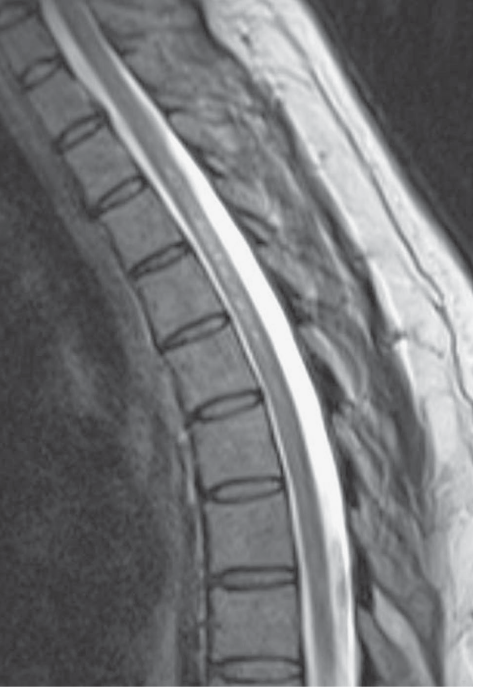
Figure: MRI of spinal cord in NMOSD demonstrating a longitudinally extensive lesion (LETM) spanning multiple vertebral segments. - Adams & Victor's Principles of Neurology, 12th Ed.
- Spinal cord: LETM (≥3 contiguous segments), centered on gray matter on axial sequences, with focal enhancement and swelling
- Optic nerve: T2-hyperintense lesion spanning >1/2 nerve length or involving chiasm (vs. spotty changes in MS)
- Area postrema: Dorsal medullary lesion - "highly confirmatory" of NMOSD
- Brain: Can have "cloud-like" asymmetric hemispheric lesions, hypothalamic lesions, periaqueductal lesions; unlike MS lesions, these often resolve completely
- Cord atrophy may appear weeks after onset
Key CSF distinction from MS: Pleocytosis with neutrophils and eosinophils; oligoclonal bands are absent in >80% of NMO patients (vs. present in ~85-95% of MS)
Diagnostic Criteria (2015 IPND)
NMOSD with AQP4-IgG (requires ALL 3):
- At least one core clinical characteristic
- Positive AQP4-IgG (cell-based assay strongly recommended)
- Exclusion of alternative diagnoses
NMOSD without AQP4-IgG or unknown status (requires ALL 3):
- At least two core clinical characteristics, with:
- At least one must be optic neuritis, LETM, or area postrema syndrome
- Dissemination in space (two or more different characteristics)
- Fulfillment of additional MRI requirements
- Negative AQP4-IgG (best available method) or unavailable
- Exclusion of alternative diagnoses
Older "Wingerchuk 2006" definite criteria (still referenced): Optic neuritis + acute myelitis + at least 2 of 3: (1) LETM ≥3 segments, (2) brain MRI not meeting MS criteria, (3) AQP4-IgG seropositive.
Clinical Course
- Typically relapsing (>90% of cases); monophasic in <10%
- AQP4-seronegative patients more likely to have a monophasic course
- Without treatment: 60% blind and >50% with permanent limb paralysis at 8 years; respiratory failure in 1/3 from cervical myelitis
- With modern therapies: 5-year survival improved from 68-75% (pre-2000) to 91-98% (as of 2017)
- Unlike MS, damage does not remit to any great extent; each attack causes cumulative, permanent disability
- AQP4-seropositive: >50% will relapse within 1 year if untreated - Harrison's 22E
Treatment
Acute Attacks
- High-dose IV methylprednisolone: 1 g/day for 5-10 days + oral prednisone taper (first line)
- Plasma exchange (5-7 exchanges, 1.5 plasma volumes/exchange): for steroid-refractory attacks
Relapse Prevention (FDA-Approved Monoclonal Antibodies)
| Drug | Mechanism | Attack Risk Reduction (AQP4+) | Dosing |
|---|---|---|---|
| Eculizumab (Soliris) | Anti-C5 complement inhibitor | 94% (add-on to IST) | 900 mg wkly x4, then 1200 mg q2wk |
| Ravulizumab | Anti-C5 complement inhibitor | 100% (add-on to IST) | Long-acting dosing |
| Inebilizumab (Uplizna) | Anti-CD19 B-cell depleter | 77-78% (monotherapy) | 300 mg IV at day 1 & 15, then q6mo |
| Satralizumab (Enspryg) | Anti-IL-6 receptor | 74-77% (mono or add-on) | 120 mg SC at wks 0,2,4 then q4wk |
Clinical practice note (Harrison's 22E): Inebilizumab or satralizumab are preferred first-line; complement inhibitors (eculizumab/ravulizumab) are second-line for nonresponders. All four are approved only for AQP4-seropositive patients.
- AQP4-seronegative patients: Empiric therapy with rituximab or mycophenolate mofetil (off-label); relapse risk is lower (~half have only a single attack)
Important safety notes:
- Eculizumab/ravulizumab: Boxed warning for life-threatening meningococcal infections - meningococcal vaccination (Men-ACWY x2 + MenB) required before starting
- Satralizumab: Monitor for transaminase elevation, neutropenia; 30% had ≥7% weight gain
- MS drugs (natalizumab, fingolimod, interferons) can worsen NMOSD - correct diagnosis before treating
NMOSD vs. MS: Key Distinctions
| Feature | NMOSD | MS |
|---|---|---|
| Antibody | AQP4-IgG (~75-90%) | None specific |
| Primary pathology | Astrocytopathy | Oligodendropathy/demyelination |
| Spinal lesion | LETM (≥3 segments), gray matter centered | Short, white matter eccentric |
| Optic neuritis | Bilateral, severe, full-length nerve | Unilateral, moderate, focal |
| CSF OCBs | <20% | 85-95% |
| Gliosis | Absent/minimal | Characteristic |
| Cavitation | Common | Rare |
| Recovery from attacks | Poor, cumulative disability | Usually remits (early disease) |
| Cerebellum | Almost never affected | Frequently affected |
| Comorbid autoimmunity | ~40% | Rare (except hypothyroidism) |
Recent Evidence (2025)
A 2025 systematic review and network meta-analysis (PMID 40773034) published in J Neurol characterized MRI lesion differences between AQP4+ NMOSD, MOGAD, and MS, further refining imaging-based diagnostic criteria. A Bayesian network meta-analysis (PMID 40775082) from August 2025 compared approved immunotherapies in NMOSD, supporting the current hierarchy of treatments above.
Sources: Harrison's Principles of Internal Medicine, 22E (2025) | Adams & Victor's Principles of Neurology, 12th Ed. | Localization in Clinical Neurology, 8th Ed.
This is a shared conversation. Sign in to Orris to start your own chat.