
Assessment and resuscitation of ac patient with acute burns. Anaestheticim
Reading File
Now let me run parallel searches across the library for burns assessment, resuscitation, and anaesthetic management.
Finding Sources
Finding Sources
Finding Sources
Good results. Now let me read the most relevant sections in parallel - Morgan & Mikhail (anaesthesia burns), Rosen's (emergency burns assessment), and Tintinalli's (burns management).
Reading File
Reading File
Reading File
Excellent data. Now let me get the anaesthetic-specific content from Morgan & Mikhail (airway management in burns, drug considerations, inhalation injury) and more from Rosen's.
Reading File
Reading File
Finding Sources
Let me get more of the anaesthetic-specific content from Morgan & Mikhail and search for succinylcholine/drug considerations in burns.
Reading File
Finding Sources
Finding Sources
Excellent data. Now let me fetch the Rule of Nines diagram and get Rosen's burns classification/assessment detail.
Loading Image
Reading File
I now have comprehensive data from multiple authoritative anaesthesia and emergency medicine textbooks. Let me compile the full answer.
Assessment and Resuscitation of the Acute Burns Patient - Anaesthetic Perspective
1. Primary Survey (ABCDE)
A - Airway (Most Critical Decision)
Early airway assessment and securing is the single most important anaesthetic intervention in burns. Thermal injury causes predictable swelling of glottic and supraglottic structures, and edema worsens rapidly - especially as IV fluid resuscitation begins.
Indications for immediate/early intubation:
- Hoarse voice or stridor
- Dyspnea or tachypnea
- Altered level of consciousness
- History of exposure in an enclosed space
- Suspected smoke inhalation
- Moderate-to-severe facial or oropharyngeal burns
- Circumferential neck burns
- Visible airway injury on laryngoscopy
- Oropharyngeal soot, singed nasal/facial hair
- Burns ≥40% TBSA
- Hemodynamic instability, dysphagia
Facial burns alone (singed hair, nasal vibrissae) are NOT a mandatory indication for intubation. A hoarse voice, dyspnea, or altered consciousness are the key triggers. - Morgan and Mikhail's Clinical Anesthesiology, 7e, p.1561
Preferred intubation method:
- Awake intubation with generous topical anaesthesia ± sedation is preferred when a difficult airway is anticipated with partial obstruction
- RSI with video laryngoscopy is acceptable if intubation is performed early (before significant edema develops)
- If "can't intubate, can't oxygenate" develops, follow the failed airway algorithm - surgical airway is rarely but definitionally indicated
No single feature adequately predicts the need for intubation; when in doubt, early intubation is encouraged. Flexible laryngoscopy can substitute for empirical intubation in mild/inconclusive cases. - Rosen's Emergency Medicine, p.835
B - Breathing
- Supplemental humidified oxygen to maintain SpO2 >92% in all suspected inhalation injury
- Lung-protective ventilation: Tidal volume 6-8 mL/kg, plateau pressures <35 cmH2O, PEEP ~8 cmH2O
Recommended initial ventilator settings (Rosen's, p.835):
| Parameter | Adult | Paediatric |
|---|---|---|
| Tidal volume | 6-8 mL/kg | 6-8 mL/kg |
| Respiratory rate | 8-12 | 12-45 |
| Plateau pressure | <35 cmH2O | <35 cmH2O |
| I:E ratio | 1:1 to 1:3 | 1:1 to 1:3 |
| PEEP | 8 cmH2O | 8 cmH2O |
- Permissive hypercapnia is acceptable
- Bronchoscopy is the gold standard to diagnose inhalation injury, determine severity, and clear debris
- Inhaled beta-agonists for bronchoconstriction
- Nebulised heparin (5,000-10,000 IU) + N-acetylcysteine reduces duration of mechanical ventilation
- Corticosteroids are not recommended empirically
C - Circulation and Haemodynamics
Burn Shock Physiology:
- Cardiac output falls by up to 50% within 30 minutes of major burn
- Mechanism: massive burn-induced vasoconstriction → normovolaemic hypoperfusion ("burn shock")
- If adequate crystalloid resuscitation is given, cardiac function normalises within 48 hours, then transitions to hyperdynamic physiology as healing begins
Vascular Access:
- Two large-bore IV catheters - through unburned tissue if at all possible
- IV access through burned tissue is acceptable if necessary
2. Burn Size Assessment - Rule of Nines
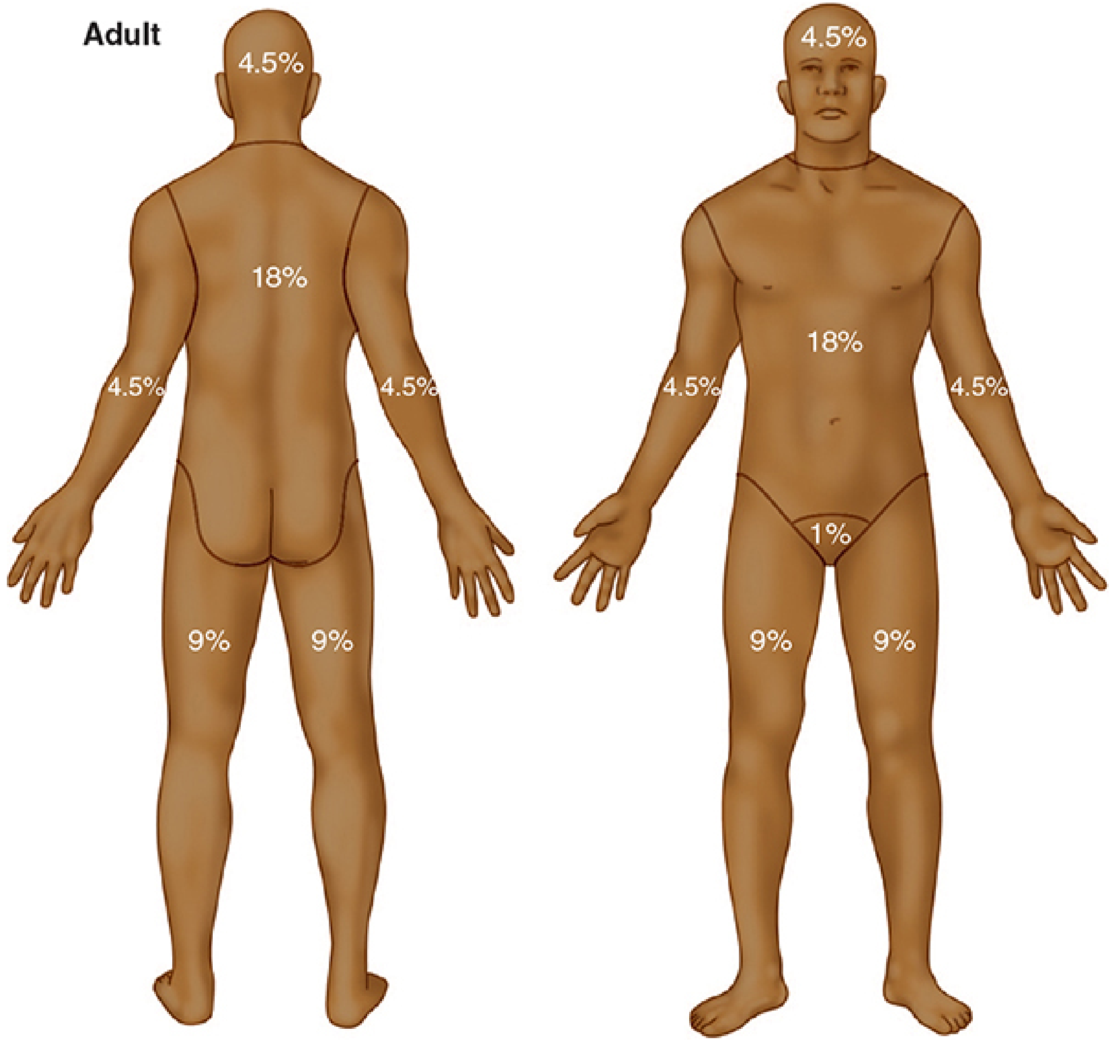
Rule of Nines for estimating burn TBSA in adults. Head = 9%, each arm = 9%, chest = 18%, back = 18%, each leg = 18%, perineum = 1%.
Important: First-degree burns (epidermal only, no blistering) are excluded from TBSA calculations used for fluid resuscitation formulae. Including them leads to fluid creep. - Morgan and Mikhail's, p.1558
Burn depth classification:
- Superficial (1st degree): Epidermal only - not included in TBSA
- Partial thickness (2nd degree): Into dermis - painful, blistered
- Full thickness (3rd degree): All skin layers destroyed - insensate, leathery
3. Fluid Resuscitation
Start IV fluid resuscitation for:
- Adults: burns >20% TBSA
- Children: burns >10% TBSA
The fluid of choice is Lactated Ringer's (LR). Avoid:
- Hypertonic saline (increased acute kidney failure risk in burns)
- Blood products during initial resuscitation (increased mortality)
- Albumin (no outcomes benefit vs. crystalloid)
- Hydroxyethyl starch
Resuscitation Formulae (first 24 hours from time of injury):
| Formula | Volume | Timing |
|---|---|---|
| Parkland | LR 4 mL/kg × %TBSA | Half in first 8h, half over next 16h |
| Modified Brooke | LR 2 mL/kg × %TBSA | Half in first 8h, half over next 16h |
| ABA (paediatric <14 yrs) | LR 3 mL/kg × %TBSA | Half in first 8h, half over next 16h |
| Infants/young children (≤30 kg) | LR 3 mL/kg × %TBSA + D5LR maintenance | — |
Time is calculated from the time of injury, not time of arrival. If a patient arrives 4 hours post-burn, the first 8-hour volume must be delivered in the remaining 4 hours. - Rosen's Emergency Medicine, p.836
Second 24 hours: Administer half the first 24-hour volume.
Monitoring Adequacy of Resuscitation:
- Urine output is the primary endpoint: target 0.5-1.0 mL/kg/h in adults; 1.0 mL/kg/h in children; 1-2 mL/kg/h in infants <1 year
- If urine output >1.0 mL/kg/h in adults, slow the infusion rate
Complications of Over-Resuscitation:
- Fluid creep: Volume administered far exceeds calculated goal (often due to miscalculation from including 1st-degree burns, or sedation-induced hypotension prompting excess fluids)
- Abdominal Compartment Syndrome (ACS): Risk with circumferential abdominal burns or >6 mL/kg/%TBSA received. Measure bladder pressure via Foley catheter; intra-abdominal pressure >20 mmHg warrants decompression
- Pneumonia and ARDS: Excess fluid promotes pulmonary edema, especially in the context of inhalation injury
4. CO and Cyanide Poisoning
Carbon Monoxide (CO):
- CO binds haemoglobin with ~250× the affinity of oxygen
- Carboxyhemoglobin (HbCO) shifts O2-Hb dissociation curve leftward (reduced O2 delivery)
- Pulse oximetry is falsely normal - it cannot distinguish HbO2 from HbCO. Always get ABG/VBG for direct HbCO measurement
- HbCO >10%: clinically significant (smokers have baseline up to 10%)
- HbCO >20%: intubate and ventilate with 100% O2
- HbCO >60%: lethal
- Hyperbaric oxygen is indicated for all significant CO poisonings and requires multiple sessions to reduce long-term neurological sequelae
Cyanide poisoning should be considered in any patient with enclosed-space fire, altered mental status, and metabolic acidosis despite adequate oxygenation. - Morgan and Mikhail's, p.1560
5. Anaesthetic Drug Considerations
Succinylcholine and the Hyperkalemia Window
This is one of the most important anaesthetic considerations in burns:
| Time from burn | Succinylcholine safety |
|---|---|
| 0-48 hours | SAFE - can be used for RSI |
| >48 hours | DANGEROUS - risk of lethal hyperkalemia |
| Up to 2 years post-burn | Risk persists |
Mechanism: Major burns (>20% TBSA) disrupt neuromuscular end-plates and cause upregulation of extrajunctional acetylcholine receptors throughout the muscle membrane. On depolarisation with succinylcholine, massive K+ efflux from all these receptors causes lethal hyperkalaemia. This vulnerability starts around 3-5 days but becomes clinically significant beyond 48 hours.
"Beyond 48 h following a major burn injury, succinylcholine administration can produce lethal hyperkalemia. This risk persists for up to 2 years following burn injury." - Morgan and Mikhail's Clinical Anesthesiology, 7e, p.1561
After 48 hours: Use non-depolarising muscle relaxants (NDMRs) - e.g. rocuronium. Note that burn patients typically require larger doses of NDMRs due to upregulated receptor numbers (pharmacodynamic resistance).
Temperature Management
- Burns patients cannot thermoregulate
- All resuscitation and care environments must be maintained near 40°C
- Use radiant warming, forced-air warming devices, and fluid warmers
Analgesia
- Multimodal analgesia is preferred (opioid + adjuncts)
- IV morphine: 2-4 mg IV in adults, 0.05 mg/kg in children <50 kg
- Caution with regional techniques in the early post-burn period - they may mask symptoms of compartment syndrome
6. Indications for Admission / Burns Unit Transfer
Major burns requiring specialist burns unit transfer:
- Partial or full thickness >10% TBSA (all ages) or >20% TBSA (adults)
- Burns involving face, hands, feet, genitalia, perineum, or major joints
- Full thickness burns >5% TBSA
- Circumferential burns of limbs or chest
- Chemical or electrical burns
- Inhalation injury
- Significant co-morbidities or extremes of age
Summary: Anaesthetic Priorities at a Glance
- Airway first - anticipate a difficult, evolving airway; intubate early if any signs of inhalation injury or airway compromise
- 100% O2 always initially - treats CO poisoning and hypoxia simultaneously
- LR resuscitation using Parkland (4 mL/kg/%TBSA) or modified Brooke (2 mL/kg/%TBSA); titrate to urine output 0.5-1 mL/kg/h
- Succinylcholine is safe in first 48 hours only - use NDMR (rocuronium) beyond 48 hours
- Pulse oximetry unreliable with CO poisoning - ABG mandatory
- Avoid fluid creep - monitor for abdominal compartment syndrome
- Warm everything - patient, IV fluids, environment
Sources: Morgan and Mikhail's Clinical Anesthesiology 7e; Rosen's Emergency Medicine; Tintinalli's Emergency Medicine; Miller's Anesthesia 10e
This is a shared conversation. Sign in to Orris to start your own chat.