
Malignant glaucoma
I'll start by reading the medical library skill, then search for malignant glaucoma content in parallel.
Reading File
Now I'll search the library and PubMed simultaneously.
Finding Sources
Searching PubMed
"malignant glaucoma" OR "aqueous misdirection"
I have two textbook sources. Let me read both simultaneously.
Reading File
Reading File
Let me get the truncated Wills Eye content and fetch the relevant image.
Reading File
Loading Image
I now have comprehensive content from both textbooks. Let me also get the follow-up section from Wills Eye.
Reading File
I now have all the information needed for a thorough answer. Here is the full review:
Malignant Glaucoma (Aqueous Misdirection Syndrome)
Definition and Pathophysiology
Malignant glaucoma, also called aqueous misdirection syndrome or ciliolenticular block, is a rare but serious form of secondary angle-closure glaucoma in which aqueous humor is misdirected posteriorly into (or behind) the vitreous rather than flowing forward into the anterior chamber through its normal route.
The core mechanism involves anterior rotation of the ciliary body and ciliary processes, which presses against the lens equator (in phakic eyes) or the anterior vitreous face (in aphakic/pseudophakic eyes). This creates a block at the level of the ciliary body-lens interface, diverting aqueous backward. The accumulated fluid in the vitreous then displaces the lens-iris diaphragm anteriorly, causing diffuse shallowing of the anterior chamber and raised IOP - even though the drainage angle may be open or a patent peripheral iridotomy (PI) is already present.
Newer theories additionally implicate: choroidal expansion, reduced conductivity of fluid through vitreous, and reduced trans-scleral fluid movement.
- Kanski's Clinical Ophthalmology, 10th ed.
- Wills Eye Manual
Predisposing Factors / Triggers
- Following incisional surgery - trabeculectomy (most classic), cataract surgery, glaucoma drainage device implantation, retinal surgery
- Eyes with small anterior segments: hyperopia, nanophthalmos
- Primary angle-closure glaucoma (PACG) - highest risk group
- Can occur spontaneously without surgery
- May be precipitated by miotics (pilocarpine) - this is a classic exam point; miotics worsen the condition by causing further anterior rotation of the ciliary body
Clinical Features
Symptoms
- May be mild initially
- Moderate pain, red eye, photophobia as IOP rises
- Characteristic myopic shift - patients notice improved unaided near vision (as the anteriorly displaced lens increases its refractive power)
Signs (Critical triad)
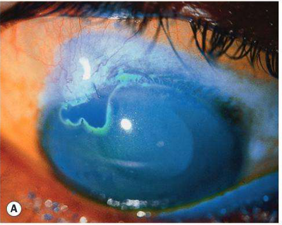
- Diffusely shallow or flat anterior chamber (uniform shallowing - both centrally and peripherally, unlike pupillary block where peripheral shallowing is more pronounced)
- Elevated IOP (though may be only mildly raised early on)
- Patent peripheral iridotomy - no iris bombé - the key distinguishing feature from pupillary block
- Negative Seidel test (no wound leak)
- No choroidal detachment on B-scan ultrasound
Differential Diagnosis
| Feature | Malignant Glaucoma | Pupillary Block | Choroidal Detachment | Wound Leak |
|---|---|---|---|---|
| AC shallowing | Diffuse (central + peripheral) | Peripheral > central (iris bombé) | Present | Present |
| IOP | High | High | Low | Low |
| Iris bombé | Absent | Present | Absent | Absent |
| Patent PI | Present | Absent/blocked | Present | Present |
| Seidel test | Negative | Negative | Negative | Positive |
Workup
- History - prior ocular surgery, miotic use
- Slit lamp exam - assess for patent PI, iris bombé (bombé rules out pupillary block)
- Gonioscopy + IOP measurement
- Dilated retinal exam (unless phakic angle closure is likely)
- B-scan ultrasound - to rule out choroidal detachment and suprachoroidal hemorrhage
- Seidel test - to exclude wound leak
Treatment
Treatment follows a stepwise escalation from medical to laser to surgical:
Step 1 - Ensure patent PI
If no PI or it is not clearly patent, pupillary block cannot be excluded - perform PI first. If signs persist with a patent PI, proceed to medical therapy.
Step 2 - Medical (First-line)
The goal is to tighten the zonules and pull the lens posteriorly, and to shrink the vitreous:
| Drug | Dose | Mechanism |
|---|---|---|
| Atropine 1% | q.i.d. topically | Dilates ciliary ring, tightens zonules, retracts lens posteriorly |
| Phenylephrine 2.5-10% | q.i.d. topically | Cycloplegic support, pupil dilation |
| Acetazolamide | 500 mg IV or 2 × 250 mg PO | Reduces aqueous production |
| Timolol 0.5% | Daily or b.i.d. | Reduces aqueous production |
| Brimonidine/Apraclonidine | b.i.d. | Reduces aqueous production |
| Mannitol 20% | 1-2 g/kg IV over 45 min | Vitreous dehydration/shrinkage - moves lens posteriorly |
Critical: Miotics are absolutely contraindicated - they worsen aqueous misdirection by causing further anterior rotation of the ciliary body.
If medical therapy breaks the attack (anterior chamber deepens, IOP normalizes): continue atropine 1% daily indefinitely to prevent recurrence.
Step 3 - Laser (if medical fails)
- Nd:YAG laser disruption of the anterior hyaloid face - fired through the iridotomy (phakic) or through the posterior capsule (pseudophakic/aphakic). The goal is to create a unicameral eye - allowing free aqueous communication between anterior chamber and vitreous cavity.
- In pseudophakic eyes: YAG posterior capsulotomy + anterior hyaloid disruption
- Cyclodiode laser (to reduce aqueous production/ciliary body ablation) may also be effective
- Argon laser to ciliary processes (less commonly used)
Step 4 - Surgical (if laser fails)
- Pars plana vitrectomy (PPV) - the definitive surgical treatment. The key is to remove sufficient anterior vitreous gel to allow free flow of aqueous to the anterior chamber.
- PPV with irido-zonulo-hyaloidectomy - vitrectomy combined with localized excision of iris, zonules, lens capsule, and anterior hyaloid face; reformation of anterior chamber. This creates a truly unicameral eye.
- Lensectomy + anterior hyaloid disruption
Note: Before vitrectomy, consider performing a sclerotomy to drain any undetected anterior choroidal detachment, which may co-exist.
Contralateral Eye
Perform peripheral iridotomy (PI) in the contralateral eye if the angle is occludable - typically within 1 week of treating the involved eye, as there is significant risk of malignant glaucoma developing in the fellow eye.
Key Distinguishing Concept
The hallmark that sets malignant glaucoma apart from all other causes of shallow AC with elevated IOP is: diffusely flat anterior chamber + raised IOP + patent PI + no choroidal detachment. The fact that the iridotomy is patent (ruling out pupillary block) yet the chamber remains flat is the diagnostic key.
Sources: Kanski's Clinical Ophthalmology: A Systematic Approach, 10th ed., p. 435 | The Wills Eye Manual, pp. 623-625
Pathogenesis of it
The classic explanation is ciliolenticular block - the ciliary processes rotate anteriorly, press against the lens equator, and redirect aqueous posteriorly into the vitreous. This is well established but newer theories have expanded on it significantly.
Searching sources...
Finding Sources
Searching PubMed
"malignant glaucoma" pathogenesis mechanism
Good. Let me now pull the deeper pathogenesis sections from Kanski - specifically the retrolenticular/classification section and look for any extended mechanistic discussion.
Reading File
Finding Sources
I have the key Wills Eye etiology paragraph (line 4603) and Kanski content. Let me fetch the Wills Eye etiology paragraph and look for any dedicated Kanski pathogenesis section on malignant glaucoma specifically.
Reading File
Finding Sources
I have all the content needed. Here is the focused pathogenesis answer:
Pathogenesis of Malignant Glaucoma
The pathogenesis involves several interacting mechanisms operating in sequence. There is a classic (anatomical/mechanical) theory and several newer theories that have refined understanding.
1. Anatomical Predisposition
The initiating vulnerability is a structurally crowded anterior segment:
- Short axial length (hyperopia, nanophthalmos)
- Small corneal diameter
- Anteriorly positioned lens with a thick lens/cornea ratio
- Shallow anterior chamber with narrow angles
- Tight relationships between the ciliary body, lens equator, and vitreous face
These features mean any additional forward rotation of the ciliary body has very little anatomical reserve before it makes contact with the lens equator.
2. The Classic Theory: Ciliolenticular Block (Ciliary Body Rotation)
This is the central, established mechanism:
Step 1 - Trigger event
A precipitating event (most commonly intraocular surgery, particularly trabeculectomy in PACG eyes, but also spontaneous) causes anterior rotation of the ciliary body and ciliary processes. The exact trigger for this rotation is incompletely understood, but anterior traction from supra-ciliary effusion, choroidal congestion, or surgical hypotony are implicated.
Step 2 - Ciliolenticular apposition
The anteriorly rotated ciliary processes come into apposition with the equator of the crystalline lens (or the anterior vitreous face in aphakic/pseudophakic eyes). This creates a functional block at the ciliary body-lens junction.
Step 3 - Posterior aqueous misdirection
Instead of flowing forward through the posterior chamber into the pupil and then the anterior chamber, aqueous is deflected posteriorly - into and behind the vitreous gel. The anterior hyaloid face is not a perfect barrier; aqueous seeps through or around it and accumulates within the vitreous body.
Step 4 - Vitreous expansion and anterior displacement
The accumulating aqueous within the vitreous increases vitreous volume, driving the entire lens-iris diaphragm anteriorly. In pseudophakic eyes, the IOL and anterior vitreous face are pushed forward instead.
Step 5 - Secondary angle closure
The anteriorly displaced lens-iris diaphragm pushes the iris against the trabecular meshwork, causing diffuse angle closure - but crucially, this is not pupillary block (the iris does not bow forward in the classic bombé pattern, and a patent PI does not relieve it).
Step 6 - Self-perpetuating cycle
The raised IOP and further ciliary body effusion worsen the anterior rotation of the ciliary processes, deepening the block. The misdirection becomes self-sustaining.
3. Why It Differs from Pupillary Block
This is pathogenically distinct and the distinction is clinically critical:
| Pupillary Block | Malignant Glaucoma | |
|---|---|---|
| Block level | Pupil (iris-lens interface) | Ciliary body-lens equator |
| Iris pattern | Bombé (peripheral bowing) | Diffuse flat, no bombé |
| PI effect | Relieves it | Does NOT relieve it |
| Driving force | Anterior-posterior pressure gradient at pupil | Posterior misdirection of aqueous into vitreous |
A patent PI equalizes the anterior-posterior chamber pressure in pupillary block. In malignant glaucoma, the block is posterior to the iris entirely - opening the iris does nothing to redirect aqueous that is already trapped behind the vitreous.
4. Newer/Additional Theories
The Wills Eye Manual explicitly notes that the ciliolenticular block theory alone does not fully explain all cases, and three additional mechanisms have been proposed:
a) Choroidal Expansion
- Uveal/choroidal congestion or effusion causes the ciliary body to expand and rotate anteriorly
- This is likely the initiating event in surgical and some spontaneous cases
- Explains why hypotony (e.g., post-trabeculectomy) - which increases uveal vasodilation and transudation - is such a potent trigger
b) Reduced Conductivity of Fluid Through the Vitreous
- Normal vitreous has some hydraulic conductivity that allows fluid to move anteriorly
- With age, vitreous syneresis (liquefaction and condensation), or in structurally abnormal eyes, the vitreous gel becomes a relative barrier to posterior-to-anterior fluid movement
- This means aqueous that enters the vitreous cannot easily move forward, and instead it accumulates and pushes the vitreous anteriorly en masse
c) Reduced Trans-scleral Fluid Movement
- Some aqueous normally exits the eye via the uveoscleral (trans-scleral) route, including across the ciliary body and sclera
- In susceptible eyes, reduced trans-scleral outflow increases posterior segment pressure, contributing to forward displacement of ocular structures and reduced ability to absorb posteriorly misdirected fluid
5. Why Miotics Make It Worse
This is a direct consequence of the pathogenesis and a classic exam point:
- Miotics (e.g., pilocarpine) cause ciliary muscle contraction
- This pulls the ciliary processes further anteriorly and inward toward the lens equator
- This tightens the ciliolenticular block rather than relieving it
- The zonules slacken, allowing the lens to move anteriorly, worsening the angle closure
Conversely, cycloplegics (atropine) are therapeutic because they relax the ciliary muscle, widen the ciliary ring, tighten the zonules, and pull the lens posteriorly - breaking the block.
6. Role of the Anterior Hyaloid Face
The anterior hyaloid is a key anatomical structure in the pathogenesis:
- It normally forms a barrier between the vitreous cavity and the posterior chamber
- In malignant glaucoma, aqueous accumulates posterior to the hyaloid
- This is why disrupting the anterior hyaloid face (by YAG laser or surgically) is curative - it creates a unicameral eye where aqueous can freely communicate between vitreous, posterior chamber, and anterior chamber, eliminating the misdirection
Summary Diagram (Conceptual)
Trigger (surgery/hypotony/spontaneous)
         ↓
Choroidal expansion → Anterior rotation of ciliary processes
         ↓
Ciliolenticular block (ciliary processes press on lens equator)
         ↓
Aqueous misdirected POSTERIORLY into vitreous
         ↓
Vitreous accumulates aqueous → reduced conductivity traps it further
         ↓
Lens-iris diaphragm displaced ANTERIORLY
         ↓
Diffuse secondary angle closure + raised IOP
         ↓
Self-perpetuating cycle (more effusion → more rotation)
- Kanski's Clinical Ophthalmology: A Systematic Approach, 10th ed., pp. 393, 435
- The Wills Eye Manual, pp. 623-624
This is a shared conversation. Sign in to Orris to start your own chat.