
anatomy of upper limb, lower limb, locomotive system
Reading File
This is a broad visual/anatomical query. Let me search the library across all three topics simultaneously.
Finding Sources
Finding Sources
Finding Sources
Excellent — Gray's Anatomy for Students and THIEME Atlas are both available. Let me now read key sections from all three topics in parallel.
Reading File
Reading File
Reading File
Let me now get the core upper limb and lower limb chapter content from Gray's, and the locomotive system content from THIEME Atlas.
Reading File
Reading File
Finding Sources
Good content coming in. Let me read more of each chapter's core anatomy sections.
Reading File
Reading File
Finding Sources
Let me read more sections: upper limb muscles/nerves/vessels and lower limb muscles/nerves/vessels, plus get the THIEME locomotor system content.
Reading File
Reading File
Finding Sources
Reading File
Reading File
Reading File
Finding Sources
Reading File
Reading File
I now have rich content across all three topics. Let me also get the THIEME block that covers bones/joints/muscles/vessels general content plus key upper limb content from the THIEME atlas.
Finding Sources
Reading File
Reading File
I now have comprehensive content to produce a detailed answer. Let me also fetch a few key images to include.
Loading Image
Loading Image
Loading Image
Loading Image
Loading Image
Anatomy of the Upper Limb, Lower Limb & Locomotive System
Sources: Gray's Anatomy for Students; THIEME Atlas of Anatomy — General Anatomy and Musculoskeletal System
I. The Locomotive (Musculoskeletal) System — Overview
The locomotor system (also called the musculoskeletal system) is divided into two functional parts:
| Component | Role |
|---|---|
| Skeleton + Skeletal Connections (bones, joints, ligaments) | Passive part — provides the rigid framework and leverage |
| Striated Skeletal Musculature | Active part — generates force to move the framework |
The limbs are subdivided into girdles (attachment to trunk) and free limbs:
- Upper limb: Shoulder girdle + free upper limb
- Lower limb: Pelvic girdle + free lower limb
— THIEME Atlas of Anatomy, General Anatomy and Musculoskeletal System, p. 43
II. Upper Limb
Regions & Gateway to the Upper Limb
The upper limb communicates with the trunk through the axillary inlet, bounded by:
- Posterior — superior margin of the scapula
- Anterior — clavicle
- Medial — lateral margin of rib I
The apex is the medial margin of the coracoid process. Large blood vessels pass over rib I; proximal brachial plexus passes through this inlet.
Regions: Shoulder → Arm → Forearm → Hand
Bones & Joints
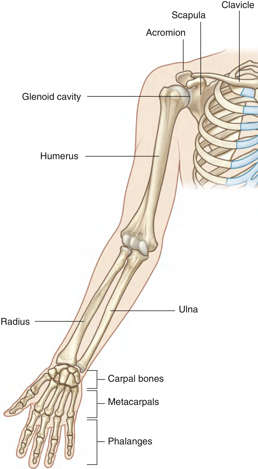
| Segment | Bones | Key Joint |
|---|---|---|
| Shoulder girdle | Clavicle + Scapula | Sternoclavicular joint; Acromioclavicular joint |
| Arm | Humerus | Glenohumeral joint (ball-and-socket) |
| Forearm | Radius (lateral), Ulna (medial) | Elbow (hinge); proximal & distal radioulnar joints |
| Wrist | 8 carpal bones in 2 rows | Radiocarpal joint; midcarpal joint |
| Hand | 5 metacarpals + phalanges (2 in thumb, 3 in each finger) | Carpometacarpal; MCP; IP joints |
Key points:
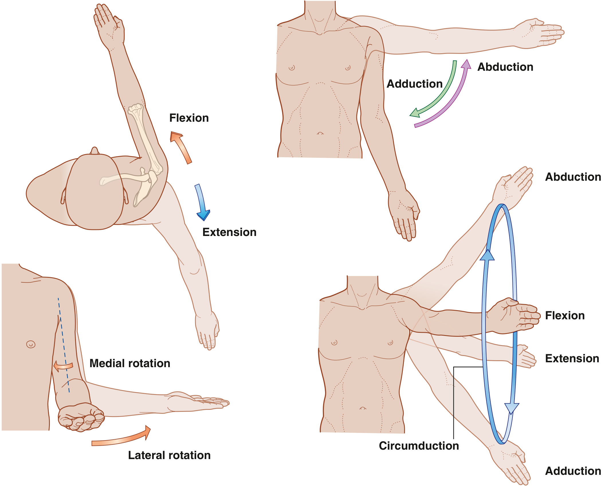
- The glenohumeral joint is a ball-and-socket that allows flexion, extension, abduction, adduction, medial/lateral rotation, and circumduction.
- The elbow is a hinge allowing flexion/extension.
- Pronation/supination occurs at the radioulnar joints — the radius rotates around the ulna.
- The wrist joint is formed between the radius, articular disc (distal to ulna), and proximal carpal row.
- The thumb's carpometacarpal joint is a saddle joint giving it greater freedom than the fingers; opposition is possible because metacarpal I is oriented at right angles to the others.
Movements of the Arm at the Glenohumeral Joint
Muscle Compartments
| Region | Compartment | Main Action | Key Muscles |
|---|---|---|---|
| Arm | Anterior | Flexion of elbow | Biceps brachii, brachialis, coracobrachialis |
| Arm | Posterior | Extension of elbow | Triceps brachii |
| Forearm | Anterior | Wrist/finger flexion, pronation | Flexor carpi radialis/ulnaris, flexor digitorum superficialis/profundus, pronator teres |
| Forearm | Posterior | Wrist/finger extension, supination | Extensor carpi radialis, extensor digitorum, supinator |
| Back/thoracic wall | — | Shoulder-trunk attachment | Trapezius, latissimus dorsi, rhomboids, pectoralis major |
Intrinsic hand muscles include the thenar muscles (opposition/flexion of thumb), hypothenar muscles (little finger), lumbricals, and interossei (fine finger movements).
Innervation — Brachial Plexus (C5–T1)
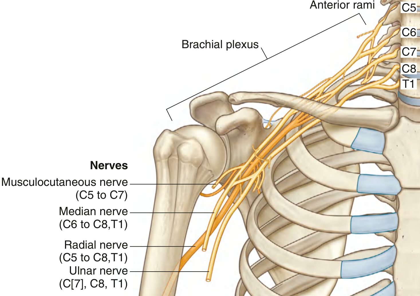
The brachial plexus (anterior rami C5–C8, T1) forms in the neck and continues through the axillary inlet:
| Nerve | Compartments Supplied | Skin Territory |
|---|---|---|
| Musculocutaneous (C5–C7) | Anterior arm | Anterolateral forearm |
| Median (C6–C8, T1) | Anterior forearm (most), thenar muscles, lateral 2 lumbricals | Palmar surface of lateral 3½ digits |
| Ulnar (C7, C8, T1) | Flexor carpi ulnaris, medial FDP, most intrinsic hand muscles | Medial 1½ digits |
| Radial (C5–C8, T1) | All posterior arm & forearm | Posterior forearm, dorsolateral hand |
| Axillary (C5–C6) | Deltoid, teres minor | Upper lateral arm |
Clinical notes on nerves related to bone:
- Axillary nerve — wraps around the surgical neck of the humerus (vulnerable in shoulder dislocation and proximal humeral fractures)
- Radial nerve — passes diagonally in the radial groove of the humerus (vulnerable in midshaft humeral fractures)
- Ulnar nerve — passes posterior to the medial epicondyle ("funny bone"; vulnerable in elbow injuries)
Dermatomes & Myotomes (Upper Limb)
| Spinal Level | Autonomous Dermatome Zone | Key Myotome Test |
|---|---|---|
| C5 | Upper lateral arm | Shoulder abduction |
| C6 | Palmar thumb pad | Elbow flexion |
| C7 | Index finger pad | Elbow extension |
| C8 | Little finger pad | Finger flexion |
| T1 | Medial elbow | Finger abduction/adduction |
Reflexes: biceps tap → C6; triceps tap → C7.
Superficial Veins
The cephalic vein (lateral), basilic vein (medial), and median cubital vein (connecting them at the elbow) lie in superficial fascia. The median cubital vein, overlying the cubital fossa, is the most common site for venepuncture.
III. Lower Limb
Regions
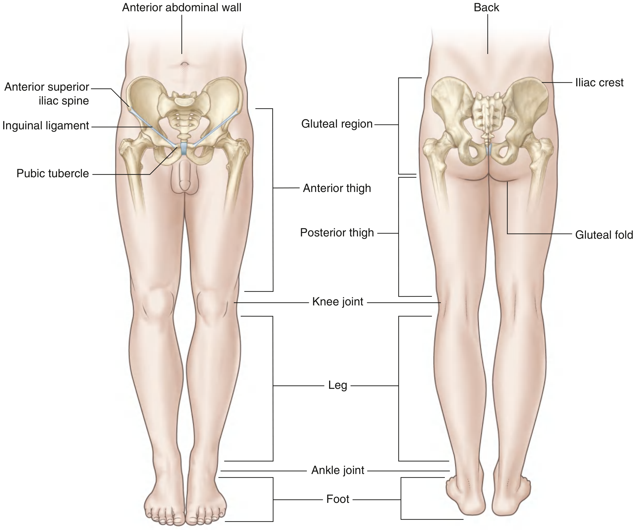
Regions: Gluteal region → Thigh → Leg → Foot
Key transitional areas:
- Femoral triangle — passageway for femoral nerve, artery, vein between abdomen and thigh (inguinal ligament forms the base)
- Popliteal fossa — diamond-shaped posterior to the knee; major vessels and nerves traverse here between thigh and leg
- Tarsal tunnel — posteromedial ankle; nerves/vessels/flexor tendons pass from leg to foot
Functions
- Weight bearing — center of gravity lies anterior to S2; vertical line falls posterior to hip, anterior to knee and ankle, allowing joints to lock passively with minimal muscle energy
- Locomotion — integration of movements at all lower limb joints during gait cycles
Bones & Joints
| Segment | Bones | Key Joint |
|---|---|---|
| Pelvic girdle | Ilium, ischium, pubis (hip bone); sacrum | Sacroiliac joint; pubic symphysis |
| Thigh | Femur | Hip joint (ball-and-socket) |
| Leg | Tibia (medial), Fibula (lateral) | Knee (hinge + some rotation); superior/inferior tibiofibular joints |
| Foot | 7 tarsals + 5 metatarsals + phalanges (2 in hallux, 3 in other toes) | Ankle (talocrural); subtalar; tarsometatarsal; MTP; IP joints |
Key foot anatomy:
- Inversion/eversion occurs at subtalar and midtarsal (transverse tarsal) joints
- Longitudinal and transverse arches are maintained by ligaments and muscles — the medial longitudinal arch is highest; arches absorb and transmit forces during walking
Muscles
| Region | Compartment | Main Action | Key Nerves |
|---|---|---|---|
| Gluteal region | — | Hip extension, abduction, external rotation | Superior gluteal (abductors/medius/minimus); Inferior gluteal (gluteus maximus) |
| Thigh — anterior | — | Hip flexion, knee extension | Femoral nerve (quadriceps, sartorius) |
| Thigh — medial | — | Hip adduction | Obturator nerve (adductors, gracilis) |
| Thigh — posterior | — | Knee flexion, hip extension | Tibial part of sciatic (hamstrings: biceps femoris, semimembranosus, semitendinosus) |
| Leg — anterior | — | Dorsiflexion, toe extension | Common fibular (deep branch) — tibialis anterior, extensor digitorum |
| Leg — lateral | — | Eversion | Common fibular (superficial) — fibularis longus/brevis |
| Leg — posterior (superficial) | — | Plantarflexion | Tibial nerve — gastrocnemius, soleus |
| Leg — posterior (deep) | — | Toe flexion, inversion | Tibial nerve — tibialis posterior, FHL, FDL |
The iliopsoas (psoas major + iliacus) — the primary hip flexor — originates on the posterior abdominal wall and descends under the inguinal ligament to attach to the lesser trochanter.
Innervation — Lumbar (L1–L4) & Sacral (L4–S3) Plexuses
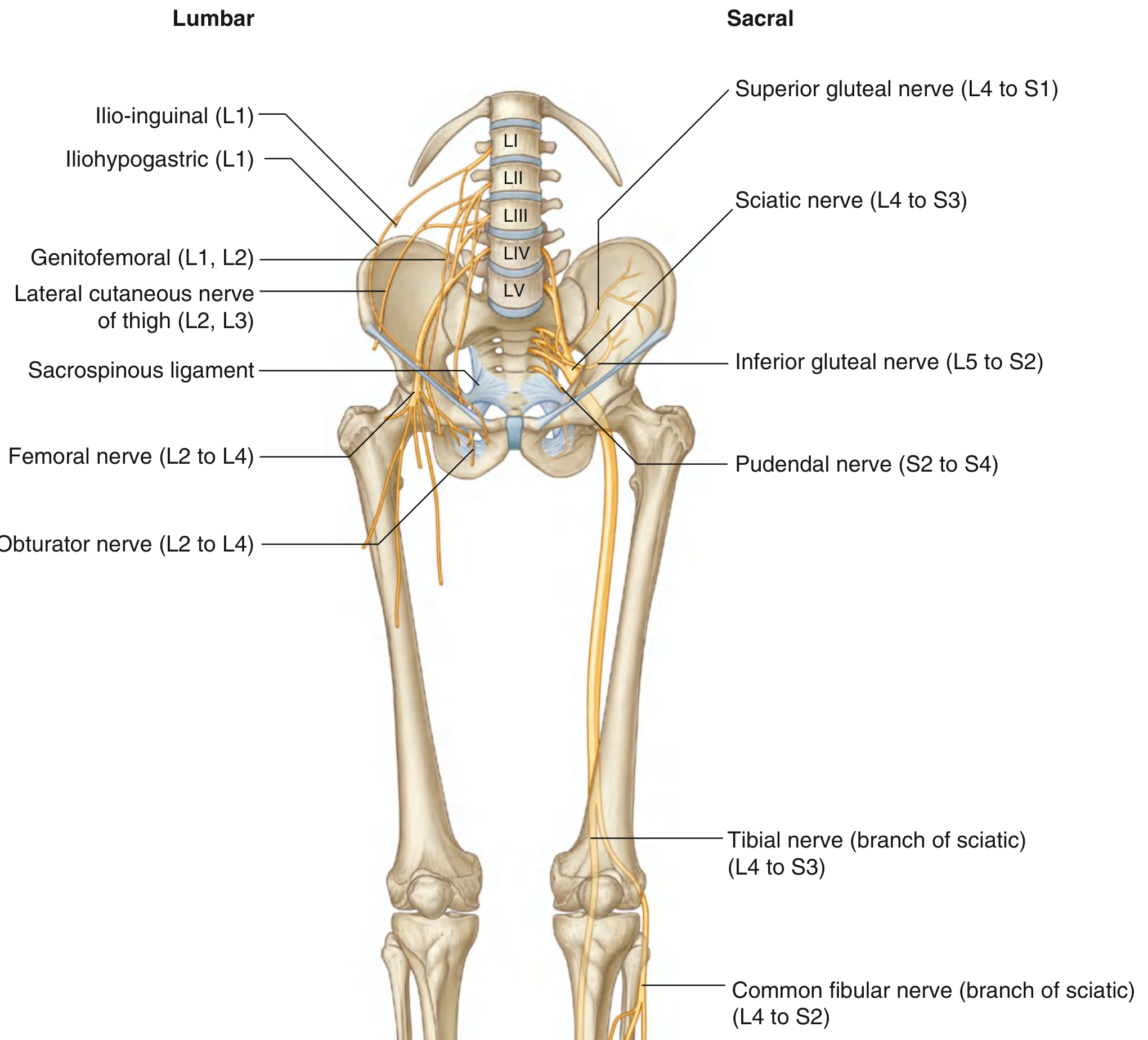
| Nerve | Origin | Motor Territory | Sensory Territory |
|---|---|---|---|
| Femoral | L2–L4 | Anterior thigh (quadriceps, sartorius) | Anterior thigh, medial leg & ankle |
| Obturator | L2–L4 | Medial thigh (adductors) | Medial thigh |
| Sciatic | L4–S3 | All muscles below the knee; hamstrings | Via tibial & common fibular branches |
| Tibial (sciatic branch) | L4–S3 | Posterior leg, sole of foot | Lateral ankle and foot |
| Common fibular (sciatic branch) | L4–S2 | Anterior & lateral leg, dorsum of foot | Lateral leg, dorsum of foot |
| Superior gluteal | L4–S1 | Gluteus medius, minimus, tensor fascia lata | — |
| Inferior gluteal | L5–S2 | Gluteus maximus | — |
Clinical note: The common fibular nerve is vulnerable at the neck of the fibula — it can be rolled against bone here, and is at risk from tight leg casts, fractures, or direct impact.
Dermatomes & Myotomes (Lower Limb)
| Spinal Level | Autonomous Zone | Key Myotome Test |
|---|---|---|
| L1 | Over inguinal ligament | Hip flexion |
| L2 | Lateral thigh | Hip flexion |
| L3 | Lower medial thigh | Knee extension |
| L4 | Medial great toe | Knee extension; ankle dorsiflexion |
| L5 | Medial surface of digit II | Knee flexion |
| S1 | Little toe | Plantarflexion |
| S2 | Back of thigh | Knee flexion; plantarflexion |
Reflexes: patellar tap → L3/L4; calcaneal tendon (Achilles) tap → S1/S2.
Superficial Veins
Two major superficial veins:
- Great saphenous vein — longest vein in the body; runs medially from foot to saphenofemoral junction at the groin
- Small saphenous vein — runs along the posterior leg and drains into the popliteal vein
These are common sites of varicose veins and are used as venous grafts in coronary bypass surgery.
Blood Supply
The external iliac artery becomes the femoral artery as it passes under the inguinal ligament, descends through the femoral triangle, enters the adductor canal, and becomes the popliteal artery behind the knee. Distally it divides into anterior and posterior tibial arteries (the posterior tibial gives off the fibular artery).
IV. Comparative Summary: Upper vs. Lower Limb
| Feature | Upper Limb | Lower Limb |
|---|---|---|
| Primary function | Manipulation, prehension, sensory exploration | Support, locomotion, balance |
| Girdle | Pectoral girdle (clavicle + scapula) | Pelvic girdle (hip bone) |
| Nerve plexus | Brachial (C5–T1) | Lumbar (L1–L4) + Sacral (L4–S3) |
| Major gateway | Axillary inlet (above rib I) | Gap below inguinal ligament |
| Proximal joint | Glenohumeral (most mobile joint in body) | Hip (less mobile but very stable) |
| Distal arch | None (flat palm) | Longitudinal + transverse arches of foot |
| Key superficial veins | Cephalic, basilic, median cubital | Great saphenous, small saphenous |
| Rotation of limb bud (embryology) | Lateral rotation → thumb anterior | Medial rotation → great toe medial |
Gray's Anatomy for Students, Chapter 6 (Lower Limb) and Chapter 7 (Upper Limb); THIEME Atlas of Anatomy — General Anatomy and Musculoskeletal System, pp. 43–44The above is a comprehensive, image-rich reference drawn directly from Gray's Anatomy for Students and the THIEME Atlas of Anatomy. Here's a quick orientation to the sections:
- Locomotive System — defines the passive (skeleton/joints) and active (muscles) subdivisions and how limbs attach to the trunk
- Upper Limb — covers bones & joints (shoulder to phalanges), muscle compartments & their actions, brachial plexus innervation (C5–T1), nerve-to-bone relationships (axillary, radial, ulnar vulnerabilities), dermatomes/myotomes, and superficial veins
- Lower Limb — covers regions, bones & joints (hip to foot arches), gluteal/thigh/leg muscle compartments, lumbar and sacral plexus innervation, dermatomes/myotomes, superficial veins, and arterial supply
- Comparison table — summarizes key structural and functional differences between the two limbs
Let me know if you want to go deeper into any specific region (e.g., brachial plexus, rotator cuff, knee joint, foot arches, or clinical correlations like nerve injury patterns).
This is a shared conversation. Sign in to Orris to start your own chat.