
Protein . and Junction complexes Physiology MBBS 1st year
Reading File
Now let me search the medical library for protein structure and junction complexes content relevant to MBBS 1st year physiology.
Finding Sources
Finding Sources
Finding Sources
Finding Sources
Finding Sources
Reading File
Good findings. Now let me read the Ganong's physiology section on intercellular connections and the Lippincott Biochemistry protein structure section in detail.
Reading File
Reading File
Excellent content found. Let me read more of the Lippincott protein structure section (secondary, tertiary, quaternary) and the Ganong junction section for gap junctions.
Reading File
Reading File
Now let me fetch the key diagrams so they display properly in the answer.
Loading Image
Loading Image
Loading Image
I now have comprehensive content from authoritative textbooks (Lippincott's Biochemistry and Ganong's Review of Medical Physiology). Let me now compose the full MBBS 1st year answer.
Proteins and Junction Complexes - MBBS 1st Year Physiology & Biochemistry
PART 1: PROTEIN STRUCTURE
(Source: Lippincott Illustrated Reviews: Biochemistry, 8th ed.)
Proteins are composed of amino acids joined by peptide bonds in a linear sequence, which then fold into a unique 3D shape that determines function. Protein complexity is analyzed across four organizational levels:
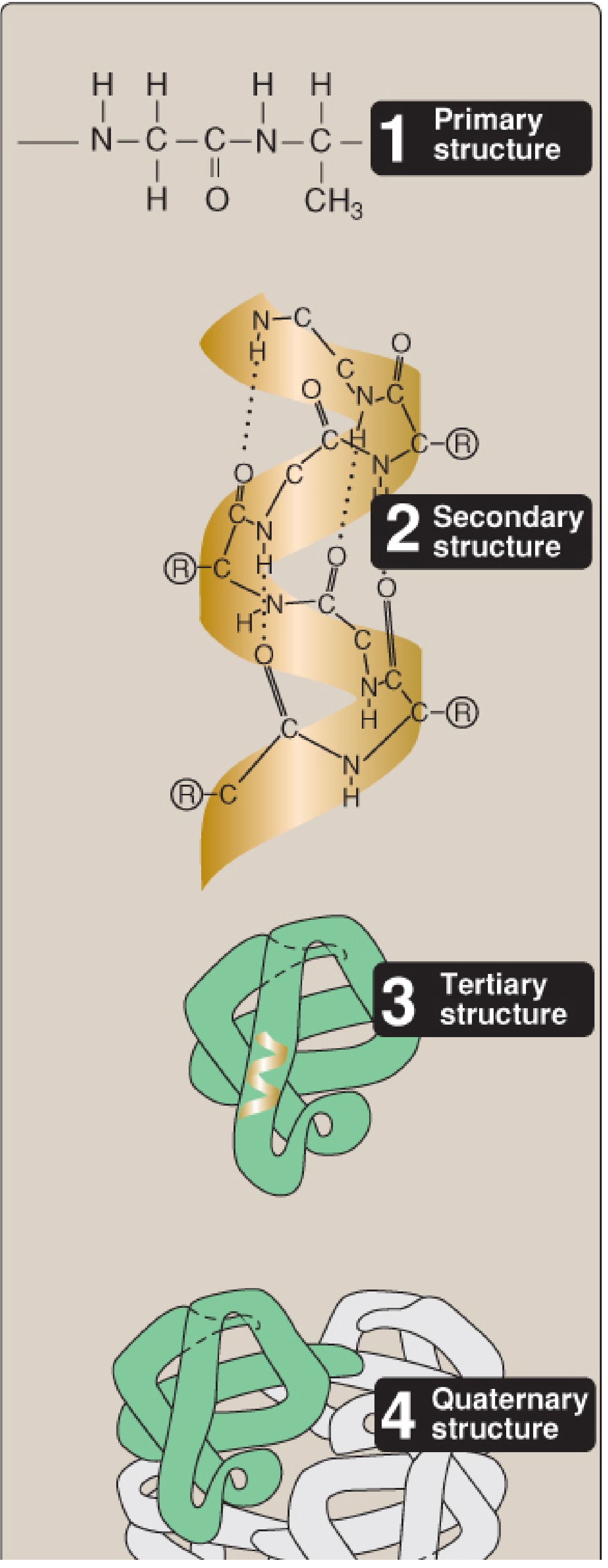
1. Primary Structure
The linear sequence of amino acids from N-terminus (left) to C-terminus (right).
- Adjacent amino acids are joined by peptide bonds (amide linkages between α-COOH of one and α-NH₂ of the next)
- Peptide bonds have partial double-bond character - they are rigid, planar, and almost always in trans configuration
- Linkage of 50+ amino acids = polypeptide / protein
- Each amino acid component is called a residue
- Abnormal primary structure (as in genetic diseases) = misfolding = loss of function (e.g., sickle cell disease)
Peptide bond properties:
- Resistant to heat and urea
- Requires strong acid/base + elevated temperature to break
- -C=O and -NH groups are uncharged (do not accept/release protons at pH 2-12)
2. Secondary Structure
Local folding of the polypeptide chain stabilized by hydrogen bonds between backbone atoms (not R groups).
A. Alpha (α)-Helix
- Right-handed coil; 3.6 amino acids per turn
- Stabilized by H-bonds between C=O of residue n and N-H of residue n+4
- H-bonds are parallel to the backbone axis
- Proline disrupts the helix (introduces a kink); Glycine adds flexibility
- Charged/bulky R-groups and β-carbon branching (e.g., valine) disfavor α-helix
B. Beta (β)-Sheet
- Two or more peptide strands aligned laterally
- H-bonds are perpendicular to the backbone
- Can be parallel (N-termini on same side) or antiparallel (N-termini alternating)
- Pleated appearance due to alternating position of α-carbons above/below the plane
- R groups extend above and below the sheet alternately
C. Beta (β)-Bends (Reverse turns)
- Reverse the direction of the polypeptide chain
- Usually 4 amino acids; often contain Proline (kink-former) and Glycine (smallest R group)
- Connect antiparallel β-strands
- Stabilized by H-bonds between 1st and 4th residue in the bend
- Found on the protein surface; often contain charged residues
3. Tertiary Structure
The overall 3D shape of a single polypeptide chain - how secondary elements pack together.
Stabilizing forces:
| Force | Description |
|---|---|
| Hydrophobic interactions | Nonpolar R groups cluster in core (strongest contributor) |
| Hydrogen bonds | Between polar R groups |
| Ionic bonds (salt bridges) | Between oppositely charged R groups |
| Disulfide bonds | Covalent S-S bonds between cysteine residues |
- Proteins are classified as fibrous (structural, water-insoluble, e.g., collagen, keratin) or globular (functional, often water-soluble, e.g., hemoglobin, enzymes)
- Motifs (super-secondary structures): recurring arrangements of secondary structures (e.g., helix-turn-helix, β-barrel)
- Domains: independently folding, functional units within a polypeptide
4. Quaternary Structure
Assembly of two or more polypeptide subunits (protomers) into a functional complex.
- Stabilized by same non-covalent forces as tertiary structure
- Example: Hemoglobin = 2α + 2β subunits (tetramer)
- Allows cooperative behavior (e.g., O₂ binding in hemoglobin)
- Homodimer = two identical subunits; Heterodimer = two different subunits
PART 2: JUNCTION COMPLEXES
(Source: Ganong's Review of Medical Physiology, 26th Edition)
Intercellular junctions fall into two broad groups:
- Junctions that fasten cells together (mechanical stability)
- Junctions that permit transfer of molecules between cells (communication)
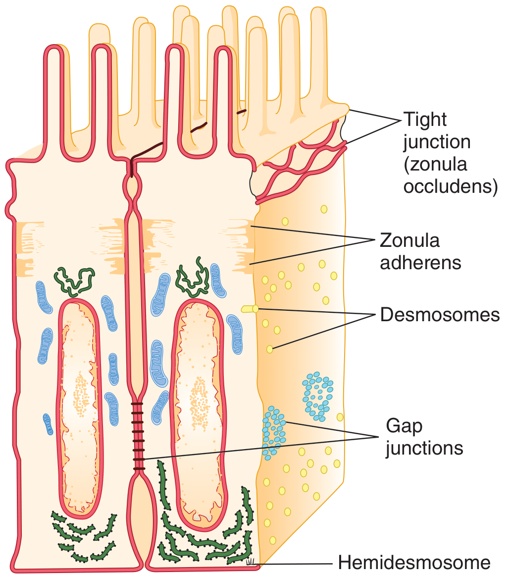
1. Tight Junction (Zonula Occludens)
- Located at the apical margins of epithelial cells (intestinal mucosa, renal tubules, choroid plexus)
- Made of ridges (half from each cell) that adhere so strongly they almost obliterate the intercellular space
- Transmembrane proteins: Occludin, Claudins, Junctional Adhesion Molecules (JAMs)
- Two main functions:
- Paracellular barrier - regulate/restrict passage of ions and solutes between cells (degree of "leakiness" depends on claudin composition)
- Fence function - prevent lateral diffusion of membrane proteins, maintaining distinct apical vs. basolateral domains (essential for directional epithelial transport)
- Also important for endothelial barrier function and endothelium-dependent vasodilation
2. Adherens Junction (Zonula Adherens)
- Continuous structure located basal to the zonula occludens in epithelial cells
- Major site of attachment for intracellular microfilaments (actin cytoskeleton)
- Contains cadherins (Ca²⁺-dependent adhesion molecules)
- Helps hold cells together and transmit mechanical forces
3. Desmosome (Macula Adherens)
- Patch-like thickenings of apposed membranes of two adjacent cells
- Intracellularly connected to intermediate filaments (keratin in epithelial cells)
- Intercellular space contains filamentous material including cadherins and other transmembrane proteins
- Provides strong mechanical links - especially important in tissues subject to mechanical stress (skin, cardiac muscle)
4. Hemidesmosome
- Looks like half a desmosome
- Attaches cells to the underlying basal lamina (not to adjacent cells)
- Connected intracellularly to intermediate filaments
- Contains integrins (not cadherins) - key difference from desmosomes
5. Focal Adhesions
- Also attach cells to their basal lamina
- Associated with actin filaments inside the cell
- Labile structures that play an important role in cell movement
6. Gap Junction (Nexus)
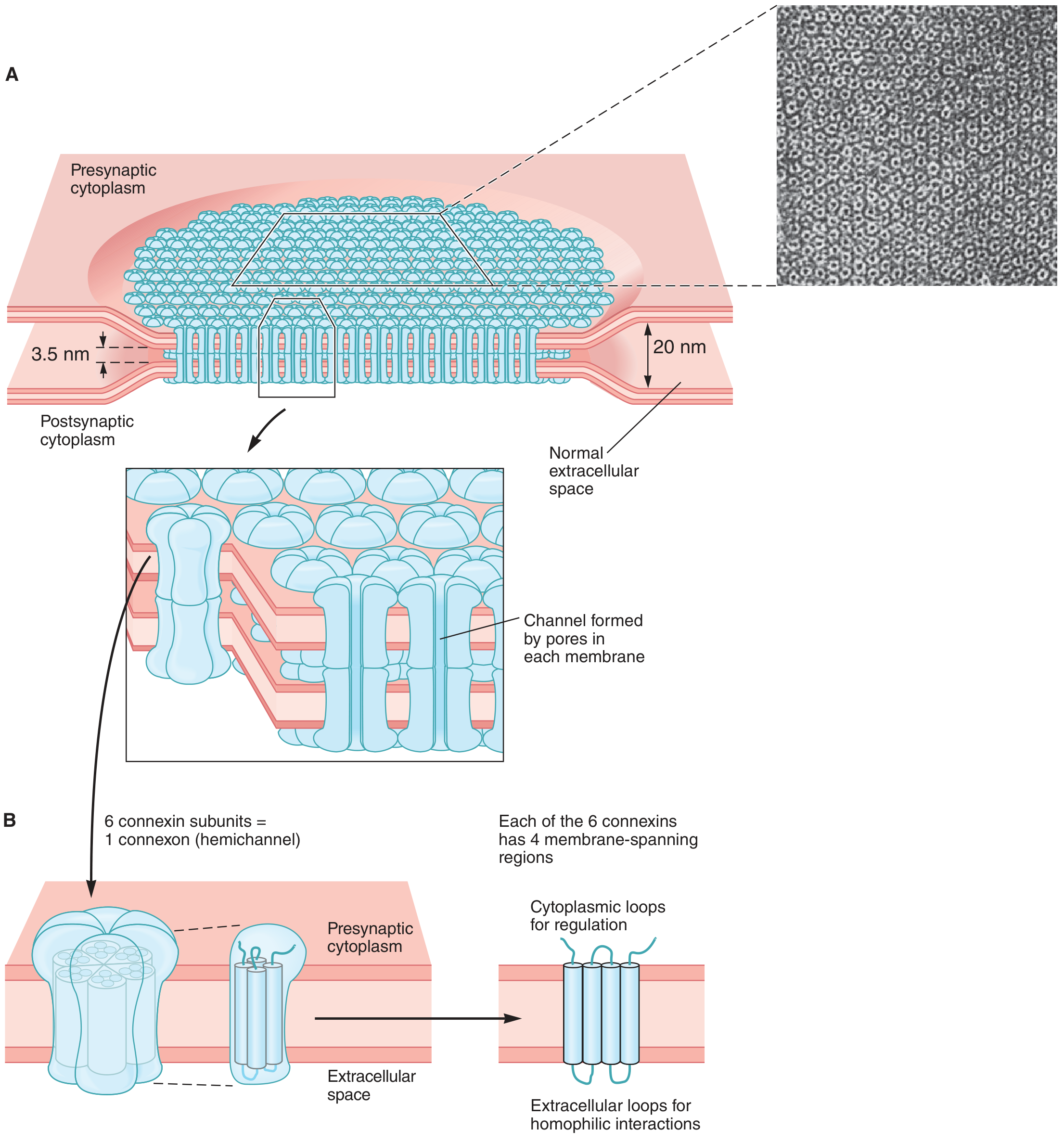
- Intercellular space is reduced to ~4 nm (vs ~20 nm normally)
- Formed by connexons from each cell aligning to form a dodecameric gap junction
- Each connexon = 6 connexin proteins surrounding a central channel
- Each connexin traverses the membrane 4 times
- Pore diameter: 0.8-1.4 nm; allows passage of molecules < 1000 Da
- Permits passage of: ions, sugars, amino acids, cAMP, IP₃, and other intracellular signaling molecules
- Functions:
- Rapid electrical coupling between cells (e.g., cardiac muscle, smooth muscle, CNS)
- Exchange of chemical messengers and metabolites
- Connexins can also form hemichannels (connexin semichannels) for regulated passage between cytoplasm and ECF
- At least 20 different connexin genes in humans
- Mutations in connexins cause selective diseases: arrhythmias, female infertility, cataracts, hearing loss (Connexin 26 mutations = autosomal recessive deafness), abnormal bone development
Quick Comparison Table
| Junction | Location | Key Proteins | Intracellular Link | Function |
|---|---|---|---|---|
| Tight (Zonula Occludens) | Apical | Claudins, Occludin, JAMs | Cytoplasmic proteins | Seal paracellular space; maintain cell polarity |
| Adherens (Zonula Adherens) | Below tight junction | Cadherins | Actin filaments | Mechanical adhesion |
| Desmosome (Macula Adherens) | Lateral | Cadherins, Desmoplakin | Intermediate filaments (keratin) | Strong mechanical anchor |
| Hemidesmosome | Basal | Integrins | Intermediate filaments | Anchor cell to basal lamina |
| Focal Adhesion | Basal | Integrins | Actin filaments | Cell movement, adhesion to ECF |
| Gap Junction (Nexus) | Lateral | Connexins (hexamers) | None | Electrical and metabolic coupling |
High-Yield Points for Exams
- Peptide bond is rigid and planar due to partial double-bond character; always in trans configuration
- α-helix: H-bonds parallel to backbone; 3.6 residues/turn; Proline breaks it
- β-sheet: H-bonds perpendicular to backbone; can be parallel or antiparallel
- Tight junctions = claudins + occludin + JAMs - control paracellular permeability
- Desmosomes use cadherins but link to intermediate filaments (not actin)
- Hemidesmosomes use integrins (not cadherins) and also link to intermediate filaments
- Gap junctions: connexon = 6 connexins; allow molecules < 1000 Da; responsible for electrical coupling in heart
- Connexin 26 (GJB2) mutation = most common cause of hereditary non-syndromic deafness
Sources:
- Lippincott Illustrated Reviews: Biochemistry, 8th ed. - Protein Structure chapter
- Ganong's Review of Medical Physiology, 26th ed. - Intercellular Connections & Gap Junctions
junction complexes with digram physiology MBBS 1st year
Finding Sources
Finding Sources
Reading File
Reading File
Reading File
Reading File
Now let me fetch all the key diagrams in parallel.
Loading Image
Loading Image
Loading Image
All three key diagrams are loaded. Now let me get the detailed table from Junqueira's Histology and read the desmosome/hemidesmosome TEM image.
Reading File
Loading Image
I now have all the content and diagrams needed for a comprehensive, diagram-rich answer. Here it is:
Junction Complexes - MBBS 1st Year Physiology
Overview
Intercellular junctions between cells fall into two broad groups:
- Junctions that fasten cells together - provide structural stability (tight junctions, adherens junctions, desmosomes, hemidesmosomes)
- Junctions that permit transfer of molecules - allow communication (gap junctions)
Diagram 1 - Position of all junctions in a polarized epithelial cell (Ganong's)
(Small intestinal mucosa showing the apico-basal arrangement)
Diagram 2 - 3D detailed view of junctional complexes (Junqueira's Histology)
(Shows all junction types with their cytoskeletal connections and basal lamina)
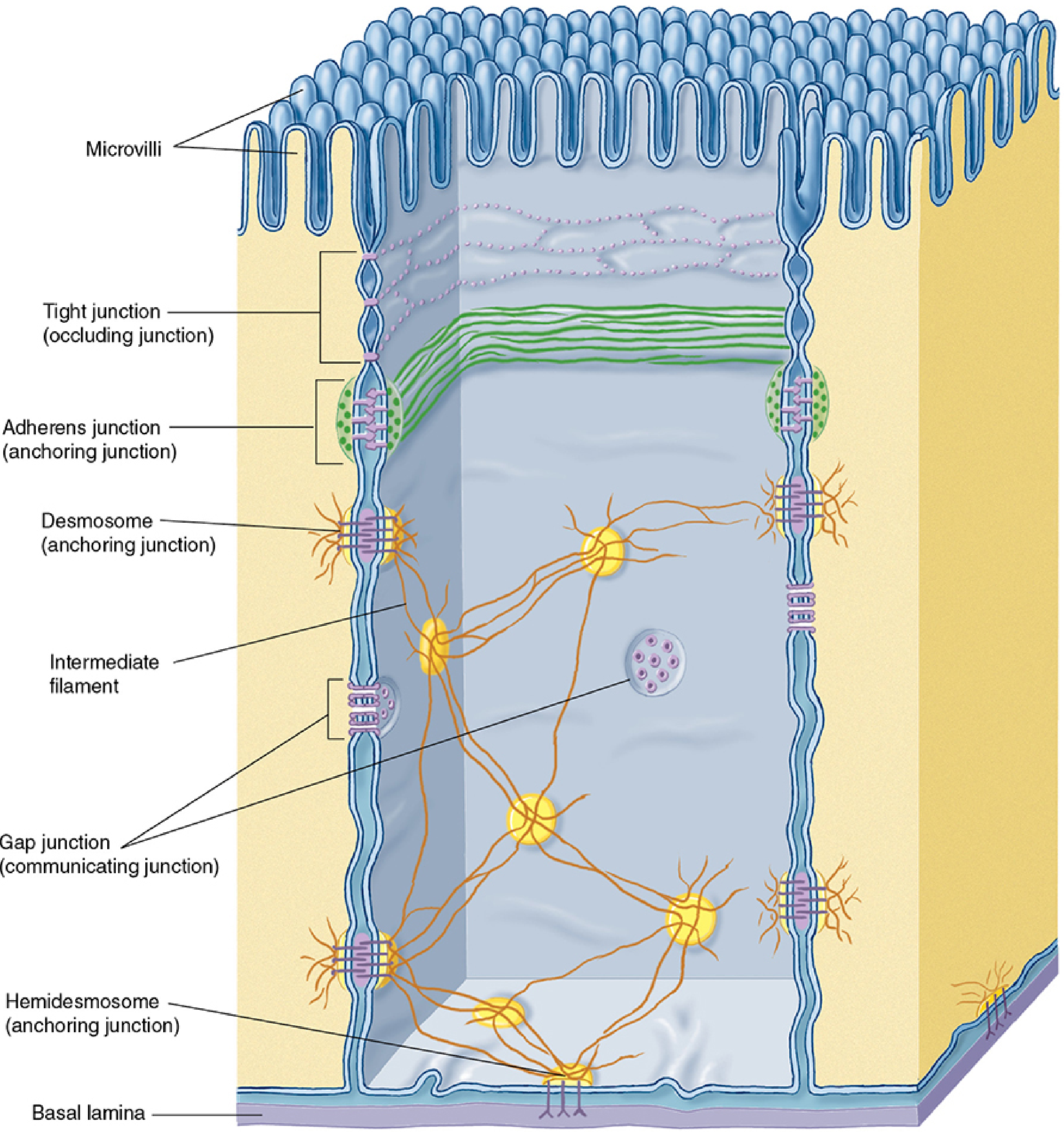
Diagram 3 - TEM (Electron Microscope) view of junctional complex
(Actual ultrastructural appearance showing MV = microvilli, TJ = tight junction, AJ = adherens junction, D = desmosome, IF = intermediate filaments)
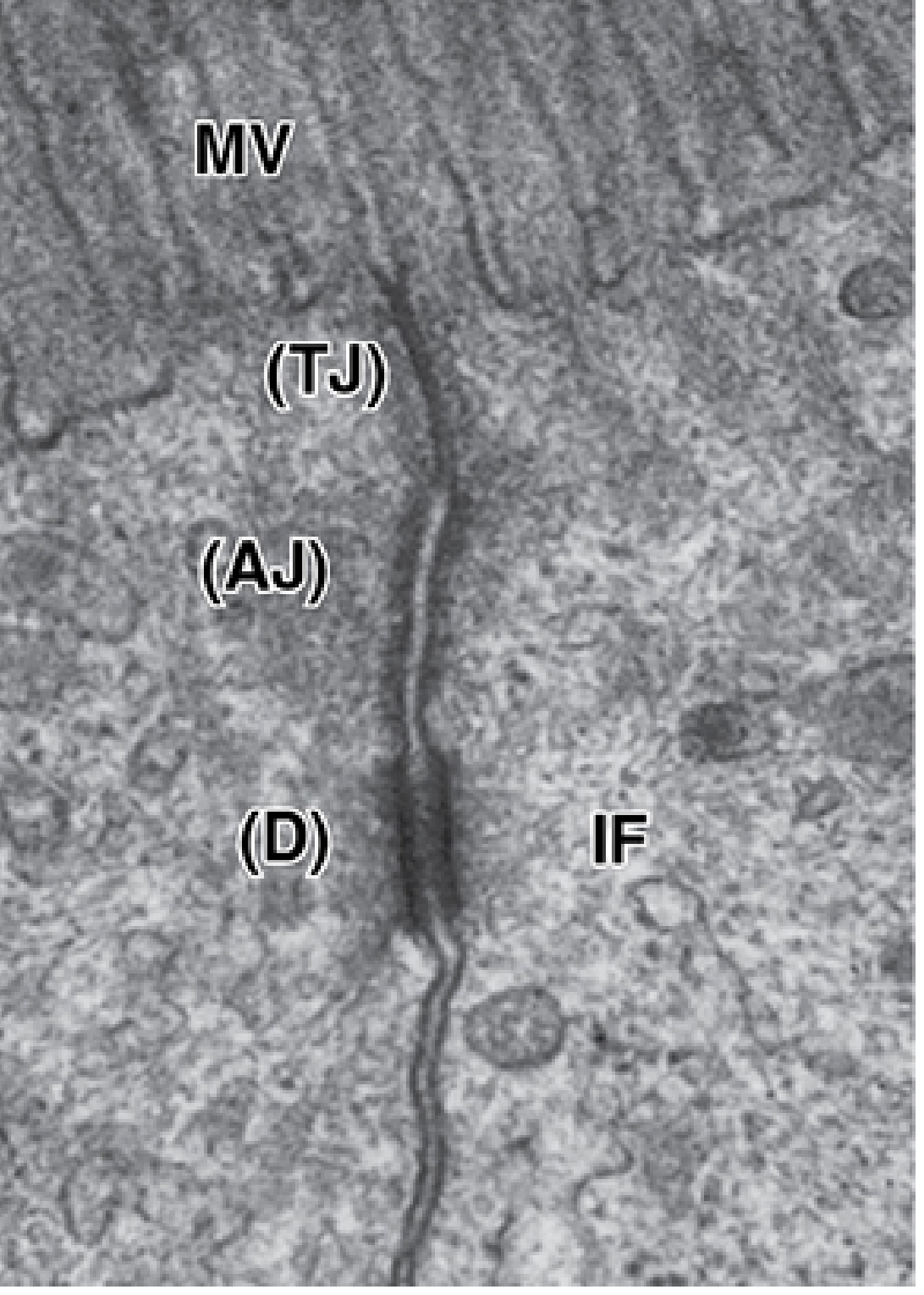
1. Tight Junction (Zonula Occludens)
Position: Most apical junction - forms a complete band encircling the cell
Structure:
- Made of ridges (half from each adjacent cell) that interdigitate and almost obliterate the intercellular space
- Three families of transmembrane proteins:
- Claudins - main barrier proteins (determine leakiness)
- Occludin
- Junctional Adhesion Molecules (JAMs)
- Linked intracellularly to actin filaments via scaffolding proteins (ZO-1, ZO-2, ZO-3)
Functions:
| Function | Detail |
|---|---|
| Paracellular barrier | Seals the space between cells; forces transport through cells (transcellular) rather than between them |
| Fence function | Prevents lateral diffusion of membrane proteins between apical and basolateral domains - maintains cell polarity |
| Variable leakiness | Leakiness depends on claudin type - proximal renal tubule = leaky (few strands); urinary bladder = tight (many strands) |
Locations: Intestinal mucosa, renal tubule walls, choroid plexus, blood-brain barrier endothelium
Clinical significance: Defects in occludin compromise the fetal blood-brain barrier → severe neurologic disorders
2. Adherens Junction (Zonula Adherens)
Position: Just below the tight junction (apical side); also forms a continuous band around the cell
Structure:
- Transmembrane proteins: E-cadherin + catenin complexes (Ca²⁺-dependent)
- Intracellularly linked to actin filaments (major attachment site for actin cytoskeleton)
Functions:
- Creates strong cell-to-cell adhesion
- Stabilizes and strengthens the nearby tight junction
- Mechanically couples the cytoskeletons of adjacent cells
- Transmits contractile forces between cells
Clinical significance: Loss of E-cadherin in carcinomas promotes tumor invasion and malignant transformation
3. Desmosome (Macula Adherens)
Position: Scattered as spot-like patches along the lateral cell surface (not a continuous band - hence "macula" = spot)
Structure:
- Apposed membrane thickenings (plaques) on the cytoplasmic face of each cell
- Transmembrane proteins: Desmogleins and Desmocollins (members of the cadherin family)
- Intracellularly linked to intermediate filaments (keratins) - both parallel to and radiating away from the membrane
- Intercellular space filled with filamentous material linking the two plaques
Functions:
- Provides the strongest mechanical anchoring between cells
- Distributes mechanical stress across the entire tissue (like rivets)
- Especially important in tissues under physical stress: skin, cardiac muscle, uterus
Clinical significance: Autoimmunity against desmoglein 1 and 3 → Pemphigus vulgaris (blistering skin disorder due to loss of epidermal cell cohesion)
4. Hemidesmosome
Position: Basal surface of epithelial cells (anchors cell to basal lamina - NOT to adjacent cells)
Structure:
- Looks like half a desmosome but is functionally different
- Transmembrane proteins: Integrins (not cadherins - key exam difference!)
- Intracellularly linked to intermediate filaments
- Extracellularly binds to laminin in the basal lamina
Function: Anchors epithelium to the underlying basal lamina
Clinical significance: Mutations in integrin-β4 → Epidermolysis bullosa (severe skin blistering disorder)
5. Focal Adhesion
Position: Basal surface (also attaches cell to basal lamina)
Structure:
- Contains integrins
- Linked intracellularly to actin filaments (not intermediate filaments)
- Labile (transient, dynamic) structures
Function: Cell movement and migration; also mediates signal transduction from ECM
6. Gap Junction (Nexus / Communicating Junction)
Position: Scattered along lateral surface; spot-like patches
Structure:
- Intercellular space reduced to ~4 nm (vs normal ~20 nm)
- Each gap junction = two connexons (one from each cell) aligned face-to-face → dodecameric structure
- Each connexon = 6 connexin proteins surrounding a central channel
- Each connexin has 4 transmembrane domains
- Pore diameter = 0.8-1.4 nm → passes molecules up to ~1000 Da
Substances that can pass: Ions (Na⁺, K⁺, Ca²⁺), sugars, amino acids, cAMP, IP₃, small signaling molecules
Functions:
- Electrical coupling - rapid propagation of action potentials (cardiac muscle, smooth muscle, CNS)
- Metabolic coupling - exchange of nutrients and metabolic intermediates
- Developmental signaling - coordination of cell growth and differentiation
- Connexin semichannels - connexons alone (hemichannels) can also allow regulated release of small molecules to the ECF
Regulation: Gap junction channels are gated (not simply passive) - closed by low pH, elevated Ca²⁺, membrane depolarization
Clinical significance of connexin mutations:
- Connexin 26 (GJB2) mutation → most common cause of autosomal recessive non-syndromic deafness
- Connexin 30 (Cx30) defect → Clouston syndrome (skin disorder)
- Connexin deletions → cardiac arrhythmias, sudden cardiac death
- Various connexins → cataracts, peripheral neuropathy, female sterility
Master Comparison Table
| Feature | Tight Junction | Adherens Junction | Desmosome | Hemidesmosome | Gap Junction |
|---|---|---|---|---|---|
| Latin name | Zonula occludens | Zonula adherens | Macula adherens | - | Nexus |
| Shape | Belt/band | Belt/band | Spot/patch | Spot/patch | Spot/patch |
| Key proteins | Claudins, Occludin, JAMs | E-cadherin, catenins | Desmogleins, Desmocollins | Integrins | Connexins |
| Cytoskeleton link | Actin filaments | Actin filaments | Intermediate filaments (keratin) | Intermediate filaments | None |
| Position | Most apical | Below tight junction | Lateral, scattered | Basal | Lateral, scattered |
| Main function | Paracellular seal + polarity | Adhesion + TJ stabilization | Strong mechanical anchor | Anchor to basal lamina | Electrical/metabolic coupling |
| Clinical disease | Blood-brain barrier defects | E-cadherin loss → carcinoma | Pemphigus vulgaris | Epidermolysis bullosa | Deafness (Cx26), arrhythmias |
High-Yield Exam Points
- The apico-basal order from top to bottom: Tight junction → Adherens junction → Desmosome → Gap junction → Hemidesmosome (mnemonic: "TADGH" - Tight, Adherens, Desmosome, Gap, Hemi)
- Only gap junctions allow direct cell-to-cell transfer of molecules
- Desmosomes and hemidesmosomes both anchor to intermediate filaments - but desmosomes use cadherins while hemidesmosomes use integrins
- Tight junctions control paracellular permeability - leakiness is determined by claudin subtype
- Connexin 26 mutation = most common inherited cause of deafness
- Gap junctions pass molecules up to 1000 Da through a pore of 0.8-1.4 nm
- The term "zonula" = belt/band (encircles the whole cell); "macula" = spot/patch (discrete points)
Sources: Ganong's Review of Medical Physiology, 26th ed. (pp. 52-54); Junqueira's Basic Histology, 17th ed. (pp. 200-204)
This is a shared conversation. Sign in to Orris to start your own chat.