
dilated cardiomyopathy echocardiography enlarged left ventricle
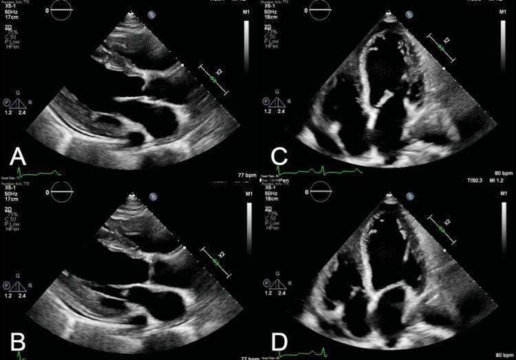
Transthoracic echocardiography (TTE) images presenting four grayscale views of a heart with severe dilated cardiomyopathy. Panels A and B display the parasternal long-axis view (PLAX) in diastole and systole, respectively. These views reveal massive dilatation of the left ventricle (LV) and left atrium (LA), with the LA significantly larger than the adjacent aortic root. The mitral valve leaflets appear prominent, and the systolic frame (B) shows restricted leaflet coaptation. Panels C and D show the apical four-chamber view (A4C) in diastole and systole. There is striking biatrial and biventricular dilatation, giving the heart a globular appearance. A comparison between the diastolic (C) and systolic (D) phases demonstrates minimal change in ventricular cavity size, indicating a severely reduced ejection fraction (EF approximately 20%) and global hypokinesis. The right ventricle (RV) is also markedly dilated, nearly reaching the size of the enlarged left ventricle. These findings are characteristic of advanced heart failure with multi-chamber enlargement.
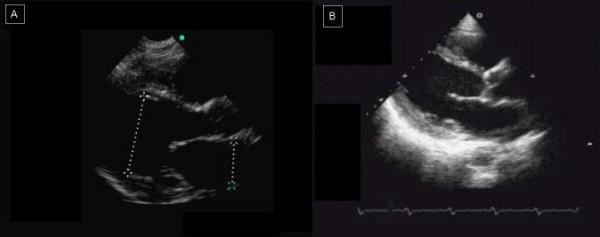
This diagnostic comparison displays two echocardiographic images (Parasternal Long-Axis View) of a patient with dilated cardiomyopathy. Image A was acquired using a Hand-Held Device (HHD), while Image B was obtained using Standard Echocardiography (SED). Both images demonstrate clear hallmarks of dilated cardiomyopathy, specifically a significantly enlarged and rounded left ventricle (LV) with thinning of the ventricular walls. Anatomical structures including the left atrium, mitral valve, and LV outflow tract are visible in both. The comparison highlights differences in imaging performance: Image A (HHD) shows lower spatial resolution with grainier texture and less defined endocardial borders, and includes calipers (dotted lines) measuring ventricular dimensions. Image B (SED) offers superior contrast and detail of the myocardium and valvular structures. This side-by-side comparison serves as an educational tool for evaluating the diagnostic utility of portable ultrasound versus high-end systems in identifying severe structural heart disease and chamber dilation.
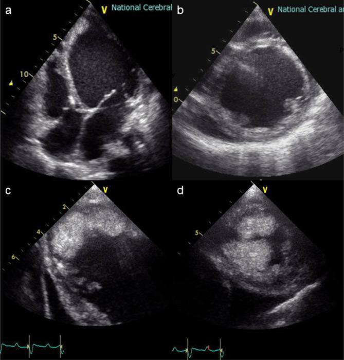
This composite diagnostic image consists of four transthoracic echocardiography (TTE) frames (a-d) showing pathological changes in the heart. Panels (a) and (b) demonstrate severe global dilation of the left ventricle (LV) with noticeable wall thinning, characteristic of advanced dilated cardiomyopathy. The right ventricle appears relatively normal in volume compared to the enlarged LV. Panels (c) and (d) focus on the apical region of the left ventricle, revealing a large, irregular, and heterogeneous echogenic mass adherent to the endocardial surface. This finding is consistent with a massive intracavitary thrombus, a common complication of severely reduced cardiac output and blood stasis in dilated heart failure. The images serve as clinical evidence for preoperative planning prior to ventricular assist device (VAD) implantation, highlighting the risk of embolic events and the necessity for thrombectomy.
Discharge Summary Explained - Mukesh Kumar (Cardiology)
Patient Overview
- Non-diabetic, hypertensive male admitted with severe shortness of breath (dyspnea), Ghabrahat (anxiety/restlessness), Chakkar (dizziness) for 3 days, and decreased appetite and urine output for 4 days.
- Department: Cardiology
- Date of Discharge: 01-08-2025
Final Diagnosis - Term by Term
1. Dilated Cardiomyopathy (DCM)
- Prevalence: Approximately 1 in 250 adults
- Causes: Genetic mutations (TTN gene in ~25% of familial cases), viral myocarditis, alcohol, medications, nutritional deficiencies, or idiopathic (no identifiable cause)
- Symptoms: Reduced exercise tolerance, dyspnea, orthopnea, edema, fatigue, and in advanced cases - low cardiac output signs like weak pulse and hypotension
- Diagnosis: Echocardiogram showing ventricular dilation and fractional shortening <25%; chest X-ray shows enlarged heart (cardiothoracic ratio >0.5) - Goldman-Cecil Medicine
2. Mild MR (Mitral Regurgitation)
3. Mild TR (Tricuspid Regurgitation)
4. Severe LV Dysfunction, LVEF ~25%
- LV = Left Ventricle - the main pumping chamber
- LVEF = Left Ventricular Ejection Fraction - the percentage of blood pumped out with each heartbeat
- Normal LVEF = 55-70%
- This patient's LVEF = 25% - severely reduced, meaning the heart is pumping only 1/4 of the blood it should
- This is classified as HFrEF (Heart Failure with Reduced Ejection Fraction)
- Prognosis note: The 5-year survival is less than 50% in patients with EF <25% without treatment - Goldman-Cecil Medicine
5. Cardiogenic Shock (Improved)
- This patient presented in cardiogenic shock - a life-threatening emergency
- It improved with treatment (likely IV inotropes, diuretics, vasopressors)
6. Congestive Heart Failure (Improved)
7. Type 1 Respiratory Failure (Improved)
- Type 1 = Hypoxic respiratory failure - low oxygen levels (PaO2 <60 mmHg) with normal or low CO2
- This happened because fluid flooded the lungs (pulmonary edema from heart failure), preventing oxygen exchange
- It improved - patient likely required supplemental oxygen, possibly non-invasive ventilation (BiPAP/CPAP) during admission
Why These Diagnoses Are Connected
Standard Treatment for This Condition (HFrEF with DCM)
| Medication Class | Examples | Benefit |
|---|---|---|
| ACE inhibitor / ARB / ARNI | Ramipril, Sacubitril-Valsartan (Entresto) | Reduces mortality, improves EF |
| Beta-blocker | Carvedilol, Bisoprolol, Metoprolol | Reduces sudden death risk |
| MRA (mineralocorticoid antagonist) | Spironolactone, Eplerenone | Reduces fluid, mortality |
| SGLT2 inhibitor | Dapagliflozin, Empagliflozin | Reduces hospitalizations/mortality |
- Diuretics (furosemide) for fluid relief
- ICD (implantable defibrillator) - indicated if LVEF remains <35% after 3 months of optimal therapy, for prevention of sudden cardiac death
- CRT (cardiac resynchronization therapy) if there is bundle branch block
- Anticoagulation if LV thrombus or atrial fibrillation is present
- In refractory cases: LVAD (ventricular assist device) or cardiac transplantation
Key Takeaways for the Family
- The patient was critically ill on admission but has improved - all three acute conditions (shock, CHF, respiratory failure) are marked "improved"
- The underlying DCM and severe LV dysfunction remain - this is a chronic condition requiring lifelong medications and regular follow-up
- Strict medication compliance is essential - stopping medications suddenly is dangerous
- Lifestyle: No alcohol, low salt diet, fluid restriction, daily weight monitoring (report gain >1-2 kg in 24 hours)
- Regular cardiology follow-up with repeat echocardiogram in 3-6 months to reassess LVEF
- Watch for warning signs: Worsening breathlessness, leg swelling, inability to lie flat, decreased urine output - these need immediate medical attention