
Brown Seqiard syndrome Sembulingam
Brown Sequard syndrome spinal cord hemisection diagram sensory motor deficits
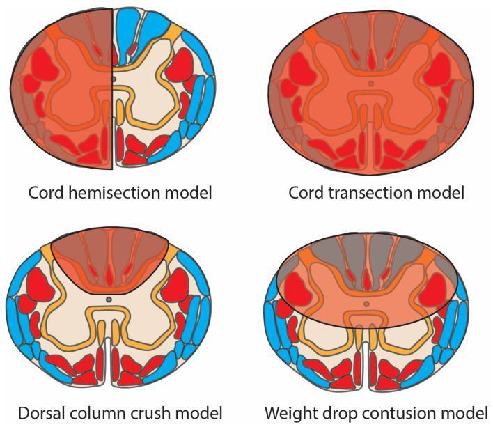
This medical schematic diagram illustrates four pre-clinical spinal cord injury (SCI) models in axial cross-section, highlighting damaged regions (translucent red overlay), motor tracts (opaque red), and sensory tracts (blue). 1. Cord Hemisection Model: Shows damage isolated to one lateral half of the spinal cord, characteristic of Brown-Séquard syndrome, affecting both motor and sensory tracts on the ipsilateral side. 2. Cord Transection Model: Depicts complete injury across the entire cord diameter, resulting in total disruption of all motor and sensory pathways. 3. Dorsal Column Crush Model: Features localized damage concentrated in the posterior (dorsal) funiculus, primarily affecting ascending sensory tracts. 4. Weight Drop Contusion Model: Displays a diffuse, central injury zone impacting the grey matter and surrounding white matter tracts, simulating clinical traumatic contusion. The diagram serves as an educational tool for comparing injury patterns across experimental models used in neurotrauma research and their resulting impact on anatomical tracts.
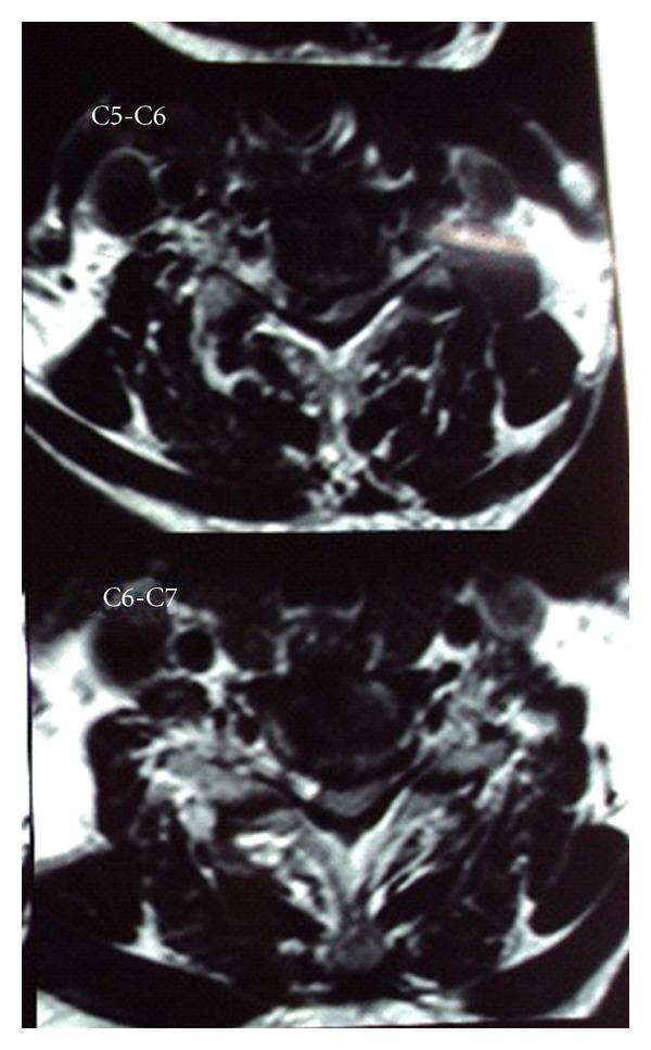
This diagnostic image consists of two axial T2-weighted magnetic resonance imaging (MRI) slices of the cervical spine, labeled at the C5-C6 and C6-C7 levels. The images demonstrate significant right paracentral disc herniation at both levels, resulting in pronounced hemicord compression. At the C5-C6 level, the herniated disc material is seen extruding into the spinal canal, causing asymmetric flattening and displacement of the spinal cord toward the left. Similar pathological findings are observed at the C6-C7 level, though the compression at C5-C6 appears more severe. The vertebral bodies and surrounding soft tissues show signs of degenerative spondylosis. The visual evidence of unilateral cord compression is a classic radiological correlate for Brown-Séquard syndrome, illustrating the mechanical etiology of ipsilateral motor loss and contralateral sensory deficits. This material is suitable for intermediate to advanced medical learners focusing on neuroradiology, orthopedics, or neurology.
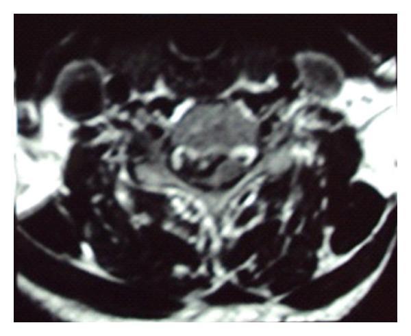
This diagnostic image is an axial T2-weighted MRI of the cervical spine at the C4-C5 level. The scan demonstrates a significant right paracentral disc herniation protruding into the spinal canal. The herniated disc material causes focal mass effect on the right side of the spinal cord, resulting in evident hemicord compression and distortion of the cord's normal elliptical contour. Despite the mechanical compression, the signal intensity within the spinal cord remains relatively uniform, with no definitive T2 hyperintensity to suggest acute myelomalacia or cord edema at this level. The vertebral body and posterior elements are visualized, though anatomical detail is slightly limited by image graininess. This clinical finding correlates with Brown-Séquard Syndrome, characterized by ipsilateral motor deficits and contralateral sensory loss due to unilateral cord compression. This image serves as a key educational example of how a focal disc protrusion can cause significant mechanical deformation of the spinal cord and lead to specific neurological deficits.
Brown-Séquard Syndrome
Definition
Anatomical Basis
| Tract | Pathway | Decussation |
|---|---|---|
| Corticospinal tract (motor) | Lateral column | Crosses in medulla (before entering cord) |
| Dorsal columns (DCML) | Posterior column | Crosses in medulla (at nucleus gracilis/cuneatus) |
| Spinothalamic tract | Anterolateral column | Crosses within 1-2 segments of entry in cord |
Classic Clinical Features
On the Ipsilateral side (same side as lesion), below the level:
- Spastic paralysis (UMN signs) - loss of voluntary motor function due to corticospinal tract damage (already crossed in medulla, so damage = ipsilateral effect)
- Loss of proprioception, vibration sense, discriminative/fine touch - dorsal column (fasciculus gracilis/cuneatus) is cut before it crosses; crosses in medulla
- Loss of "discrete light touch" - principal pathway (dorsal column) is transected
On the Contralateral side (opposite side), 2-6 segments below the level:
- Loss of pain and temperature sensation - spinothalamic tract fibers that entered the cord at the lesion level have already crossed to the other side; they are now running in the cord on the opposite side of the lesion
At the level of lesion (ipsilateral):
- LMN signs (flaccid paralysis, loss of reflexes) at the exact segment, due to anterior horn/root damage
- Band of hyperesthesia just above the lesion (due to irritation)
Summary Table
| Feature | Side | Level |
|---|---|---|
| Spastic paralysis (UMN) | Ipsilateral | Below lesion |
| Loss of proprioception + vibration + fine touch | Ipsilateral | Below lesion |
| Loss of pain + temperature | Contralateral | 2-6 segments below lesion |
| Flaccid paralysis (LMN) | Ipsilateral | At lesion level |
Mechanism Explained (Guyton & Hall)
"All motor functions are blocked on the side of the transection in all segments below the level of the transection. Yet, only some of the modalities of sensation are lost on the transected side, and others are lost on the opposite side. The sensations of pain, heat, and cold - served by the spinothalamic pathway - are lost on the opposite side of the body in all dermatomes two to six segments below the level of the transection. By contrast, sensations transmitted only in the dorsal and dorsolateral columns - kinesthetic and position sensations, vibration sensation, discrete localization, and two-point discrimination - are lost on the same side of the transection."
- Guyton & Hall Textbook of Medical Physiology
Causes
- Penetrating/stab injuries (most classic cause)
- Spinal cord tumors (extrinsic compression)
- Cervical disc herniation (right paracentral disc can compress one half)
- Spinal epidural hematoma
- Multiple sclerosis
- Radiation myelopathy
- Spinal cord ischemia/infarct
Important Notes for Physiology Exams (Sembulingam context)
- Pure hemisection is rare in clinical practice - most cases are "Brown-Séquard plus" (incomplete patterns)
- The contralateral pain/temp loss begins 2-6 segments below the lesion (not at the level) - this is because spinothalamic fibers travel 1-2 segments up the cord before crossing
- Crude touch is preserved on both sides - bilateral because spinothalamic (contralateral) AND some ipsilateral projections carry crude touch
- Brown-Séquard syndrome has the best prognosis among all incomplete spinal cord syndromes
- The syndrome demonstrates the principle of dissociated sensory loss - a cardinal concept in neuroanatomy