
ITP flowchart pathology
ITP immune thrombocytopenic purpura pathology flowchart pathogenesis
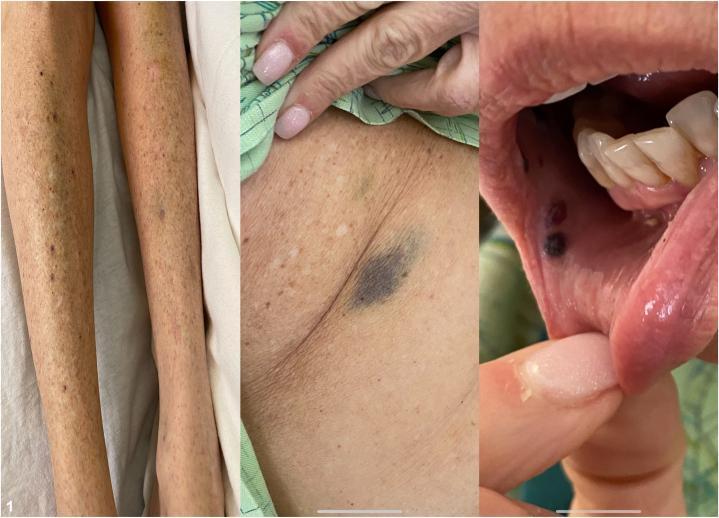
This composite clinical photograph illustrates multisite cutaneous and mucosal manifestations of drug-induced immune thrombocytopenic purpura (ITP). The left panel shows the lower extremities with a diffuse, symmetric distribution of pinpoint, non-blanching red-to-purple petechiae and larger, confluent purpuric macules. The center panel displays a close-up of the axillary region, highlighting a prominent, deep-purple ecchymosis alongside scattered petechiae, indicative of significant dermal hemorrhage. The right panel demonstrates mucosal involvement with 'wet purpura'—specifically, dark, hemorrhagic bullae and macules located on the buccal mucosa and inner lip. These findings collectively characterize a systemic bleeding diathesis, clinically relevant for identifying severe thrombocytopenia (platelet counts typically <10,000/uL) and assessing the risk of internal hemorrhage in a dermatology or hematology context.
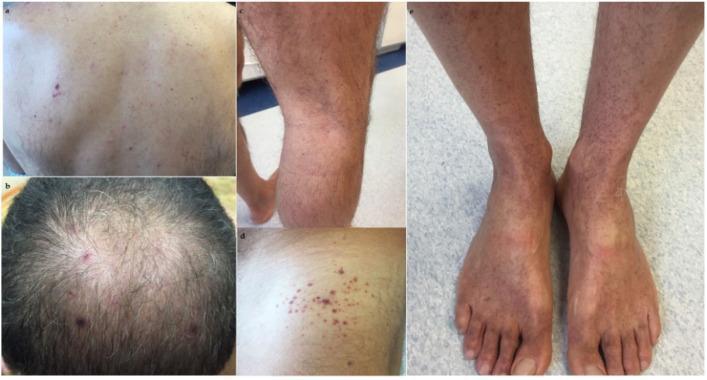
A series of clinical photographs documenting the dermatological manifestations of immune thrombocytopenic purpura (ITP). The images show widespread petechiae—small (1-3 mm), non-blanching, reddish-purple macules caused by capillary hemorrhage—distributed across several anatomical regions. On the back and scalp, the petechiae are relatively sparse and discrete. In contrast, the lower extremities, including the shins and the dorsal surfaces of the feet, demonstrate a higher density of lesions, appearing more confluent and clustered. The morphology is consistent with thrombocytopenia-induced microhemorrhages under the skin. The color variation from bright red to deep purple suggests varying stages of extravasated blood. This visual evidence serves as a clinical marker for evaluating systemic coagulation disorders and platelet dysfunction in a medical education context.
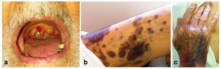
This composite clinical photograph displays dermatologic and mucosal manifestations of severe thrombocytopenia, likely secondary to immune thrombocytopenic purpura (ITP) in the context of COVID-19. Panel (a) shows the oral cavity with prominent hemorrhagic bullae (wet purpura) on the soft palate and oropharynx, presenting as blood-filled, tense blisters. Panel (b) illustrates the lower extremity featuring multifocal, scattered ecchymoses and petechiae, varying in color from reddish-purple to dark brown, reflecting different stages of extravasated blood. Panel (c) demonstrates confluent purpura on the dorsum of the hand, where the skin appears extensively bruised and darkened. These visual findings are classic indicators of a high risk for life-threatening bleeding due to profoundly low platelet counts. The images provide critical educational examples of mucocutaneous bleeding signs that necessitate urgent hematological evaluation.
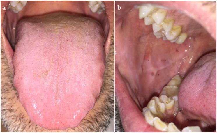
Two-panel clinical photograph illustrating the oral cavity following treatment for immune thrombocytopenic purpura (ITP). Image (a) presents a dorsal view of the tongue, demonstrating a pink, moist surface with normal filiform and fungiform papillae distribution. There is evidence of resolving ecchymosis, appearing as faint, darker spots near the lateral borders, indicating remission of previous hemorrhagic lesions. Image (b) shows the right buccal mucosa and dental arches. The mucosa is predominantly light pink and healthy, with minimal residual hyperpigmented macules likely representing resolving petechiae or ecchymoses. The adjacent teeth show mild extrinsic staining and dental calculus, while the gingival margins appear stable. Collectively, these images document the clinical resolution of oral manifestations of a bleeding disorder—specifically the healing of traumatic-origin ecchymotic areas and mucosal hemorrhages—following corticosteroid therapy.
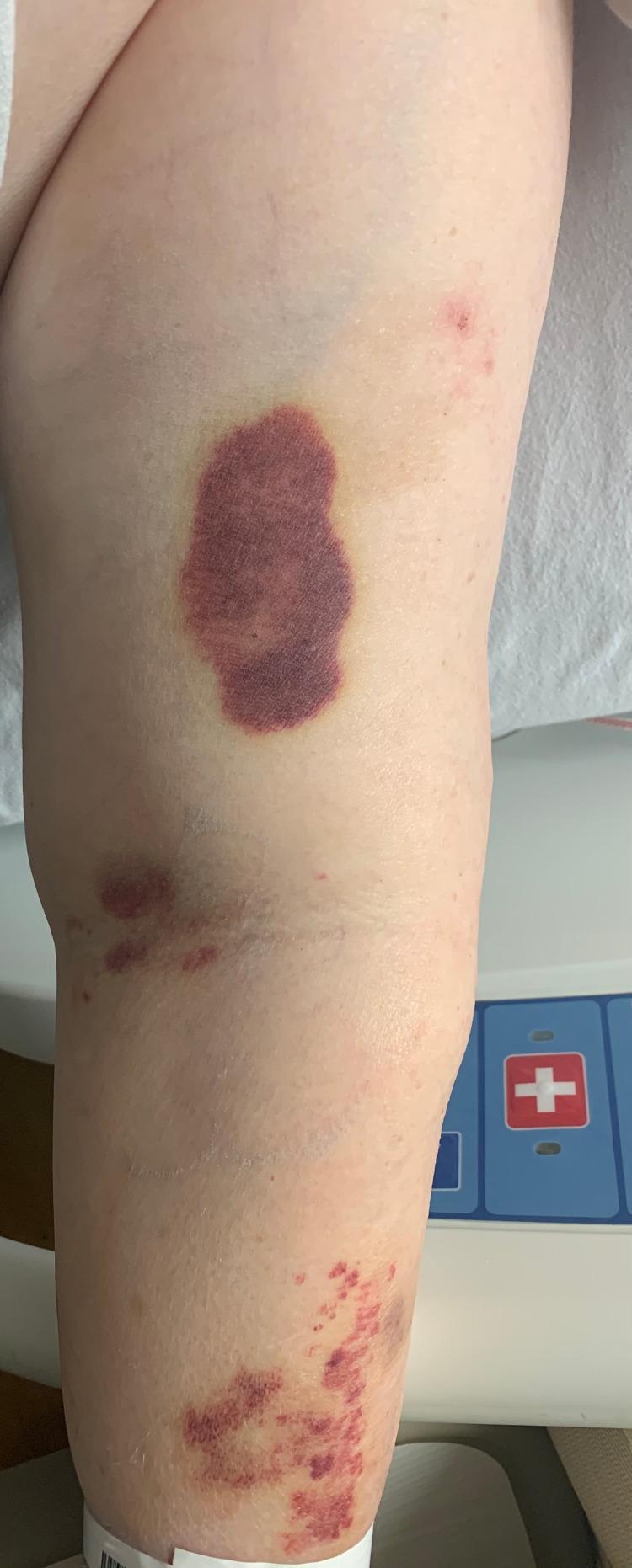
This clinical photograph displays skin lesions on a patient's extremity, consistent with a diagnosis of Immune Thrombocytopenic Purpura (ITP). The primary finding is a large, well-demarcated, oval-shaped ecchymosis (bruise) approximately 5-7 cm in length, located on the proximal limb. The lesion is deep purple to violaceous in color, indicating a recent subcutaneous hemorrhage, with a peripheral yellow-green hue suggesting hemoglobin degradation and the start of resolution. Distal to the primary ecchymosis, there are scattered clusters of petechiae (pinpoint red-purple spots) and smaller, irregular purpuric patches. These cutaneous manifestations are characteristic of a significant bleeding diathesis or severe thrombocytopenia. The distribution shows a mix of confluent ecchymoses proximally and more discrete, non-blanching petechiae distally. This image is an educational example of how a severely low platelet count manifests visually as spontaneous soft tissue bleeding and dermal hemorrhage.
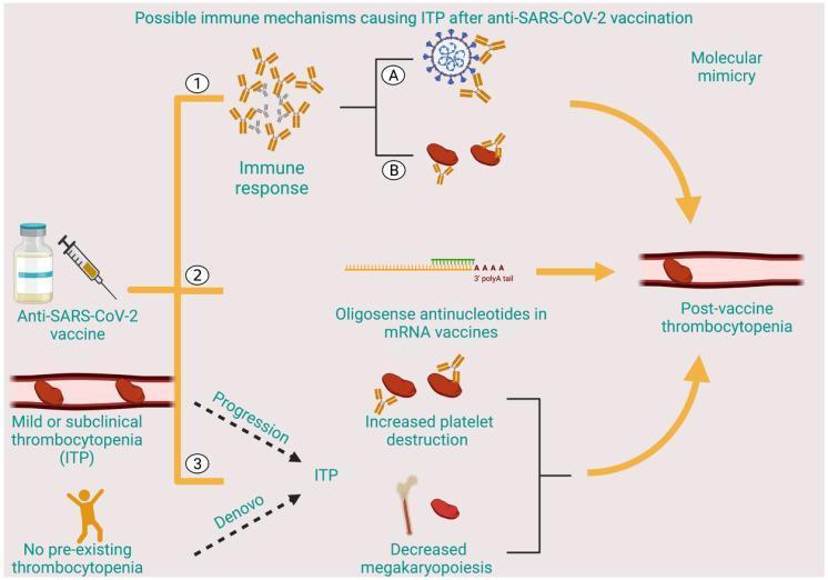
A pathophysiology diagram illustrating three proposed immune mechanisms linking anti-SARS-CoV-2 vaccination to immune thrombocytopenic purpura (ITP). Pathway 1 outlines an immune response where antibodies generated against the SARS-CoV-2 spike protein (A) cross-react with platelet antigens (B) through molecular mimicry, leading to thrombocytopenia. Pathway 2 depicts the potential role of oligosense antinucleotides in mRNA vaccines, identified by an RNA sequence with a 3' polyA tail, which can directly cause post-vaccine thrombocytopenia. Pathway 3 illustrates the development of ITP through two clinical scenarios: the progression of pre-existing mild/subclinical ITP or de novo development in patients with no prior history. These clinical pathways converge on two cellular mechanisms: increased peripheral platelet destruction by antibodies and decreased megakaryopoiesis within the bone marrow. The final common outcome across all pathways is post-vaccine thrombocytopenia within the vasculature. This medical illustration is intended for educational purposes regarding hematological adverse events and vaccine-related autoimmunity.
ITP (Immune Thrombocytopenic Purpura) - Pathology Flowchart
Definition
PATHOGENESIS FLOWCHART
TRIGGER (Primary: unknown / Secondary: infection, drugs, autoimmune disease)
            │
            ▼
   LOSS OF IMMUNE TOLERANCE
   (failure of self-tolerance mechanisms; T-reg cell dysfunction)
            │
            ▼
   AUTO-REACTIVE T HELPER CELLS ACTIVATED
   (recognize platelet surface antigens presented by APCs)
            │
     ┌──────┴──────────────────┐
     ▼                         ▼
B CELL ACTIVATION         T CELL–MEDIATED
(by Tfh cells)            DIRECT CYTOTOXICITY
     │                         │
     ▼                         ▼
IgG AUTOANTIBODY PRODUCTION   CD8+ T cells directly lyse
targeting platelet GPs:        antibody-coated platelets
  • GPIIb/IIIa (CD41/CD61)
  • GPIb/IX (CD42a/b)
     │
     ├──────────────────────────────────┐
     ▼                                  ▼
ANTIBODY-COATED PLATELETS        MEGAKARYOCYTES IN BONE MARROW
circulate in bloodstream         also targeted by autoantibodies
     │                           → impaired platelet production
     ▼                           (paradoxical thrombocytopenia
SPLENIC MACROPHAGES              despite increased megakaryocytes)
recognize IgG Fc regions via
FcγRIIA & FcγRIII receptors
     │
     ▼
PHAGOCYTOSIS & DESTRUCTION
of antibody-opsonized platelets
in the RETICULOENDOTHELIAL SYSTEM
(spleen >> liver)
     │
     ▼
THROMBOCYTOPENIA
(Platelet count <100,000/µL; severe <30,000/µL)
     │
     ├──────────────────────────────────────┐
     ▼                                      ▼
BLEEDING MANIFESTATIONS               COMPENSATORY RESPONSE
• Skin: petechiae, purpura, ecchymoses  Bone marrow increases
• Mucosa: wet purpura (oral bullae)     megakaryopoiesis
• GI/GU: internal bleeding             (megakaryocytes appear
• CNS: intracranial hemorrhage          normal to increased in number
  (rare, life-threatening)              but functionally impaired)
KEY IMMUNOLOGICAL MECHANISMS
| Mechanism | Detail |
|---|---|
| Primary autoantigen | GPIIb/IIIa (most common); GPIb/IX |
| Autoantibody class | IgG (occasionally IgM) |
| Platelet clearance site | Spleen (primary), liver |
| Clearance receptor | FcγRIIA on splenic macrophages |
| T cell role | Th cells drive autoantibody production; CD8+ T cells cause direct platelet lysis |
| Bone marrow | Megakaryocytes increased but platelet output impaired - autoantibodies suppress thrombopoiesis |
CLASSIFICATION (IWG 2009 Terminology)
| Phase | Duration | Platelet count |
|---|---|---|
| Newly diagnosed | 0-3 months | <100,000/µL |
| Persistent | 3-12 months | <100,000/µL |
| Chronic | >12 months | <100,000/µL |
| Severe | Any | <30,000/µL + bleeding |
| Refractory | Post-splenectomy failure | <30,000/µL |
BONE MARROW FINDINGS
- Megakaryocytes: normal to increased in number
- Megakaryocyte morphology: often immature (dwarf megakaryocytes)
- Erythroid and myeloid series: normal (helps distinguish from aplastic anemia/leukemia)
- Bone marrow biopsy is not always required - performed selectively to rule out other causes
PERIPHERAL BLOOD SMEAR
- Decreased platelets (thrombocytopenia)
- Platelets that are present may be large (megathrombocytes) - reflecting increased marrow turnover
- No red cell fragmentation (distinguishes from TTP/HUS)
- No blasts (distinguishes from leukemia)
SECONDARY ITP - COMMON ASSOCIATIONS
HIV infection ──────────────┐
HCV infection ───────────────┤
H. pylori ───────────────────┤──► SECONDARY ITP
SLE / Antiphospholipid Ab ───┤     (treat underlying cause first)
Lymphoproliferative disease ─┤
Drug-induced (quinine, etc.) ┘
PATHOLOGICAL IMAGE - ITP Clinical Manifestations
TREATMENT RATIONALE (based on pathophysiology)
| Mechanism targeted | Treatment |
|---|---|
| Reduce autoantibody production | Corticosteroids (1st line), Rituximab (anti-CD20) |
| Block Fc receptor-mediated clearance | IVIG, Anti-D immunoglobulin |
| Stimulate platelet production | TPO receptor agonists (eltrombopag, romiplostim) |
| Remove primary site of destruction | Splenectomy (2nd line) |
| Eradicate trigger (secondary ITP) | H. pylori eradication, antiviral therapy (HCV/HIV) |
- [Henry's Clinical Diagnosis and Management by Laboratory Methods](Henry's Clinical Diagnosis), Table 41.2 & section on Immune Thrombocytopenias, p. 966-970
- [Janeway's Immunobiology 10e](Janeway's Immunobiology), Section 15-9: Autoantibodies against blood cells, p. 617