
RENAL CAUSES OF OEDEMA IN DETAIL WITH CAUSE PATHOPHYSIOLOGY AND WHICH INVESTIGATION AND WHY THAT INVESTIGATION IS NEEDED
Renal Causes of Oedema - Detailed Overview
| Renal Cause | Key Mechanism | Oedema Type |
|---|---|---|
| Nephrotic Syndrome | Proteinuria → hypoalbuminaemia → low oncotic pressure + primary Na retention | Underfill and/or Overfill |
| Acute Nephritic Syndrome (AGN) | Reduced GFR + primary Na/water retention | Overfill |
| Acute Kidney Injury (AKI) | Failure to excrete Na and water | Overfill |
| Chronic Kidney Disease (CKD) | Reduced GFR → Na and water retention | Overfill |
1. Nephrotic Syndrome
Causes
- Minimal Change Disease (MCD) - most common in children
- Focal Segmental Glomerulosclerosis (FSGS)
- Membranous Nephropathy
- Mesangial proliferative GN
- Diabetes mellitus (diabetic nephropathy)
- Systemic Lupus Erythematosus
- Amyloidosis
- Drugs (gold, penicillamine, NSAIDs, heroin)
- Infections (HBV, HCV, malaria, HIV)
Pathophysiology
- Massive proteinuria (>3.5 g/day in adults; >40 mg/m²/hr in children) leads to urinary albumin losses
- Reduced hepatic synthesis cannot compensate → hypoalbuminaemia (serum albumin <3.5 g/dL, severe when <2 g/dL)
- Falling plasma oncotic pressure (normally 25-28 mmHg) → Starling forces shift: net fluid flux from intravascular into interstitial space
- Reduction in effective arterial blood volume (EABV) → arterial underfilling
- Underfilling activates: RAAS (↑ renin, ↑ aldosterone), ADH (↑ AVP), sympathetic nervous system, catecholamines
- Secondary renal Na and water retention → worsens interstitial oedema
- Plasma volume is contracted (typical of MCD)
- In other forms of nephrotic syndrome, the kidney itself primarily retains Na - independent of plasma oncotic pressure
- Micropuncture studies show pronounced NaCl reabsorption in the distal nephron and thick ascending limb (TAL)
- The proteinuric kidney has enhanced Na+ reabsorption in collecting ducts and a diminished response to ANP
- Filtered serine proteases (from heavy proteinuria) cleave and activate ENaC (epithelial Na channel) in the collecting duct
- Plasma volume is expanded, RAAS is suppressed, ANP is elevated
- Most patients with non-MCD nephrotic syndrome fall in this group
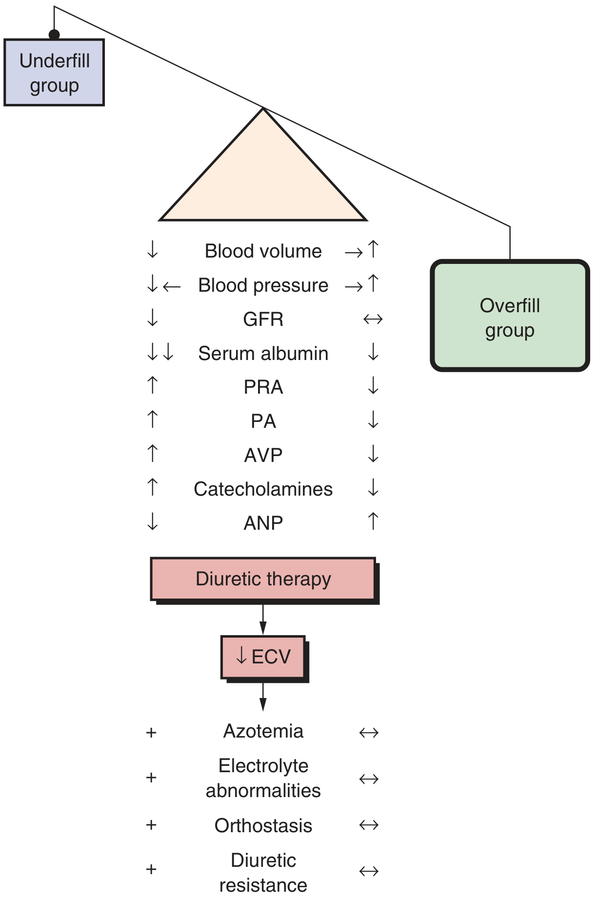
The diagram below (from Brenner & Rector's The Kidney) shows the contrast between underfill and overfill groups - note that in underfill, blood volume, BP, GFR, and PRA are all low, while serum albumin drops sharply; in overfill, BP and ANP are elevated and RAAS is suppressed:
- Dyslipidaemia: Increased hepatic lipoprotein synthesis triggered by low plasma oncotic pressure
- Hypoalbuminaemia reduces furosemide binding to plasma proteins, enlarging its volume of distribution → diuretic resistance
- Hyperaldosteronism (in underfill group) reinforces NaCl reabsorption at distal nephron and collecting duct
Investigations for Nephrotic Syndrome and Why
| Investigation | Why It Is Needed |
|---|---|
| Urine dipstick + microscopy | Detects heavy proteinuria (3+ or 4+); confirms absence of RBC casts (which suggest nephritis, not nephrosis) |
| 24-hour urine protein OR Spot urine protein:creatinine ratio (PCR) | Quantifies proteinuria - confirms nephrotic range (>3.5 g/day or PCR >300-350 mg/mmol); PCR >2 mg/mg in children. This defines the syndrome and monitors response to treatment |
| Serum albumin | Confirms hypoalbuminaemia; severity guides risk of complications (thrombosis, infection); albumin <2 g/dL = severe |
| Serum cholesterol and triglycerides (fasting lipid profile) | Hyperlipidaemia is a diagnostic feature; increased cardiovascular risk |
| Serum creatinine and eGFR | Assesses baseline renal function; rising creatinine suggests superimposed AKI or progressive disease |
| Serum electrolytes | Na, K - hyponatraemia (dilutional) common; hyperkalaemia can occur with RAAS activation or ACEi/ARB treatment |
| Serum complement (C3, C4, CH50) | Low complement suggests secondary causes: lupus nephritis (low C3 and C4), MPGN (low C3), post-streptococcal GN (low C3 transiently) |
| ANA, anti-dsDNA, ANCA | Screen for lupus nephritis (ANA/anti-dsDNA), vasculitis (ANCA) as secondary causes |
| HBsAg, anti-HCV, HIV serology | Viral infections cause secondary nephrotic syndrome - membranous nephropathy with HBV, MPGN with HCV |
| Blood glucose / HbA1c | Diabetic nephropathy is a leading secondary cause |
| Serum protein electrophoresis (SPEP) and Bence-Jones protein (urine) | Exclude myeloma/amyloidosis as secondary causes, especially in older patients |
| Renal ultrasound | Assesses kidney size and echogenicity; large kidneys suggest infiltrative disease (amyloid, DM) or early nephrotic syndrome; small kidneys suggest chronicity |
| Renal biopsy | Definitive - identifies the specific glomerular lesion (MCD, FSGS, MN, MPGN); guides treatment. In adults, biopsy is generally done upfront. In children with typical steroid-sensitive NS, it is deferred unless atypical features are present (age <1 year, hypocomplementaemia, macroscopic haematuria) |
| Urine sodium (FENa) | FENa <0.2% suggests volume-contracted (underfill) nephrotic syndrome; helps guide whether albumin or diuretics alone are appropriate |
| Coagulation screen (PT, PTT, fibrinogen) | Hypercoagulable state is common in nephrotic syndrome due to urinary loss of anticoagulant proteins (protein C, S, antithrombin III) and elevated procoagulant factors |
| Chest X-ray | Detects pleural effusion (transudate), pulmonary oedema from fluid overload |
2. Acute Nephritic Syndrome (Glomerulonephritis)
Causes
- Post-streptococcal GN (PSGN) - prototypical cause, especially in children
- IgA nephropathy
- Lupus nephritis
- ANCA-associated vasculitis (GPA, MPA)
- Anti-GBM disease (Goodpasture syndrome)
- MPGN, infective endocarditis-associated GN
Pathophysiology
- Inflammatory injury to glomeruli → diffuse proliferative changes → reduction in the number of functioning nephrons
- Sudden fall in GFR → the kidneys cannot excrete the normal daily load of Na and water
- Primary renal sodium retention occurs (the kidney is the primary culprit, not arterial underfilling) - through incompletely understood mechanisms that may involve:
- ENaC activation by filtered proteases
- Suppressed tubular response to ANP
- Na and water retention → expansion of extracellular fluid volume → ↑ capillary hydrostatic pressure → fluid transudation into interstitium
- Plasma volume is expanded (RAAS is suppressed, ANP is elevated - unlike underfill NS)
- Hypertension (volume-dependent) is a hallmark
- Oedema tends to be periorbital (facial, especially in morning) and peripheral
- Haematuria (RBC casts, dysmorphic RBCs) and subnephrotic proteinuria accompany the oedema - this distinguishes it from nephrotic syndrome
- Robbins' Pathologic Basis of Disease: "Nephritic syndrome is caused by inflammatory glomerular disease and is dominated by the acute onset of either grossly visible or microscopic hematuria, diminished GFR, mild to moderate proteinuria, and hypertension."
Investigations for Acute Nephritic Syndrome and Why
| Investigation | Why It Is Needed |
|---|---|
| Urine dipstick + microscopy | Detects haematuria (microscopic or macroscopic), RBC casts (pathognomonic of glomerular bleeding), granular casts, and subnephrotic proteinuria |
| 24-hour urine protein / PCR | Confirms subnephrotic proteinuria (<3.5 g/day), distinguishing nephritis from nephrosis |
| Serum creatinine and eGFR | Quantifies degree of AKI; rapidly progressive GN (RPGN) causes fast deterioration over days-weeks |
| Serum electrolytes, bicarbonate | Hyperkalaemia and metabolic acidosis common with oliguria |
| Serum complement: C3, C4 | PSGN: low C3, normal C4 (alternative pathway); Lupus: low C3 and C4 (classical pathway); Normal complement in IgA/ANCA/anti-GBM |
| ASO titre (Anti-Streptolysin O) + Anti-DNase B | Elevated in PSGN (evidence of preceding streptococcal infection); ASO may be negative in skin infection, making Anti-DNase B more sensitive |
| ANA, anti-dsDNA | Diagnose lupus nephritis |
| ANCA (cANCA/PR3, pANCA/MPO) | Diagnose ANCA-associated vasculitis (GPA, MPA); pauci-immune crescentic GN |
| Anti-GBM antibodies | Diagnose Goodpasture syndrome (anti-GBM disease) |
| Serum IgA | Elevated in ~50% of IgA nephropathy patients |
| Blood cultures | If infective endocarditis-associated GN is suspected (fever, murmur, embolic phenomena) |
| Renal ultrasound | Rules out obstruction; assesses echogenicity, kidney size; confirms bilateral disease |
| Renal biopsy | Definitive for diagnosis and prognosis; light microscopy (diffuse endocapillary proliferation in PSGN), immunofluorescence (IgA deposits in IgA-N; C3 in PSGN; IgG + C3 linear in anti-GBM), electron microscopy (sub-epithelial "humps" in PSGN, mesangial deposits in IgA-N) |
| Throat/skin swab | Identify streptococcal infection in suspected PSGN |
3. Acute Kidney Injury (AKI)
Causes
- Pre-renal: volume depletion, sepsis, cardiogenic shock
- Intra-renal: acute tubular necrosis (ATN), glomerulonephritis, interstitial nephritis, contrast nephropathy
- Post-renal: obstruction
Pathophysiology of Oedema
- AKI = rapid decline in GFR within hours to days
- Failure of the kidney to excrete sodium and water is the primary mechanism
- Oliguria or anuria → Na and water accumulate → ECF volume expansion
- ↑ capillary hydrostatic pressure → peripheral oedema, pulmonary oedema
- This is pure primary renal sodium retention - RAAS is typically suppressed in established AKI (RAAS may be activated in early pre-renal phase)
- Comprehensive Clinical Nephrology, 7th Ed: "Patients with AKI have limited ability to excrete sodium and water. Primary sodium retention by the kidney characterises AKI."
Investigations for AKI and Why
| Investigation | Why It Is Needed |
|---|---|
| Serum creatinine (serial) | Confirms and tracks AKI; rise of ≥26.5 µmol/L in 48h or ≥1.5x baseline in 7 days = AKI by KDIGO criteria |
| Serum urea (BUN) | BUN:creatinine ratio >20:1 suggests pre-renal; <10:1 suggests intrinsic renal (ATN) |
| Serum electrolytes | Hyperkalaemia is life-threatening in AKI; hyponatraemia from water retention; hyperphosphataemia |
| Urine sodium and FENa | FENa <1% = pre-renal (tubules avidly reabsorb Na); FENa >2% = intrinsic renal (tubular injury prevents reabsorption). Critical for distinguishing cause |
| Urine osmolality | Pre-renal: >500 mOsm/kg (concentrated); ATN: ~300 mOsm/kg (isosthenuric) |
| Urine microscopy | Granular "muddy brown" casts = ATN; RBC casts = GN; WBC casts = pyelonephritis/interstitial nephritis |
| Renal ultrasound (urgent) | Rules out post-renal obstruction (hydronephrosis) - a treatable cause; assesses kidney size |
| Chest X-ray | Pulmonary oedema from fluid overload in oliguric AKI |
| ECG | Hyperkalaemia can cause peaked T waves, widened QRS, ventricular fibrillation |
| Urine protein:creatinine ratio | Significant proteinuria suggests glomerular cause of AKI |
| Complement, ANCA, ANA, anti-GBM | If GN suspected as cause of AKI |
4. Chronic Kidney Disease (CKD) / End-Stage Renal Disease
Causes
- Diabetic nephropathy (most common globally)
- Hypertensive nephrosclerosis
- Chronic GN
- Polycystic kidney disease
- Reflux nephropathy / recurrent pyelonephritis
Pathophysiology of Oedema
- Progressive loss of nephrons → declining GFR
- With advanced CKD (stages G4-G5): GFR <30 mL/min → the remaining nephrons cannot excrete the daily Na load
- Na and water retention → ECF volume expansion → peripheral oedema and hypertension
- Mechanism: "Advanced CKD may lead to sodium and water retention by GFR reduction" - Comprehensive Clinical Nephrology, 7th Ed
- In the absence of significant proteinuria, frank oedema is less common until GFR falls substantially
- If concurrent nephrotic-range proteinuria (e.g., diabetic CKD), hypoalbuminaemia adds to the oncotic pressure deficit
- Hypertension from volume overload + increased systemic vascular resistance is universal in advanced CKD
- Renal artery stenosis (renovascular disease) can cause severe sodium and water retention with flash pulmonary oedema - particularly in bilateral stenosis - through ischaemic activation of RAAS
Investigations for CKD and Why
| Investigation | Why It Is Needed |
|---|---|
| Serum creatinine and eGFR (CKD-EPI formula) | Stages CKD (G1-G5); guides management intensity |
| Urine albumin:creatinine ratio (ACR) | Quantifies albuminuria (the "A" in CKD staging: A1 <30, A2 30-300, A3 >300 mg/g); higher ACR = worse prognosis and contributes to oedema |
| Serum electrolytes | Hyperkalaemia, metabolic acidosis (low bicarbonate), hyponatraemia |
| Full blood count | Normocytic anaemia from reduced EPO production (cardinal feature of CKD) |
| Calcium, phosphate, PTH | CKD-mineral bone disorder (CKD-MBD): hypocalcaemia, hyperphosphataemia, secondary hyperparathyroidism |
| Blood glucose / HbA1c | Identify diabetic aetiology; monitor glycaemic control |
| Fasting lipids | Dyslipidaemia worsens cardiovascular risk; also contributes to progression |
| Renal ultrasound | Small, echogenic kidneys = advanced CKD; asymmetric kidney size may suggest renovascular disease or reflux nephropathy; hydronephrosis = obstruction |
| Chest X-ray | Pulmonary oedema / cardiomegaly from chronic fluid overload and uraemic cardiomyopathy |
| Renal biopsy | If cause is unclear and kidney size is adequate (not applicable in very small kidneys) |
| Doppler renal artery ultrasound | If renovascular disease (renal artery stenosis) suspected as cause of flash pulmonary oedema or refractory hypertension |
Summary: Starling Forces in Renal Oedema
| Mechanism | Cause | Starling Change |
|---|---|---|
| Low plasma oncotic pressure | Nephrotic syndrome (proteinuria → hypoalbuminaemia) | Oncotic pressure ↓ → net filtration into interstitium ↑ |
| Increased capillary hydrostatic pressure | AKI, CKD, nephritic syndrome (Na retention → volume overload) | Hydrostatic pressure ↑ → net filtration into interstitium ↑ |
| Both combined | Severe nephrotic syndrome with renal impairment | Compounded oedema |
"Expansion of ECF volume usually results from kidney sodium and water retention. Generalized edema results from an apparent increase in the interstitial fluid volume, most often in response to HF, cirrhosis with ascites, and nephrotic syndrome." - Comprehensive Clinical Nephrology, 7th Edition
CAN U EXPLAIN IN CHART FORM MD LEVEL
ls -la /tmp/workspace/ 2>/dev/null || mkdir -p /tmp/workspace && echo "created"
HI
CAN U EXPLAIN IN CHART FORM MD LEVEL IN CHAT ONLY
🫘 RENAL CAUSES OF OEDEMA — MD LEVEL CHART
CHART 1 — OVERVIEW: Classification of Renal Oedema
┌─────────────────────────────────────────────────────────────────────┐
│              RENAL CAUSES OF OEDEMA                                 │
├─────────────────────────┬───────────────────────────────────────────┤
│  MECHANISM              │  CONDITIONS                               │
├─────────────────────────┼───────────────────────────────────────────┤
│ ↓ Plasma Oncotic P      │ Nephrotic Syndrome (Hypoalbuminaemia)     │
├─────────────────────────┼───────────────────────────────────────────┤
│ ↑ Capillary Hydrostatic │ Acute Nephritic Syndrome                  │
│   Pressure (Na/H2O      │ Acute Kidney Injury (AKI)                 │
│   retention)            │ Chronic Kidney Disease (CKD)              │
├─────────────────────────┼───────────────────────────────────────────┤
│ BOTH combined           │ Severe nephrotic + renal impairment       │
│                         │ Diabetic nephropathy                      │
└─────────────────────────┴───────────────────────────────────────────┘
CHART 2 — NEPHROTIC SYNDROME
2A. Causes
┌──────────────────────────┬────────────────────────────────────────────────────┐
│ PRIMARY (Idiopathic)     │ SECONDARY                                          │
├──────────────────────────┼────────────────────────────────────────────────────┤
│ Minimal Change Disease   │ Diabetes mellitus (Kimmelstiel-Wilson lesion)      │
│ (MCD) — #1 in children   │ SLE (Class V lupus nephritis)                      │
│                          │                                                    │
│ Focal Segmental          │ Amyloidosis (AL/AA)                                │
│ Glomerulosclerosis       │ HBV → Membranous nephropathy                       │
│ (FSGS) — #1 in adults    │ HCV → MPGN                                         │
│                          │ HIV → FSGS (collapsing)                            │
│ Membranous Nephropathy   │ Malaria → quartan nephropathy                     │
│ (MN)                     │                                                    │
│                          │ Drugs: Gold, Penicillamine, NSAIDs,                │
│ MPGN                     │ Heroin, Captopril                                  │
│                          │ Myeloma / Lymphoma                                 │
└──────────────────────────┴────────────────────────────────────────────────────┘
2B. Pathophysiology — TWO Mechanisms
MECHANISM A: UNDERFILL (Classic — typical of MCD)
━━━━━━━━━━━━━━━━━━━━━━━━━━━━━━━━━━━━━━━━━━━━━━━━━━━━━
Massive Proteinuria (>3.5 g/day)
        ↓
Urinary albumin loss + ↓ Hepatic synthesis
        ↓
Hypoalbuminaemia (serum albumin <3.5 g/dL)
        ↓
↓ Plasma Oncotic Pressure (COP)
        ↓
Starling forces disturbed → Fluid leaks into interstitium
        ↓
↓ Intravascular volume → ARTERIAL UNDERFILLING
        ↓
    ┌───────────────────────────────────────┐
    │ ↑ RAAS (↑ Renin, ↑ Aldosterone)      │
    │ ↑ ADH (AVP)                           │
    │ ↑ Sympathetic NS / Catecholamines     │
    │ ↓ ANP                                 │
    └───────────────────────────────────────┘
        ↓
Secondary renal Na + H2O retention → worsens oedema
        ↓
OEDEMA (dependent: ankles, face, ascites, pleural effusion)

Clinical markers: ↓ BP, ↓ blood volume, ↑ PRA, ↑ aldosterone
━━━━━━━━━━━━━━━━━━━━━━━━━━━━━━━━━━━━━━━━━━━━━━━━━━━━━


MECHANISM B: OVERFILL (Primary renal Na retention — non-MCD)
━━━━━━━━━━━━━━━━━━━━━━━━━━━━━━━━━━━━━━━━━━━━━━━━━━━━━
Proteinuric kidney
        ↓
Filtered serine proteases → cleave & activate ENaC
(Epithelial Na Channel) in Collecting Duct
        ↓
↑ NaCl reabsorption in Distal Nephron + TAL
        ↓
Diminished tubular response to ANP
        ↓
Primary Na + H2O retention (independent of albumin)
        ↓
↑ Plasma volume → ↑ Capillary hydrostatic pressure
        ↓
OEDEMA

Clinical markers: ↑ BP, ↑ blood volume, ↓ PRA, ↓ aldosterone, ↑ ANP
━━━━━━━━━━━━━━━━━━━━━━━━━━━━━━━━━━━━━━━━━━━━━━━━━━━━━
2C. Underfill vs Overfill — Side-by-Side
┌──────────────────────┬──────────────────────┬──────────────────────┐
│ PARAMETER            │ UNDERFILL            │ OVERFILL             │
├──────────────────────┼──────────────────────┼──────────────────────┤
│ Typical cause        │ MCD                  │ FSGS, MN, MPGN       │
│ Plasma volume        │ ↓                    │ ↑                    │
│ Blood pressure       │ ↓ (or normal)        │ ↑                    │
│ Serum albumin        │ ↓↓ (very low)        │ ↓                    │
│ PRA (Renin)          │ ↑                    │ ↓                    │
│ Aldosterone          │ ↑                    │ ↓                    │
│ AVP                  │ ↑                    │ ↓                    │
│ Catecholamines       │ ↑                    │ ↓                    │
│ ANP                  │ ↓                    │ ↑                    │
│ GFR                  │ ↓                    │ Normal / ↔           │
│ FENa                 │ <0.2% (avid Na ret.) │ Higher               │
│ Diuretic response    │ Poor (risk of AKI)   │ Better               │
│ Risk of diuretics    │ Orthostasis, AKI     │ ↔ (safe)             │
└──────────────────────┴──────────────────────┴──────────────────────┘
2D. Investigations for Nephrotic Syndrome
┌────────────────────────────────────┬──────────────────────────────────────────────────────┐
│ INVESTIGATION                      │ RATIONALE (WHY ORDERED)                              │
├────────────────────────────────────┼──────────────────────────────────────────────────────┤
│ Urine dipstick + microscopy        │ Confirms heavy proteinuria (3+/4+); no RBC casts     │
│                                    │ (distinguishes from nephritis)                       │
├────────────────────────────────────┼──────────────────────────────────────────────────────┤
│ 24h urine protein /                │ Defines nephrotic range: >3.5 g/day (adult)          │
│ Spot urine PCR                     │ PCR >300 mg/mmol = nephrotic; monitors treatment     │
├────────────────────────────────────┼──────────────────────────────────────────────────────┤
│ Serum albumin                      │ Confirms hypoalbuminaemia; <2 g/dL = severe risk     │
│                                    │ of thrombosis, infection, anasarca                   │
├────────────────────────────────────┼──────────────────────────────────────────────────────┤
│ Fasting lipid profile              │ Hyperlipidaemia = diagnostic feature of NS           │
│                                    │ (↑ hepatic lipoproteins due to ↓ oncotic pressure)   │
├────────────────────────────────────┼──────────────────────────────────────────────────────┤
│ Serum creatinine + eGFR            │ Baseline renal function; rising Cr = AKI or RPGN    │
├────────────────────────────────────┼──────────────────────────────────────────────────────┤
│ Serum electrolytes                 │ Dilutional hyponatraemia; hyperkalaemia (RAAS/ACEi)  │
├────────────────────────────────────┼──────────────────────────────────────────────────────┤
│ Urine sodium / FENa                │ FENa <0.2% = underfill (volume contracted)           │
│                                    │ → guide albumin vs diuretic-only therapy             │
├────────────────────────────────────┼──────────────────────────────────────────────────────┤
│ Complement: C3, C4, CH50           │ Low C3+C4 → Lupus nephritis (classical pathway)      │
│                                    │ Low C3 only → MPGN / PSGN (alternative pathway)     │
│                                    │ Normal → MCD, FSGS, MN                               │
├────────────────────────────────────┼──────────────────────────────────────────────────────┤
│ ANA, anti-dsDNA, ANCA              │ Screen for SLE (ANA/anti-dsDNA), vasculitis (ANCA)   │
├────────────────────────────────────┼──────────────────────────────────────────────────────┤
│ HBsAg, anti-HCV, HIV               │ Viral secondary causes; guides specific treatment    │
├────────────────────────────────────┼──────────────────────────────────────────────────────┤
│ Blood glucose / HbA1c              │ Diabetic nephropathy = #1 secondary cause            │
├────────────────────────────────────┼──────────────────────────────────────────────────────┤
│ SPEP + urine Bence Jones protein   │ Rule out myeloma/amyloid (esp. >40 yrs, unexplained) │
├────────────────────────────────────┼──────────────────────────────────────────────────────┤
│ Coagulation screen (PT, aPTT,      │ Loss of antithrombin III, protein C/S → DVT/PE risk  │
│ fibrinogen, antithrombin III)      │ Guides prophylactic anticoagulation decision         │
├────────────────────────────────────┼──────────────────────────────────────────────────────┤
│ Renal ultrasound                   │ Large kidneys = DM/amyloid/infiltration              │
│                                    │ Small kidneys = chronicity; baseline anatomy          │
├────────────────────────────────────┼──────────────────────────────────────────────────────┤
│ RENAL BIOPSY                       │ GOLD STANDARD — identifies exact lesion              │
│                                    │ (MCD, FSGS, MN, MPGN); guides steroid/IS therapy    │
│                                    │ Adults: do upfront; Children (typical): defer        │
└────────────────────────────────────┴──────────────────────────────────────────────────────┘
CHART 3 — ACUTE NEPHRITIC SYNDROME
3A. Causes
┌──────────────────────────────┬──────────────────────────────────────────┐
│ CONDITION                    │ TYPICAL PATTERN                          │
├──────────────────────────────┼──────────────────────────────────────────┤
│ Post-streptococcal GN (PSGN) │ Prototype; children; low C3, normal C4   │
│ IgA Nephropathy              │ Synpharyngitic haematuria; normal C3/C4  │
│ Lupus Nephritis (III/IV)     │ Low C3 + C4; ANA+, anti-dsDNA+          │
│ ANCA vasculitis (GPA, MPA)   │ RPGN; pauci-immune; ANCA+               │
│ Anti-GBM disease             │ RPGN; linear IgG on IF; anti-GBM Ab+    │
│ MPGN                         │ Low C3; tram-track on LM                 │
│ Infective endocarditis GN    │ Fever, murmur, emboli, ↓ C3             │
└──────────────────────────────┴──────────────────────────────────────────┘
3B. Pathophysiology
Inflammatory glomerular injury
        ↓
Diffuse endocapillary proliferation
(neutrophil/monocyte infiltration + mesangial proliferation)
        ↓
↓ Number of functioning nephrons
        ↓
ACUTE ↓ GFR (oliguria)
        ↓
PRIMARY RENAL Na + H2O RETENTION
(ENaC activation by filtered proteases; ↓ ANP response)
        ↓
ECF volume expansion
        ↓
↑ Capillary hydrostatic pressure
        ↓
OEDEMA (periorbital → peripheral → pulmonary)
   +
HYPERTENSION (volume-dependent)
   +
HAEMATURIA + RBC CASTS (hallmark of glomerular inflammation)
   +
SUBNEPHROTIC PROTEINURIA (<3.5 g/day)

KEY: RAAS suppressed; plasma volume EXPANDED; ANP ↑
     (Opposite of underfill nephrotic syndrome)
3C. Investigations
┌──────────────────────────────┬───────────────────────────────────────────────────────────┐
│ INVESTIGATION                │ RATIONALE                                                 │
├──────────────────────────────┼───────────────────────────────────────────────────────────┤
│ Urine microscopy             │ RBC casts = PATHOGNOMONIC of glomerular bleeding          │
│                              │ Dysmorphic RBCs (acanthocytes) confirm glomerular origin  │
├──────────────────────────────┼───────────────────────────────────────────────────────────┤
│ 24h urine protein / PCR      │ Confirms SUBnephrotic range (<3.5 g/day)                 │
│                              │ Distinguishes nephritis from nephrosis                   │
├──────────────────────────────┼───────────────────────────────────────────────────────────┤
│ Serum creatinine + eGFR      │ Severity of AKI; RPGN = rapid fall over days/weeks       │
├──────────────────────────────┼───────────────────────────────────────────────────────────┤
│ Serum electrolytes           │ Hyperkalaemia (life-threatening in oliguria)              │
│                              │ Metabolic acidosis (↓ HCO3)                              │
├──────────────────────────────┼───────────────────────────────────────────────────────────┤
│ Complement C3, C4            │ ↓C3 only → PSGN, MPGN (alternative pathway)              │
│                              │ ↓C3 + ↓C4 → Lupus (classical pathway)                   │
│                              │ Normal → IgA-N, ANCA, anti-GBM                           │
├──────────────────────────────┼───────────────────────────────────────────────────────────┤
│ ASO titre + Anti-DNase B     │ Evidence of prior streptococcal infection (PSGN)          │
│                              │ Anti-DNase B more sensitive for skin infections           │
├──────────────────────────────┼───────────────────────────────────────────────────────────┤
│ ANA, anti-dsDNA              │ Diagnose SLE nephritis                                   │
├──────────────────────────────┼───────────────────────────────────────────────────────────┤
│ ANCA (PR3/cANCA, MPO/pANCA)  │ GPA (PR3+), MPA (MPO+); pauci-immune crescentic GN       │
├──────────────────────────────┼───────────────────────────────────────────────────────────┤
│ Anti-GBM antibody            │ Goodpasture syndrome; linear IgG on biopsy IF            │
├──────────────────────────────┼───────────────────────────────────────────────────────────┤
│ Serum IgA                    │ Elevated in ~50% of IgA nephropathy                      │
├──────────────────────────────┼───────────────────────────────────────────────────────────┤
│ Blood cultures               │ If infective endocarditis suspected                      │
├──────────────────────────────┼───────────────────────────────────────────────────────────┤
│ Throat / skin swab           │ Identify Group A Strep in suspected PSGN                 │
├──────────────────────────────┼───────────────────────────────────────────────────────────┤
│ Renal ultrasound             │ Kidney size, echogenicity; rule out obstruction           │
├──────────────────────────────┼───────────────────────────────────────────────────────────┤
│ RENAL BIOPSY (urgent in RPGN)│ LM + IF + EM; identifies crescents, deposits, linearity  │
│                              │ Guides: steroids, cyclophosphamide, plasmapheresis        │
└──────────────────────────────┴───────────────────────────────────────────────────────────┘
CHART 4 — ACUTE KIDNEY INJURY (AKI)
4A. Pathophysiology of Oedema
CAUSE: Pre-renal / Intrinsic / Post-renal
        ↓
Rapid ↓ GFR (hours to days)
        ↓
Failure to excrete daily Na + H2O load
        ↓
OLIGURIA / ANURIA
        ↓
ECF volume overload
        ↓
↑ Capillary hydrostatic pressure
        ↓
┌─────────────────────────────────────────┐
│ Peripheral oedema (pitting, bilateral)  │
│ Pulmonary oedema (↑ JVP, crackles)     │
│ Hypertension                            │
│ Pleural effusion                        │
└─────────────────────────────────────────┘
RAAS: activated in pre-renal phase → suppressed in established ATN
4B. Investigations
┌──────────────────────────────┬───────────────────────────────────────────────────────────┐
│ INVESTIGATION                │ RATIONALE                                                 │
├──────────────────────────────┼───────────────────────────────────────────────────────────┤
│ Serum creatinine (serial)    │ KDIGO AKI criteria: ↑ ≥26.5 µmol/L in 48h OR            │
│                              │ ≥1.5x baseline in 7 days                                 │
├──────────────────────────────┼───────────────────────────────────────────────────────────┤
│ BUN:Creatinine ratio         │ >20:1 = pre-renal (avid urea reabsorption)               │
│                              │ <10:1 = intrinsic (tubular injury = less reabsorption)   │
├──────────────────────────────┼───────────────────────────────────────────────────────────┤
│ Urine Na + FENa              │ FENa <1% = pre-renal (tubules intact, avidly retain Na)  │
│                              │ FENa >2% = ATN (tubular injury → can't retain Na)        │
│                              │ Critical for distinguishing cause and guiding fluid Rx   │
├──────────────────────────────┼───────────────────────────────────────────────────────────┤
│ Urine osmolality             │ Pre-renal: >500 mOsm/kg (concentrated urine)             │
│                              │ ATN: ~300 mOsm/kg (isosthenuric — tubules dysfunctional) │
├──────────────────────────────┼───────────────────────────────────────────────────────────┤
│ Urine microscopy             │ Muddy brown granular casts = ATN (hallmark)              │
│                              │ RBC casts = GN; WBC casts = interstitial nephritis        │
│                              │ Eosinophiluria = AIN (drug-induced)                      │
├──────────────────────────────┼───────────────────────────────────────────────────────────┤
│ Serum K+, HCO3, PO4          │ Hyperkalaemia (→ ECG changes/VF), acidosis, hyperPO4    │
│ ECG                          │ Peaked T waves → widened QRS → sine wave → VF            │
├──────────────────────────────┼───────────────────────────────────────────────────────────┤
│ Urine PCR                    │ Significant proteinuria → glomerular cause of AKI        │
├──────────────────────────────┼───────────────────────────────────────────────────────────┤
│ Renal ultrasound (URGENT)    │ Rule out obstruction (hydronephrosis) — treatable!        │
│                              │ Bilateral kidneys: size, echogenicity                    │
├──────────────────────────────┼───────────────────────────────────────────────────────────┤
│ Chest X-ray                  │ Pulmonary oedema (bat-wing), pleural effusions, CCF       │
├──────────────────────────────┼───────────────────────────────────────────────────────────┤
│ Complement, ANCA, ANA,       │ If GN-related AKI / RPGN suspected                       │
│ anti-GBM                     │                                                           │
└──────────────────────────────┴───────────────────────────────────────────────────────────┘
CHART 5 — CHRONIC KIDNEY DISEASE (CKD)
5A. Pathophysiology of Oedema
Progressive nephron loss (DM, HTN, GN, PKD, reflux)
        ↓
↓ GFR (Stage G4: <30; Stage G5: <15 mL/min/1.73m²)
        ↓
Remaining nephrons CANNOT excrete daily Na load
        ↓
PRIMARY Na + H2O retention
(GFR reduction; ↓ tubular secretory capacity)
        ↓
ECF volume expansion
        ↓
OEDEMA + HYPERTENSION
(volume-dependent + ↑ systemic vascular resistance)

IF concurrent nephrotic-range proteinuria (e.g., diabetic CKD):
        ↓ albumin also contributes → worsens oedema

SPECIAL: Bilateral renal artery stenosis
        ↓
Ischaemic activation of RAAS
        ↓
Severe Na+H2O retention → FLASH PULMONARY OEDEMA
(hallmark presentation of renovascular disease)
5B. Investigations
┌──────────────────────────────────┬─────────────────────────────────────────────────────────┐
│ INVESTIGATION                    │ RATIONALE                                               │
├──────────────────────────────────┼─────────────────────────────────────────────────────────┤
│ Serum creatinine + eGFR          │ CKD staging (G1-G5); G4/G5 = risk of frank oedema      │
│ (CKD-EPI formula)                │ Monitors disease progression                           │
├──────────────────────────────────┼─────────────────────────────────────────────────────────┤
│ Urine ACR                        │ Stages albuminuria: A1(<30), A2(30-300), A3(>300 mg/g) │
│                                  │ A3 = heavy loss → hypoalbuminaemia → adds to oedema    │
│                                  │ Combined GFR+ACR staging predicts prognosis             │
├──────────────────────────────────┼─────────────────────────────────────────────────────────┤
│ Serum electrolytes (K, HCO3, Na) │ Hyperkalaemia, metabolic acidosis, hyponatraemia       │
├──────────────────────────────────┼─────────────────────────────────────────────────────────┤
│ Full blood count                 │ Normocytic normochromic anaemia (↓ EPO from CKD)       │
├──────────────────────────────────┼─────────────────────────────────────────────────────────┤
│ Ca, PO4, PTH, Vit D              │ CKD-MBD: ↓Ca, ↑PO4, ↑PTH, ↓active Vit D              │
├──────────────────────────────────┼─────────────────────────────────────────────────────────┤
│ Blood glucose / HbA1c            │ Diagnose/monitor DM as primary cause                   │
├──────────────────────────────────┼─────────────────────────────────────────────────────────┤
│ Fasting lipid profile            │ Dyslipidaemia → cardiovascular risk (leading death CKD) │
├──────────────────────────────────┼─────────────────────────────────────────────────────────┤
│ Renal ultrasound                 │ Small echogenic kidneys = advanced CKD/fibrosis        │
│                                  │ Asymmetric size → renovascular / reflux nephropathy    │
│                                  │ Hydronephrosis → obstructive uropathy                  │
├──────────────────────────────────┼─────────────────────────────────────────────────────────┤
│ Chest X-ray                      │ Pulmonary oedema, cardiomegaly (uraemic cardiomyopathy) │
├──────────────────────────────────┼─────────────────────────────────────────────────────────┤
│ Doppler renal artery US          │ Renovascular disease → ↑ resistive index, stenosis     │
│                                  │ Esp. if flash pulmonary oedema or refractory HTN        │
├──────────────────────────────────┼─────────────────────────────────────────────────────────┤
│ Renal biopsy                     │ If cause unclear AND kidneys are adequate size          │
│                                  │ NOT in small fibrotic kidneys (high risk, low yield)   │
└──────────────────────────────────┴─────────────────────────────────────────────────────────┘
CHART 6 — MASTER COMPARISON TABLE (All 4 Conditions)
┌────────────────────┬──────────────────┬──────────────────┬──────────────┬──────────────────┐
│ FEATURE            │ NEPHROTIC        │ NEPHRITIC (AGN)  │ AKI          │ CKD              │
├────────────────────┼──────────────────┼──────────────────┼──────────────┼──────────────────┤
│ Oedema type        │ Underfill/Overfill│ Overfill         │ Overfill     │ Overfill         │
│ Proteinuria        │ >3.5 g/day       │ <3.5 g/day       │ Variable     │ Variable (ACR)   │
│ Haematuria         │ Absent           │ PRESENT (macro/  │ Variable     │ Variable         │
│                    │                  │ microscopic)     │              │                  │
│ RBC casts          │ Absent           │ PRESENT          │ Absent (ATN) │ Absent           │
│ Serum albumin      │ ↓↓               │ Normal/mild ↓    │ Normal/↓     │ Normal to ↓      │
│ Serum creatinine   │ Normal/↑         │ ↑ (AKI)          │ ↑↑ (rapid)  │ ↑ (chronic)      │
│ Blood pressure     │ ↓ (underfill) or │ ↑ (hypertension  │ ↑            │ ↑ (universal in  │
│                    │ ↑ (overfill)     │ is hallmark)     │              │ advanced CKD)    │
│ Complement         │ Normal (primary) │ ↓C3 (PSGN/MPGN) │ Normal       │ Normal           │
│                    │ ↓ (secondary SLE)│ ↓C3+C4 (Lupus)  │              │                  │
│ Cholesterol        │ ↑↑               │ Normal           │ Normal       │ ↑                │
│ RAAS               │ ↑ (underfill) or │ ↓ (suppressed)   │ ↑ (pre-renal)│ Variable         │
│                    │ ↓ (overfill)     │                  │ ↓ (ATN)      │                  │
│ Key investigation  │ Renal biopsy     │ Renal biopsy     │ FENa + urine │ eGFR + ACR       │
│                    │ (definitive)     │ (urgent in RPGN) │ microscopy   │ + renal US       │
│ Urine finding      │ Oval fat bodies, │ RBC casts,       │ Granular     │ Variable;        │
│                    │ fatty casts,     │ dysmorphic RBCs  │ muddy brown  │ waxy/granular    │
│                    │ free fat         │                  │ casts (ATN)  │ casts (advanced) │
└────────────────────┴──────────────────┴──────────────────┴──────────────┴──────────────────┘
CHART 7 — STARLING FORCES SUMMARY
┌─────────────────────────────────────────────────────────────────────────┐
│         STARLING EQUATION: NET FILTRATION FORCE                         │
│                                                                         │
│  Jv = Kf [(Pc - Pi) - σ(πc - πi)]                                     │
│                                                                         │
│  Pc = capillary hydrostatic pressure                                    │
│  Pi = interstitial hydrostatic pressure                                 │
│  πc = plasma oncotic pressure (albumin-dependent)                       │
│  πi = interstitial oncotic pressure                                     │
├──────────────────────────┬──────────────────────────────────────────────┤
│ RENAL CONDITION          │ PRIMARY STARLING DISTURBANCE                 │
├──────────────────────────┼──────────────────────────────────────────────┤
│ Nephrotic syndrome       │ ↓ πc (↓ albumin → ↓ oncotic pressure)       │
│ (underfill)              │ → net filtration out of capillary ↑          │
├──────────────────────────┼──────────────────────────────────────────────┤
│ Nephrotic syndrome       │ ↑ Pc (Na retention → volume overload)        │
│ (overfill)               │ + ↓ πc                                       │
├──────────────────────────┼──────────────────────────────────────────────┤
│ Nephritic / AKI / CKD   │ ↑ Pc (primary Na + H2O retention →           │
│                          │ volume expansion → ↑ hydrostatic pressure)   │
└──────────────────────────┴──────────────────────────────────────────────┘
Sources: Brenner & Rector's The Kidney; Comprehensive Clinical Nephrology 7e; Robbins Pathologic Basis of Disease; Goldman-Cecil Medicine; NKF Primer on Kidney Diseases 8e
CORELATE WITH HOEMOPATHIC MEDICINE IN MODERATE DETAIL
🫘 RENAL OEDEMA — CORRELATION WITH HOMEOPATHIC MEDICINE
IMPORTANT CLINICAL NOTE
Homeopathic prescribing is based on the totality of symptoms - constitutional, mental, general, and local. The medicines below are matched to the pathophysiological picture of each renal condition plus their characteristic keynotes. This is not a replacement for conventional nephrology management.
CHART 1 — NEPHROTIC SYNDROME
Pathophysiology Recap:
┌──────────────────┬──────────────────────────────────────────────────────────────────┐
│ MEDICINE         │ CORRELATION WITH NEPHROTIC SYNDROME                             │
├──────────────────┼──────────────────────────────────────────────────────────────────┤
│ APIS MELLIFICA   │ CHIEF REMEDY for nephrotic oedema                               │
│                  │ - Pitting oedema: puffy, watery, transparent swelling           │
│                  │ - Periorbital oedema (bag-like swelling under eyes) - hallmark  │
│                  │ - Anasarca: oedema of face, eyelids, extremities, abdomen       │
│                  │ - Albuminuria with scanty urine                                 │
│                  │ - No thirst (characteristic even with oedema)                   │
│                  │ - Oedema < heat, > cold application                             │
│                  │ - Corresponds to underfill nephrotic picture (low volume)        │
│                  │ - Potency: 30C to 200C                                          │
├──────────────────┼──────────────────────────────────────────────────────────────────┤
│ ARSENICUM ALBUM  │ - Nephrotic syndrome with profound weakness + restlessness      │
│                  │ - Waxy, pale, anaemic face with puffy oedema                    │
│                  │ - Heavy albuminuria (urine like "whey water")                   │
│                  │ - Burning oedema relieved by warmth                             │
│                  │ - Associated dyslipidaemia picture (pale, waxy, lipid facies)   │
│                  │ - Ascites + pleural effusion in severe nephrotic state          │
│                  │ - Anasarca with extreme prostration                             │
│                  │ - Great thirst for sips of cold water                           │
│                  │ - Potency: 30C, 200C                                            │
├──────────────────┼──────────────────────────────────────────────────────────────────┤
│ ACETIC ACID      │ - Great wasting + anaemia with ENORMOUS anasarca               │
│                  │ - Profuse albuminuria                                            │
│                  │ - Intense thirst for large quantities of water                  │
│                  │ - Pale, waxy, emaciated face                                    │
│                  │ - Ascites with oedema of lower extremities                      │
│                  │ - Corresponds to advanced nephrotic with hypoalbuminaemia       │
│                  │ - Potency: 30C                                                  │
├──────────────────┼──────────────────────────────────────────────────────────────────┤
│ LYCOPODIUM       │ - Right-sided predominance                                      │
│ CLAVATUM         │ - Nephrotic with hepatic/digestive involvement                  │
│                  │ - Albuminuria + sediment (brick-dust, reddish)                  │
│                  │ - Oedema worse in the evening, around ankles                    │
│                  │ - Dyslipidaemia (hyperlipidaemia) correlation                   │
│                  │ - Bloating, flatulence, liver congestion co-existing            │
│                  │ - Corresponds to secondary nephrotic (amyloid, myeloma cases)  │
│                  │ - Potency: 30C, 200C, 1M                                        │
├──────────────────┼──────────────────────────────────────────────────────────────────┤
│ PHOSPHORUS       │ - Membranous nephropathy picture                                │
│                  │ - Heavy albuminuria with fatty casts (oval fat bodies in urine) │
│                  │ - Waxy, pale patient with haemorrhagic tendency                 │
│                  │ - Lipiduria (correlates with fatty/waxy casts of nephrotic NS)  │
│                  │ - Great thirst for cold drinks                                  │
│                  │ - Oedema of face + lower limbs                                  │
│                  │ - Corresponds to MN and FSGS with lipiduria                     │
│                  │ - Potency: 30C, 200C                                            │
├──────────────────┼──────────────────────────────────────────────────────────────────┤
│ HELLEBORUS NIGER │ - Anasarca with scanty dark urine                               │
│                  │ - Oedema extending to whole body                                │
│                  │ - Pericardial + pleural effusion                                │
│                  │ - Suppressed urine / near-oliguric state                        │
│                  │ - Mental sluggishness, stupor (uraemic tendency)                │
│                  │ - Advanced nephrotic with CNS involvement                       │
│                  │ - Potency: 30C                                                  │
└──────────────────┴──────────────────────────────────────────────────────────────────┘
CHART 2 — ACUTE NEPHRITIC SYNDROME (Glomerulonephritis)
Pathophysiology Recap:
┌──────────────────┬──────────────────────────────────────────────────────────────────┐
│ MEDICINE         │ CORRELATION WITH NEPHRITIC SYNDROME                             │
├──────────────────┼──────────────────────────────────────────────────────────────────┤
│ CANTHARIS        │ CHIEF REMEDY for acute inflammatory renal disease               │
│                  │ - Violent, burning inflammation of kidneys and bladder          │
│                  │ - Haematuria (bloody, smoky urine = nephritis hallmark)         │
│                  │ - Nephritis with burning, cutting pain in renal region          │
│                  │ - Oliguria → urine passed in drops with burning                 │
│                  │ - Oedema from inflammatory renal damage                         │
│                  │ - Post-infectious nephritis (PSGN picture)                     │
│                  │ - Tenesmus of bladder + constant urge                           │
│                  │ - Potency: 30C (acute), 200C                                   │
├──────────────────┼──────────────────────────────────────────────────────────────────┤
│ TEREBINTHINA     │ - Haematuria with smoky, dark, turbid urine (tea/cola coloured) │
│ (Turpentine)     │ - Renal region sensitive to touch                               │
│                  │ - Burning in kidneys + oedema                                   │
│                  │ - PSGN presentation: haematuria + periorbital oedema           │
│                  │ - Corresponding to "coffee-ground" or smoky urine of nephritis  │
│                  │ - Oliguria, strangury                                            │
│                  │ - Potency: 30C                                                  │
├──────────────────┼──────────────────────────────────────────────────────────────────┤
│ MERCURIUS CORR.  │ - Acute nephritis with profuse albuminuria + haematuria         │
│                  │ - Tenesmus of bladder (constant urge, never satisfied)          │
│                  │ - Violent burning + cutting kidney pain                         │
│                  │ - Hot sweats that don't relieve                                 │
│                  │ - Aggravated at night                                           │
│                  │ - Corresponds to aggressive nephritis (RPGN-like picture)       │
│                  │ - Potency: 30C                                                  │
├──────────────────┼──────────────────────────────────────────────────────────────────┤
│ BELLADONNA       │ - ACUTE phase: sudden onset, violent, explosive                 │
│                  │ - Acute nephritis with hypertension (hot, flushed, bounding     │
│                  │   pulse) - volume-dependent HTN of nephritic syndrome          │
│                  │ - Haematuria, suppressed urine                                  │
│                  │ - Hot, congested, pulsating renal region                        │
│                  │ - Periorbital and facial oedema with redness                    │
│                  │ - Post-streptococcal GN picture (follows sore throat)           │
│                  │ - Potency: 30C (very acute phase)                               │
├──────────────────┼──────────────────────────────────────────────────────────────────┤
│ DIGITALIS        │ - Nephritis with cardiac involvement                            │
│                  │ - Volume overload → cardiac failure complicating nephritis      │
│                  │ - Slow, irregular, very weak pulse with oedema                  │
│                  │ - Oliguria with dark urine                                      │
│                  │ - Generalised oedema from combined cardiac + renal failure      │
│                  │ - Corresponds to nephritic syndrome complicated by pulmonary    │
│                  │   oedema or cardiorenal syndrome                                │
│                  │ - Potency: 30C, 200C                                            │
└──────────────────┴──────────────────────────────────────────────────────────────────┘
CHART 3 — ACUTE KIDNEY INJURY (AKI)
Pathophysiology Recap:
┌──────────────────┬──────────────────────────────────────────────────────────────────┐
│ MEDICINE         │ CORRELATION WITH AKI                                             │
├──────────────────┼──────────────────────────────────────────────────────────────────┤
│ ARSENICUM ALBUM  │ - Pre-renal AKI from dehydration/shock/toxin picture            │
│                  │ - Extreme restlessness + prostration + anxiety                  │
│                  │ - Burning thirst (sips frequently)                              │
│                  │ - Oliguria with albuminuria                                      │
│                  │ - Oedema + ascites as sequelae                                  │
│                  │ - AKI from nephrotoxic agents (arsenic, heavy metals)           │
│                  │ - Corresponds to oliguric AKI with systemic collapse            │
│                  │ - Potency: 30C, 200C                                            │
├──────────────────┼──────────────────────────────────────────────────────────────────┤
│ CUPRUM           │ - AKI from severe cramping illness (cholera-like, vomiting,     │
│ METALLICUM       │   diarrhoea → pre-renal AKI)                                    │
│                  │ - Spasmodic vomiting + cramps → volume depletion → AKI          │
│                  │ - Suppressed urine following cramps                             │
│                  │ - Oedema as recovery phase sequela                              │
│                  │ - Potency: 30C                                                  │
├──────────────────┼──────────────────────────────────────────────────────────────────┤
│ OPIUM            │ - Oliguric/anuric AKI: complete suppression of urine            │
│                  │ - Painless retention/suppression                                │
│                  │ - Stuporous, drowsy patient                                     │
│                  │ - No desire to urinate; no complaints despite serious illness   │
│                  │ - Oedema from fluid accumulation with anuric state              │
│                  │ - Potency: 30C, 200C                                            │
├──────────────────┼──────────────────────────────────────────────────────────────────┤
│ CAMPHORA         │ - AKI in collapse/shock state (septic shock pre-renal AKI)      │
│                  │ - Ice-cold body but patient wants to uncover                    │
│                  │ - Rapid failing circulation → renal shutdown                    │
│                  │ - Potency: 30C (acute emergency)                                │
├──────────────────┼──────────────────────────────────────────────────────────────────┤
│ APIS MELLIFICA   │ - Post-inflammatory AKI (nephritis → AKI)                       │
│                  │ - Oedema + suppressed/scanty urine                              │
│                  │ - Stinging, burning sensations                                  │
│                  │ - No thirst with fluid accumulation                             │
│                  │ - Potency: 30C, 200C                                            │
└──────────────────┴──────────────────────────────────────────────────────────────────┘
CHART 4 — CHRONIC KIDNEY DISEASE (CKD)
Pathophysiology Recap:
┌──────────────────┬──────────────────────────────────────────────────────────────────┐
│ MEDICINE         │ CORRELATION WITH CKD / URAEMIA                                  │
├──────────────────┼──────────────────────────────────────────────────────────────────┤
│ ARSENICUM ALBUM  │ - Advanced CKD with uraemic debility                             │
│                  │ - Profound weakness, restlessness, anxiety out of proportion    │
│                  │ - Oedema + ascites + pleural effusion (anasarca)                │
│                  │ - Waxy, pale, uraemic facies                                    │
│                  │ - Oliguria progressing to anuria                                │
│                  │ - Albuminuria (heavy) with waxy casts in urine                  │
│                  │ - Hypertension + vomiting + diarrhoea (uraemic GI features)     │
│                  │ - Potency: 30C, 200C                                            │
├──────────────────┼──────────────────────────────────────────────────────────────────┤
│ LYCOPODIUM       │ - CKD from long-standing renal disease                          │
│ CLAVATUM         │ - Brick-dust/reddish sediment in urine                          │
│                  │ - Right-sided renal affinity                                    │
│                  │ - Hypertensive CKD with dyspepsia and bloating                 │
│                  │ - Diabetic CKD correlation: sweet urine + metabolic picture     │
│                  │ - Oedema worse in evening                                       │
│                  │ - Progressive renal failure with hepatic compromise             │
│                  │ - Potency: 200C, 1M (constitutional)                            │
├──────────────────┼──────────────────────────────────────────────────────────────────┤
│ PLUMBUM MET.     │ - CKD from nephrosclerosis / hypertensive nephropathy           │
│                  │ - Progressive renal atrophy with hypertension                  │
│                  │ - Arteriosclerosis → ↓ renal perfusion → CKD progression       │
│                  │ - Oedema with colic-type pain, constipation                    │
│                  │ - Wasting + pallor + uraemic pallor                             │
│                  │ - Interstitial nephritis from chronic toxin exposure            │
│                  │ - Corresponds to CKD G4-G5 with hypertension                   │
│                  │ - Potency: 30C, 200C                                            │
├──────────────────┼──────────────────────────────────────────────────────────────────┤
│ URANIUM NITRICUM │ - SPECIFIC remedy for DIABETIC nephropathy / CKD               │
│                  │ - Glycosuria + albuminuria simultaneously                       │
│                  │ - Corresponds perfectly to diabetic CKD (DM → nephropathy)     │
│                  │ - Oedema with great emaciation                                  │
│                  │ - Excessive thirst, increased appetite, polyuria initially      │
│                  │ - Then oliguria as nephropathy advances                         │
│                  │ - Potency: 3X, 6X, 30C                                         │
├──────────────────┼──────────────────────────────────────────────────────────────────┤
│ DIGITALIS        │ - CKD with cardiorenal syndrome                                 │
│                  │ - Oedema from combined cardiac + renal failure                  │
│                  │ - Slow, weak, irregular pulse                                   │
│                  │ - Generalised oedema with dyspnoea (pulmonary oedema)          │
│                  │ - Oliguria, dark scanty urine                                   │
│                  │ - Potency: 30C, 200C                                            │
├──────────────────┼──────────────────────────────────────────────────────────────────┤
│ KALI              │ - CKD with uraemic neuropathy + oedema                         │
│ CARBONICUM       │ - Characteristic "bag-like" swelling between upper eyelid       │
│                  │   and eyebrow (pathognomonic Kali carb oedema)                 │
│                  │ - Back pain + weakness + oedema                                 │
│                  │ - Worse at 3 AM, cold, draught                                 │
│                  │ - Hypertension with renal oedema                               │
│                  │ - Potency: 200C, 1M                                            │
└──────────────────┴──────────────────────────────────────────────────────────────────┘
CHART 5 — MASTER CORRELATION TABLE
┌────────────────────┬───────────────────────────────────────────────────────────────────────┐
│ HOMOEOPATHIC RX    │ PRIMARY RENAL OEDEMA INDICATION + KEY DIFFERENTIATING SYMPTOMS        │
├────────────────────┼───────────────────────────────────────────────────────────────────────┤
│ APIS MELLIFICA     │ Nephrotic oedema - watery, pitting, periorbital; NO thirst; < heat   │
├────────────────────┼───────────────────────────────────────────────────────────────────────┤
│ ARSENICUM ALB.     │ Any renal oedema with prostration, restlessness, burning; sips water  │
├────────────────────┼───────────────────────────────────────────────────────────────────────┤
│ ACETIC ACID        │ Massive anasarca + extreme wasting + thirst; advanced nephrotic       │
├────────────────────┼───────────────────────────────────────────────────────────────────────┤
│ CANTHARIS          │ Nephritis - haematuria + burning + cutting pain + strangury           │
├────────────────────┼───────────────────────────────────────────────────────────────────────┤
│ TEREBINTHINA       │ Smoky/dark haematuria + burning + renal oedema; PSGN picture         │
├────────────────────┼───────────────────────────────────────────────────────────────────────┤
│ PHOSPHORUS         │ Nephrotic with fatty/waxy casts + lipiduria + haemorrhagic tendency  │
├────────────────────┼───────────────────────────────────────────────────────────────────────┤
│ LYCOPODIUM         │ Right-sided CKD/nephrotic; brick-dust urine; dyslipidaemia; diabetic │
├────────────────────┼───────────────────────────────────────────────────────────────────────┤
│ PLUMBUM MET.       │ Hypertensive nephrosclerosis → CKD; vascular-renal failure; colic    │
├────────────────────┼───────────────────────────────────────────────────────────────────────┤
│ URANIUM NITRICUM   │ Diabetic nephropathy specifically; glycosuria + albuminuria together  │
├────────────────────┼───────────────────────────────────────────────────────────────────────┤
│ KALI CARBONICUM    │ CKD oedema with UNIQUE bag-like eyelid swelling; worse 3AM; backache │
├────────────────────┼───────────────────────────────────────────────────────────────────────┤
│ HELLEBORUS NIGER   │ Advanced nephrotic anasarca + stupor + suppressed urine              │
├────────────────────┼───────────────────────────────────────────────────────────────────────┤
│ DIGITALIS          │ Cardiorenal oedema; slow weak pulse; pulmonary oedema + oliguria     │
├────────────────────┼───────────────────────────────────────────────────────────────────────┤
│ OPIUM              │ Complete urinary suppression; painless; stuporous; anuric AKI        │
├────────────────────┼───────────────────────────────────────────────────────────────────────┤
│ BELLADONNA         │ Acute nephritis with sudden HTN; hot flushed face; post-strep        │
├────────────────────┼───────────────────────────────────────────────────────────────────────┤
│ MERCURIUS CORR.    │ Aggressive nephritis; haematuria + tenesmus; RPGN-like picture       │
└────────────────────┴───────────────────────────────────────────────────────────────────────┘
CHART 6 — KEYNOTE DIFFERENTIATOR: OEDEMA TYPE
┌─────────────────────────────────────────────────────────────────────────────┐
│            TYPE OF RENAL OEDEMA → BEST HOMOEOPATHIC MEDICINE                │
├──────────────────────────────────────┬──────────────────────────────────────┤
│ OEDEMA CHARACTER                     │ INDICATED MEDICINE                   │
├──────────────────────────────────────┼──────────────────────────────────────┤
│ Watery, transparent, pitting, puffy  │ APIS MELLIFICA                       │
│ (no thirst, < heat)                  │                                      │
├──────────────────────────────────────┼──────────────────────────────────────┤
│ Burning oedema, restlessness,        │ ARSENICUM ALBUM                      │
│ pallor, sips water                   │                                      │
├──────────────────────────────────────┼──────────────────────────────────────┤
│ Massive anasarca + wasting + thirst  │ ACETIC ACID                          │
├──────────────────────────────────────┼──────────────────────────────────────┤
│ Bag-like swelling upper eyelid       │ KALI CARBONICUM                      │
│ (between eyelid and eyebrow)         │                                      │
├──────────────────────────────────────┼──────────────────────────────────────┤
│ Oedema + fatty waxy skin + lipiduria │ PHOSPHORUS                           │
├──────────────────────────────────────┼──────────────────────────────────────┤
│ Oedema + haematuria + burning        │ CANTHARIS / TEREBINTHINA             │
├──────────────────────────────────────┼──────────────────────────────────────┤
│ Oedema + brick-dust sediment +       │ LYCOPODIUM                           │
│ right side + evening worse           │                                      │
├──────────────────────────────────────┼──────────────────────────────────────┤
│ Oedema + stupor + suppressed urine   │ HELLEBORUS / OPIUM                   │
├──────────────────────────────────────┼──────────────────────────────────────┤
│ Oedema + diabetic picture            │ URANIUM NITRICUM                     │
│ (glycosuria + albuminuria)           │                                      │
├──────────────────────────────────────┼──────────────────────────────────────┤
│ Oedema + hypertension + vascular     │ PLUMBUM METALLICUM                   │
│ nephrosclerosis                      │                                      │
└──────────────────────────────────────┴──────────────────────────────────────┘
CHART 7 — NOSODES & ORGAN REMEDIES (Supportive)
┌──────────────────────┬────────────────────────────────────────────────────────┐
│ MEDICINE             │ ROLE IN RENAL OEDEMA                                   │
├──────────────────────┼────────────────────────────────────────────────────────┤
│ SOLIDAGO VIRG.       │ Kidney tonic; scanty, dark, offensive urine; renal     │
│ (Goldenrod)          │ congestion; oedema with backache; CKD support          │
├──────────────────────┼────────────────────────────────────────────────────────┤
│ BERBERIS VULGARIS    │ Radiating renal pain; renal calculi; brick-dust urine  │
│                      │ Secondary obstruction-related CKD oedema               │
├──────────────────────┼────────────────────────────────────────────────────────┤
│ CANTHARIS Q          │ Mother tincture - used as drainage remedy in            │
│ (tincture)           │ acute nephritis (under homoeopathic physician care)    │
├──────────────────────┼────────────────────────────────────────────────────────┤
│ MEDORRHINUM          │ Nosode for chronic glomerulonephritis background;       │
│ (Nosode)             │ recurrent nephrotic episodes; worse at night, seashore │
├──────────────────────┼────────────────────────────────────────────────────────┤
│ SYPHILINUM           │ Nosode for destructive renal pathology, RPGN-like;     │
│ (Nosode)             │ worse at night; CKD with progressive destruction       │
└──────────────────────┴────────────────────────────────────────────────────────┘
KEY PRESCRIBING PRINCIPLE (Homoeopathic)
┌────────────────────────────────────────────────────────────────┐
│          PRESCRIBING HIERARCHY IN RENAL OEDEMA                 │
├────────────────────────────────────────────────────────────────┤
│ 1. ACUTE / EMERGENCY → Symptom similarity (Cantharis, Apis,   │
│    Arsenicum, Belladonna) — LOW potency 30C, frequent doses    │
│                                                                │
│ 2. SUB-ACUTE / CHRONIC → Constitutional remedy                │
│    (Lycopodium, Arsenicum, Phosphorus, Plumbum) —             │
│    200C to 1M, infrequent doses                               │
│                                                                │
│ 3. SUPPORTIVE → Organ/drainage remedies (Solidago,            │
│    Berberis) as mother tincture or low potency                │
│                                                                │
│ 4. MIASMATIC BACKGROUND → Nosodes (Medorrhinum, Syphilinum)  │
│    when case is stuck / chronic / relapsing                   │
└────────────────────────────────────────────────────────────────┘
Clinical note: In active nephrotic syndrome, AKI, or rapidly progressive GN, conventional management (steroids, immunosuppression, dialysis) remains primary. Homoeopathy is integrated as a complementary modality, prescribed on individualised totality of symptoms by a qualified homoeopathic physician.
MAKE ME UNDERSTAND THE CAUSE OF EACH CONDITION LIKE CELL LEVEL CAUSE LIKE MECHANISM
🔬 CELL-LEVEL MECHANISMS — RENAL CAUSES OF OEDEMA
━━━━━━━━━━━━━━━━━━━━━━━━━━━━━━━━━━━━━━━━
CONDITION 1: NEPHROTIC SYNDROME
━━━━━━━━━━━━━━━━━━━━━━━━━━━━━━━━━━━━━━━━
STEP 1 — The Normal Glomerular Filtration Barrier (What Gets Damaged)
NORMAL GLOMERULAR FILTRATION BARRIER (3 layers):
┌─────────────────────────────────────────────────────────────┐
│  LAYER 1: Fenestrated Endothelium                           │
│  - Pores 70-100 nm; coated with glycocalyx (negative charge)│
│  - Repels negatively charged albumin (charge barrier)       │
│                                                             │
│  LAYER 2: Glomerular Basement Membrane (GBM)               │
│  - Type IV collagen + laminin + heparan sulfate proteoglycans│
│  - Heparan sulfate = negatively charged → REPELS albumin    │
│  - Size barrier (blocks proteins >69 kDa = albumin)         │
│                                                             │
│  LAYER 3: Podocytes (Visceral Epithelial Cells)            │
│  - Foot processes interdigitate → form SLIT DIAPHRAGM       │
│  - Key proteins: NEPHRIN, PODOCIN, CD2AP, α-Actinin-4      │
│  - NEPHRIN is the MASTER protein of the slit diaphragm      │
│  - Loss of nephrin → slit diaphragm dissolves → proteinuria │
└─────────────────────────────────────────────────────────────┘
STEP 2 — Cell-Level Mechanism by Disease Type
A. MINIMAL CHANGE DISEASE (MCD)
TRIGGER: T-lymphocyte dysfunction
(Atopy, NSAIDs, Hodgkin lymphoma, viral infection)
        ↓
Abnormal T-cell clone activated
        ↓
Secretion of a circulating PERMEABILITY FACTOR
(likely IL-13, IL-4, VEGF, or unidentified lymphokine)
        ↓
Permeability factor reaches glomerular podocytes
        ↓
PODOCYTE FOOT PROCESS EFFACEMENT
(foot processes flatten/retract — seen ONLY on Electron Microscopy)
        ↓
┌──────────────────────────────────────────────────┐
│ Nephrin and Podocin are REDISTRIBUTED            │
│ (not destroyed — that's why MCD is reversible)   │
│ Slit diaphragm gaps widen                        │
│ Charge barrier partially disrupted              │
└──────────────────────────────────────────────────┘
        ↓
Albumin passes through → MASSIVE PROTEINURIA (>3.5 g/day)
        ↓
GBM intact; mesangium normal (no immune deposits)
        ↓
LIGHT MICROSCOPY: NORMAL (hence "minimal change")
ELECTRON MICROSCOPY: Diffuse foot process effacement
IMMUNOFLUORESCENCE: Negative (no deposits)
B. FOCAL SEGMENTAL GLOMERULOSCLEROSIS (FSGS)
TRIGGER: Circulating permeability factor (FSGS factor = suPAR)
OR Genetic mutation (NPHS1, NPHS2, TRPC6, INF2, WT1)
OR Secondary (HIV, heroin, obesity, hyperfiltration)
        ↓
Podocyte INJURY (irreversible in FSGS)
        ↓
┌──────────────────────────────────────────────────────────────┐
│ PODOCYTE LOSS mechanisms:                                    │
│ 1. suPAR (soluble urokinase plasminogen activator receptor) │
│    activates β3-integrin on podocytes → cytoskeletal        │
│    disruption → foot process effacement → detachment        │
│ 2. Genetic mutations → defective nephrin/podocin → slit     │
│    diaphragm structurally unstable                          │
│ 3. Podocytes detach from GBM → bare GBM exposed            │
└──────────────────────────────────────────────────────────────┘
        ↓
Parietal epithelial cells (Bowman's capsule) adhere to
exposed GBM → SYNECHIAE formation
        ↓
Mesangial matrix expansion + hyalinosis deposits
(focal = some glomeruli; segmental = part of each glomerulus)
        ↓
Progressive glomerulosclerosis → nephron loss
        ↓
Heavy proteinuria + progressive renal failure
        ↓
LM: Focal segmental sclerosis + hyalinosis
EM: Foot process effacement (extensive)
IF: IgM + C3 in sclerotic segments (nonspecific trapping)
C. MEMBRANOUS NEPHROPATHY (MN)
PRIMARY MN TRIGGER:
Auto-antibody against M-TYPE PHOSPHOLIPASE A2 RECEPTOR (PLA2R)
(expressed on podocyte surface — target antigen)
        ↓
Anti-PLA2R IgG4 antibody binds PLA2R on podocyte surface
        ↓
IN SITU immune complex formation on SUBEPITHELIAL side
(between podocyte foot process and GBM)
        ↓
┌──────────────────────────────────────────────────────────────┐
│ Complement activation via LECTIN/ALTERNATIVE pathway        │
│ (IgG4 does NOT activate classical complement efficiently)   │
│ → C5b-9 (Membrane Attack Complex) formed                   │
│ → MAC inserts into podocyte membrane                        │
│ → Podocyte injury: ROS production, proteases released       │
│ → GBM remodelling: basement membrane grows OVER deposits    │
│ → "SPIKE and DOME" pattern on EM/silver stain               │
└──────────────────────────────────────────────────────────────┘
        ↓
Podocyte dysfunction → slit diaphragm disruption
        ↓
PROTEINURIA (gradual onset, non-selective)
        ↓
LM: Thickened GBM, spikes on silver stain (Stage II-III)
EM: Subepithelial electron-dense deposits + spikes
IF: Granular IgG + C3 along GBM (classic "lumpy-bumpy")
Serology: Anti-PLA2R antibody POSITIVE (70-80% primary MN)
D. DIABETIC NEPHROPATHY
TRIGGER: Chronic hyperglycaemia
        ↓
CELL-LEVEL MECHANISMS (multiple parallel):

1. NON-ENZYMATIC GLYCATION (AGES)
   Glucose + GBM proteins → Advanced Glycation End Products
   AGEs cross-link collagen IV → GBM thickening
   AGEs bind RAGE receptors → pro-inflammatory signalling
        ↓
2. POLYOL PATHWAY ACTIVATION
   Glucose → Sorbitol (aldose reductase) → Fructose
   Depletes NADPH → oxidative stress in mesangial cells
        ↓
3. PROTEIN KINASE C (PKC) ACTIVATION
   Diacylglycerol (DAG) accumulates in hyperglycaemia
   → PKC-β activation → ↑ VEGF → ↑ TGF-β
   → TGF-β → mesangial matrix expansion
   → VEGF → ↑ glomerular permeability
        ↓
4. HAEMODYNAMIC CHANGES
   Hyperglycaemia → Afferent arteriole vasodilation
   (via prostaglandins, NO) → ↑ Glomerular pressure
   Efferent arteriole vasoconstriction (Angiotensin II)
   → INTRAGLOMERULAR HYPERTENSION
   → Mechanical stress on podocytes + GBM
        ↓
RESULT:
┌──────────────────────────────────────────────┐
│ GBM thickening                               │
│ Mesangial matrix expansion                   │
│ Kimmelstiel-Wilson nodules (pathognomonic)  │
│ Podocyte loss (glucotoxicity)               │
│ Loss of charge barrier (heparan sulfate ↓)  │
│ → MICROALBUMINURIA → MACROALBUMINURIA        │
│ → Progressive decline in GFR               │
└──────────────────────────────────────────────┘
        ↓
LM: Diffuse + nodular glomerulosclerosis (K-W nodules)
EM: GBM thickening, mesangial expansion
IF: Linear IgG + albumin along GBM (non-immune trapping)
━━━━━━━━━━━━━━━━━━━━━━━━━━━━━━━━━━━━━━━━
CONDITION 2: ACUTE NEPHRITIC SYNDROME
━━━━━━━━━━━━━━━━━━━━━━━━━━━━━━━━━━━━━━━━
POST-STREPTOCOCCAL GN (Prototype)
TRIGGER: Group A β-haemolytic Streptococcus
(pharyngitis or skin infection — latency 1-3 weeks)
        ↓
STEP 1 — ANTIGEN DEPOSITION
Streptococcal antigens:
• SPEB (Streptococcal pyrogenic exotoxin B) — nephritogenic
• Streptokinase
• NAPlr (Nephritis-associated plasmin receptor)
        ↓
These antigens deposit in SUBENDOTHELIAL + MESANGIAL space
OR act as planted antigens in GBM
        ↓
STEP 2 — IMMUNE COMPLEX FORMATION
Anti-streptococcal IgG antibodies arrive
→ In situ immune complex formation in glomerulus
OR circulating immune complexes deposit in glomerulus
        ↓
STEP 3 — COMPLEMENT ACTIVATION (Alternative + Lectin pathway)
Immune complexes activate C3 convertase
→ C3a + C5a (ANAPHYLATOXINS) generated
→ C3a → mast cell degranulation → vascular permeability ↑
→ C5a → CHEMOTAXIS of NEUTROPHILS + MONOCYTES
        ↓
STEP 4 — GLOMERULAR INFLAMMATION
┌─────────────────────────────────────────────────────────────┐
│ Neutrophils infiltrate glomerular capillaries               │
│ → Release proteases (elastase, cathepsin G)                │
│    → GBM degradation → haematuria (RBCs escape → RBC casts)│
│ → Release ROS → oxidative endothelial damage               │
│ → Release PAF (platelet activating factor) → thrombosis    │
│                                                             │
│ Monocytes/Macrophages → cytokine release (IL-1, TNF-α)     │
│ → Mesangial cell proliferation                             │
│ → Endothelial cell swelling + proliferation                │
│ → CAPILLARY LUMEN NARROWING → ↓ GFR                       │
└─────────────────────────────────────────────────────────────┘
        ↓
STEP 5 — CONSEQUENCE
↓ GFR → oliguria → PRIMARY Na+H2O retention
Volume overload → oedema + hypertension

LM: Diffuse endocapillary proliferative GN
    (hypercellular glomeruli, neutrophil infiltration)
EM: "HUMPS" — large subepithelial electron-dense deposits
IF: Granular "starry sky" C3 + IgG along GBM (lumpy-bumpy)
Serology: ↓C3 (consumed), ASO+, Anti-DNase B+
IgA NEPHROPATHY (Most common GN worldwide)
TRIGGER: Mucosal infection (URTI, GI) → IgA overproduction
        ↓
STEP 1 — ABERRANT IgA1 GLYCOSYLATION
Normally: IgA1 O-glycans on hinge region are galactosylated
In IgA-N: GALACTOSE DEFICIENT IgA1 (Gd-IgA1) produced
(B cells in tonsils/Peyer's patches produce abnormal IgA)
        ↓
STEP 2 — AUTO-ANTIBODY FORMATION
Gd-IgA1 is recognised as "foreign" by immune system
→ IgG and IgA auto-antibodies form against Gd-IgA1
→ IMMUNE COMPLEXES formed in circulation
        ↓
STEP 3 — MESANGIAL DEPOSITION
Gd-IgA1 complexes have HIGH AFFINITY for mesangial cells
(mesangial cells express IgA receptors — FcαRI/CD89 and TfR1)
→ Immune complexes deposit in MESANGIUM
        ↓
STEP 4 — MESANGIAL CELL ACTIVATION
┌─────────────────────────────────────────────────────────────┐
│ IgA binds mesangial receptors                               │
│ → Mesangial cell proliferation                             │
│ → Release of: IL-6, TGF-β, TNF-α, PDGF, ROS              │
│ → Complement activation (alternative pathway): C3 + C4d   │
│ → Mesangial matrix expansion                               │
│ → Podocyte damage (bystander injury via cytokines)         │
│ → Capillary wall injury → haematuria + proteinuria         │
└─────────────────────────────────────────────────────────────┘
        ↓
LM: Mesangial hypercellularity + matrix expansion
IF: Dominant IgA + C3 in mesangium (PATHOGNOMONIC)
EM: Mesangial + paramesangial electron-dense deposits
Clinical: Synpharyngitic haematuria (within 24-48h of URTI)
ANCA-ASSOCIATED VASCULITIS (GPA/MPA)
TRIGGER: Environmental antigen (silica, infection)
+ genetic susceptibility (HLA-DPB1)
        ↓
Neutrophil priming by infection/cytokines (TNF-α, IL-18)
        ↓
Primed neutrophils EXPRESS ANCA ANTIGENS on surface:
• PR3 (Proteinase 3) → cANCA → GPA
• MPO (Myeloperoxidase) → pANCA → MPA
        ↓
ANCA (IgG) binds PR3/MPO on neutrophil surface
        ↓
NEUTROPHIL ACTIVATION + ADHESION to endothelium
        ↓
┌─────────────────────────────────────────────────────────────┐
│ Activated neutrophils:                                      │
│ → Respiratory burst → massive ROS production               │
│ → Degranulation → release proteases, elastase             │
│ → NET formation (Neutrophil Extracellular Traps)           │
│ → Endothelial necrosis → FIBRINOID NECROSIS of vessel wall │
│                                                             │
│ This is PAUCI-IMMUNE (no immune deposits on IF)            │
│ Necrosis → Bowman's space filled with fibrin               │
│ Parietal cells proliferate → CRESCENT FORMATION           │
└─────────────────────────────────────────────────────────────┘
        ↓
Crescentic GN → Rapid ↓ GFR over days-weeks (RPGN)
        ↓
LM: Fibrinoid necrosis + cellular crescents (>50% glomeruli)
IF: PAUCI-IMMUNE (no/minimal deposits — key differentiator)
EM: No electron-dense deposits
Serology: cANCA/PR3+ (GPA) or pANCA/MPO+ (MPA)
━━━━━━━━━━━━━━━━━━━━━━━━━━━━━━━━━━━━━━━━
CONDITION 3: ACUTE KIDNEY INJURY (AKI)
━━━━━━━━━━━━━━━━━━━━━━━━━━━━━━━━━━━━━━━━
ACUTE TUBULAR NECROSIS (ATN) — Most common intrinsic AKI
TRIGGER: Ischaemia (hypotension, sepsis) OR Nephrotoxin
(aminoglycosides, contrast, myoglobin, cisplatin)

PHASE 1 — INITIATION (Ischaemic Injury)
        ↓
Renal ischaemia → ↓ O2 delivery to tubular cells
(Proximal tubule + Thick Ascending Limb most vulnerable
→ HIGHEST O2 demand + LEAST O2 supply = outer medulla)
        ↓
ATP DEPLETION in tubular epithelial cells
        ↓
┌──────────────────────────────────────────────────────────────┐
│ Na-K-ATPase dysfunction (needs ATP)                         │
│ → Na accumulates inside cell → cell swelling               │
│                                                             │
│ Ca2+ influx (ATP-dependent Ca pumps fail)                  │
│ → ↑ Intracellular Ca2+ → activates:                        │
│   • Phospholipases (membrane damage)                       │
│   • Proteases (cytoskeletal disruption)                    │
│   • Endonucleases (DNA fragmentation → apoptosis)          │
│                                                             │
│ Cytoskeletal disruption (actin depolymerisation)           │
│ → LOSS OF BRUSH BORDER (proximal tubule)                   │
│ → LOSS OF POLARITY of Na-K-ATPase                         │
│   (normally only on basolateral side → redistributes to    │
│    apical side → tubular Na reabsorption fails)            │
│                                                             │
│ Cell detachment from tubular basement membrane             │
│ → TUBULAR CELL SHEDDING into lumen                        │
│ → Forms TUBULAR CASTS → luminal obstruction               │
└──────────────────────────────────────────────────────────────┘
        ↓
PHASE 2 — EXTENSION (Vascular + Inflammatory)
        ↓
Endothelial injury → ↑ vasoconstriction mediators:
↑ Endothelin-1, ↑ Thromboxane A2, ↓ NO, ↓ PGI2
→ Afferent arteriole vasoconstriction → ↓ GFR
        ↓
Leucocyte-endothelial adhesion
(ICAM-1 upregulated, P-selectin expressed)
→ Neutrophil + Macrophage infiltration into interstitium
→ IL-1β, TNF-α, IL-18, IL-6 released
→ Further tubular cell injury + apoptosis
        ↓
TUBULOGLOMERULAR FEEDBACK (TGF)
Macula densa senses ↑ luminal Na (tubules can't reabsorb)
→ Afferent arteriole constriction → ↓↓ GFR (protective but harmful here)
        ↓
RESULT:
Oliguria / Anuria → Na + H2O cannot be excreted → OEDEMA
FENa >2% (tubules can't reabsorb Na — Na-K-ATPase lost polarity)
Urine osmolality ~300 mOsm (isosthenuric — tubule concentrating ability lost)
Muddy brown granular casts in urine (shed tubular cells)
━━━━━━━━━━━━━━━━━━━━━━━━━━━━━━━━━━━━━━━━
CONDITION 4: CHRONIC KIDNEY DISEASE (CKD)
━━━━━━━━━━━━━━━━━━━━━━━━━━━━━━━━━━━━━━━━
FINAL COMMON PATHWAY OF ALL CKD CAUSES
INITIAL INJURY (any cause: DM, HTN, GN, obstruction...)
        ↓
NEPHRON LOSS
        ↓
STEP 1 — HYPERFILTRATION INJURY (Remnant Nephron Theory)
┌─────────────────────────────────────────────────────────────┐
│ Surviving nephrons HYPERTROPHY                              │
│ → Afferent arteriole dilates (prostaglandins, NO)          │
│ → ↑ Intraglomerular pressure (GLOMERULAR HYPERTENSION)     │
│ → ↑ Single nephron GFR                                     │
│                                                             │
│ Angiotensin II preferentially constricts EFFERENT          │
│ arteriole → worsens intraglomerular pressure               │
│                                                             │
│ Mechanical stretch → podocyte detachment                   │
│ → Proteinuria → further nephron loss                       │
│ → VICIOUS CYCLE of nephron loss                           │
└─────────────────────────────────────────────────────────────┘
        ↓
STEP 2 — TGF-β DRIVEN FIBROSIS (KEY molecular mediator)
        ↓
Proteinuria + Angiotensin II + Hypoxia
→ ALL activate TGF-β1 in tubular cells + mesangial cells
        ↓
┌─────────────────────────────────────────────────────────────┐
│ TGF-β1 effects at cell level:                               │
│ → Mesangial cell: ↑ matrix production (collagen I, III, IV)│
│ → Tubular cells: EPITHELIAL-MESENCHYMAL TRANSITION (EMT)  │
│   (tubular cells transform into myofibroblasts)            │
│ → Myofibroblasts: produce COLLAGEN → TUBULOINTERSTITIAL    │
│   FIBROSIS (hallmark of CKD progression)                  │
│ → Peritubular capillary loss → tubular hypoxia → more EMT │
│   (self-perpetuating loop)                                 │
│ → ↓ Number of functional tubules → ↓ Na excretory capacity│
└─────────────────────────────────────────────────────────────┘
        ↓
STEP 3 — RAAS ACTIVATION (perpetuates damage)
        ↓
↓ Renal perfusion → ↑ Renin (JGA cells)
→ ↑ Angiotensin II → ↑ Aldosterone
→ ↑ Na+ reabsorption (DCT + Collecting Duct)
→ Volume expansion → OEDEMA + HTN
→ Ang II also directly causes: podocyte apoptosis,
  mesangial proliferation, TGF-β ↑, oxidative stress
        ↓
STEP 4 — LOSS OF TUBULAR FUNCTION
        ↓
As nephrons are lost + fibrosis advances:
→ ↓ Na excretory capacity
→ ↓ Acid excretion → metabolic acidosis
→ ↓ K excretion → hyperkalaemia
→ ↓ EPO production (peritubular fibroblasts fail) → anaemia
→ ↓ 1α-hydroxylase (proximal tubule) → ↓ active Vit D
  → ↓ Ca absorption → ↑ PTH → CKD-MBD
        ↓
OEDEMA = combination of:
1. ↓ GFR (can't filter Na load)
2. ↑ Aldosterone-driven Na reabsorption
3. ↓ ANP responsiveness in advanced CKD
MASTER CELL-LEVEL SUMMARY TABLE
┌──────────────────┬──────────────────────────────┬───────────────────────────────┐
│ CONDITION        │ PRIMARY CELL INJURED          │ KEY MOLECULAR EVENT           │
├──────────────────┼──────────────────────────────┼───────────────────────────────┤
│ MCD              │ Podocyte foot processes       │ T-cell lymphokine → Nephrin   │
│                  │                              │ redistribution → effacement   │
├──────────────────┼──────────────────────────────┼───────────────────────────────┤
│ FSGS             │ Podocyte (irreversible loss)  │ suPAR → β3-integrin → actin   │
│                  │                              │ cytoskeleton collapse         │
├──────────────────┼──────────────────────────────┼───────────────────────────────┤
│ Membranous       │ Podocyte (MAC injury)         │ Anti-PLA2R IgG4 → C5b-9 MAC  │
│ Nephropathy      │                              │ → ROS → GBM spike & dome      │
├──────────────────┼──────────────────────────────┼───────────────────────────────┤
│ Diabetic         │ Mesangial cell + Podocyte +  │ AGEs + PKC + TGF-β →          │
│ Nephropathy      │ Endothelium                  │ GBM thickening + K-W nodules  │
├──────────────────┼──────────────────────────────┼───────────────────────────────┤
│ PSGN             │ Endothelial + Mesangial cells │ Immune complexes → C5a →      │
│                  │ (neutrophil influx)           │ neutrophil infiltration →     │
│                  │                              │ GBM destruction → RBC casts   │
├──────────────────┼──────────────────────────────┼───────────────────────────────┤
│ IgA Nephropathy  │ Mesangial cells               │ Gd-IgA1 → mesangial receptor │
│                  │                              │ → IL-6/TGF-β → proliferation  │
├──────────────────┼──────────────────────────────┼───────────────────────────────┤
│ ANCA Vasculitis  │ Glomerular endothelium        │ ANCA → neutrophil activation  │
│                  │ (no deposits — pauci-immune)  │ → NET + ROS → fibrinoid       │
│                  │                              │ necrosis → crescents          │
├──────────────────┼──────────────────────────────┼───────────────────────────────┤
│ AKI (ATN)        │ Proximal tubular cells        │ ATP depletion → Na-K-ATPase  │
│                  │ + Thick ascending limb        │ failure → Ca influx →         │
│                  │                              │ cytoskeletal collapse →        │
│                  │                              │ cell detachment → tubular cast│
├──────────────────┼──────────────────────────────┼───────────────────────────────┤
│ CKD              │ Podocyte + Tubular cells +   │ TGF-β → EMT → fibrosis +     │
│                  │ Myofibroblasts               │ hyperfiltration → RAAS loop   │
└──────────────────┴──────────────────────────────┴───────────────────────────────┘
GLOMERULAR FILTRATION BARRIER — CELL-BY-CELL DAMAGE MAP
            BLOOD SIDE
               ↓
   ┌───────────────────────────┐
   │  ENDOTHELIAL CELL         │ ← Damaged in: PSGN, ANCA, DM (AGEs)
   │  (fenestrated + glycocalyx│   Mechanism: Immune complexes, ANCA,
   │   = charge barrier)       │   neutrophil proteases, oxidative stress
   └───────────────────────────┘
               ↓
   ┌───────────────────────────┐
   │  GLOMERULAR BASEMENT      │ ← Damaged in: DM (thickened), MN (spikes)
   │  MEMBRANE                 │   PSGN (destroyed by neutrophil elastase)
   │  (Type IV collagen +      │   Mechanism: AGEs, MAC, proteases
   │   heparan sulfate)        │
   └───────────────────────────┘
               ↓
   ┌───────────────────────────┐
   │  PODOCYTE                 │ ← Damaged in: ALL nephrotic syndromes
   │  (foot processes +        │   MCD: effacement (reversible)
   │   slit diaphragm          │   FSGS: detachment (irreversible)
   │   NEPHRIN-PODOCIN)        │   MN: MAC injury (subepithelial)
   └───────────────────────────┘
               ↓
           URINE SIDE
        PROTEINURIA if any layer fails
This cell-level cascade is what every renal condition exploits differently — the endpoint is always either slit diaphragm disruption (nephrotic) or capillary lumen occlusion by inflammation (nephritic) or tubular cell energy failure (AKI) or fibroblast-driven scarring (CKD). Each mechanism determines the clinical picture, the urine findings, and the treatment target.
MAIN TO EACH CONDIION
🔬 THE ONE MAIN MECHANISM — EACH CONDITION
1. MINIMAL CHANGE DISEASE
THE MAIN MECHANISM:
━━━━━━━━━━━━━━━━━━━━━━━━━━━━━━━━━━━━━━━━━━━━━━━━━━━

T-CELL DYSFUNCTION
        ↓
Secretes CIRCULATING PERMEABILITY FACTOR
(IL-13 / lymphokine — not yet fully identified)
        ↓
Reaches PODOCYTE FOOT PROCESSES
        ↓
NEPHRIN protein REDISTRIBUTED (not destroyed)
Slit diaphragm gaps WIDEN
        ↓
FOOT PROCESS EFFACEMENT
(visible ONLY on Electron Microscopy)
        ↓
MASSIVE PROTEINURIA → Hypoalbuminaemia → OEDEMA

━━━━━━━━━━━━━━━━━━━━━━━━━━━━━━━━━━━━━━━━━━━━━━━━━━━
ONE LINE:
T-cell lymphokine → nephrin redistribution
→ foot process effacement → proteinuria
━━━━━━━━━━━━━━━━━━━━━━━━━━━━━━━━━━━━━━━━━━━━━━━━━━━
WHY REVERSIBLE: Nephrin is not destroyed, only displaced
→ steroids restore T-cell function → nephrin returns
→ foot processes reform → proteinuria resolves
━━━━━━━━━━━━━━━━━━━━━━━━━━━━━━━━━━━━━━━━━━━━━━━━━━━
2. FOCAL SEGMENTAL GLOMERULOSCLEROSIS (FSGS)
THE MAIN MECHANISM:
━━━━━━━━━━━━━━━━━━━━━━━━━━━━━━━━━━━━━━━━━━━━━━━━━━━

suPAR (soluble Urokinase Plasminogen Activator Receptor)
circulates in blood
        ↓
Binds β3-INTEGRIN on podocyte surface
        ↓
ACTIN CYTOSKELETON inside podocyte COLLAPSES
(the internal scaffolding of the foot process breaks down)
        ↓
Foot process DETACHES from GBM
        ↓
BARE GBM exposed
        ↓
Parietal epithelial cells (Bowman's capsule) adhere
→ SYNECHIAE (adhesion scars)
→ Mesangial matrix fills in → FOCAL SEGMENTAL SCLEROSIS
        ↓
PROTEINURIA (heavy, non-selective) + progressive GFR loss

━━━━━━━━━━━━━━━━━━━━━━━━━━━━━━━━━━━━━━━━━━━━━━━━━━━
ONE LINE:
suPAR → β3-integrin → actin collapse
→ podocyte detachment → scarring
━━━━━━━━━━━━━━━━━━━━━━━━━━━━━━━━━━━━━━━━━━━━━━━━━━━
WHY IRREVERSIBLE (unlike MCD):
Podocyte LOST — cannot regenerate
→ Bare GBM → permanent scar → steroid resistance
━━━━━━━━━━━━━━━━━━━━━━━━━━━━━━━━━━━━━━━━━━━━━━━━━━━
3. MEMBRANOUS NEPHROPATHY
THE MAIN MECHANISM:
━━━━━━━━━━━━━━━━━━━━━━━━━━━━━━━━━━━━━━━━━━━━━━━━━━━

Auto-antibody (IgG4) produced against
PLA2R (Phospholipase A2 Receptor)
on the PODOCYTE SURFACE
        ↓
Anti-PLA2R IgG4 binds PLA2R
→ IN SITU immune complex forms on
  SUBEPITHELIAL side of GBM
        ↓
Complement activated (Lectin/Alternative pathway)
→ C5b-9 = MEMBRANE ATTACK COMPLEX (MAC) formed
        ↓
MAC inserts into PODOCYTE MEMBRANE
        ↓
Podocyte produces ROS + proteases
GBM matrix deposited OVER the complex
→ "SPIKE AND DOME" pattern
        ↓
Slit diaphragm disrupted → PROTEINURIA

━━━━━━━━━━━━━━━━━━━━━━━━━━━━━━━━━━━━━━━━━━━━━━━━━━━
ONE LINE:
Anti-PLA2R IgG4 → subepithelial immune complex
→ C5b-9 MAC → podocyte injury → spike & dome
━━━━━━━━━━━━━━━━━━━━━━━━━━━━━━━━━━━━━━━━━━━━━━━━━━━
KEY TEST: Anti-PLA2R antibody (positive in 70-80%)
━━━━━━━━━━━━━━━━━━━━━━━━━━━━━━━━━━━━━━━━━━━━━━━━━━━
4. DIABETIC NEPHROPATHY
THE MAIN MECHANISM:
━━━━━━━━━━━━━━━━━━━━━━━━━━━━━━━━━━━━━━━━━━━━━━━━━━━

CHRONIC HYPERGLYCAEMIA
        ↓
Glucose + GBM proteins
→ ADVANCED GLYCATION END PRODUCTS (AGEs)
        ↓
AGEs:
• Cross-link Type IV collagen → GBM THICKENING
• Bind RAGE receptors → ↑ TGF-β + VEGF + ROS
        ↓
TGF-β → Mesangial matrix expansion
→ KIMMELSTIEL-WILSON NODULES (pathognomonic)
        ↓
PARALLEL: PKC activation → ↑ VEGF → ↑ permeability
PARALLEL: Haemodynamic → afferent vasodilation
+ efferent vasoconstriction (Ang II)
→ INTRAGLOMERULAR HYPERTENSION
→ Mechanical stress → podocyte loss
        ↓
Loss of heparan sulfate charge barrier → albumin leaks
Microalbuminuria → Macroalbuminuria → Nephrotic range

━━━━━━━━━━━━━━━━━━━━━━━━━━━━━━━━━━━━━━━━━━━━━━━━━━━
ONE LINE:
AGEs → TGF-β + intraglomerular HTN
→ GBM thickening + K-W nodules + podocyte loss
━━━━━━━━━━━━━━━━━━━━━━━━━━━━━━━━━━━━━━━━━━━━━━━━━━━
5. POST-STREPTOCOCCAL GN (Nephritic Syndrome)
THE MAIN MECHANISM:
━━━━━━━━━━━━━━━━━━━━━━━━━━━━━━━━━━━━━━━━━━━━━━━━━━━

Streptococcal antigen (SPEB / NAPlr)
deposits in SUBENDOTHELIAL + MESANGIAL space
        ↓
Anti-streptococcal IgG antibody arrives
→ IN SITU IMMUNE COMPLEX forms
        ↓
COMPLEMENT ACTIVATED (Alternative pathway)
→ C3a + C5a generated
        ↓
C5a = powerful NEUTROPHIL CHEMOATTRACTANT
        ↓
NEUTROPHILS flood into glomerular capillaries
        ↓
┌─────────────────────────────────────────────┐
│ Neutrophils release:                        │
│ • Elastase + Cathepsin G → GBM destruction │
│ • ROS → endothelial damage                 │
│ → RBCs escape GBM → HAEMATURIA + RBC CASTS│
│ → Capillary lumen NARROWS                  │
└─────────────────────────────────────────────┘
        ↓
↓↓ GFR → Oliguria
→ PRIMARY Na + H2O retention → OEDEMA + HTN

━━━━━━━━━━━━━━━━━━━━━━━━━━━━━━━━━━━━━━━━━━━━━━━━━━━
ONE LINE:
Strep antigen → immune complex → C5a
→ neutrophil infiltration → GBM destruction
→ ↓ GFR → Na retention → oedema
━━━━━━━━━━━━━━━━━━━━━━━━━━━━━━━━━━━━━━━━━━━━━━━━━━━
BIOPSY EM HALLMARK: Subepithelial "HUMPS"
━━━━━━━━━━━━━━━━━━━━━━━━━━━━━━━━━━━━━━━━━━━━━━━━━━━
6. IgA NEPHROPATHY
THE MAIN MECHANISM:
━━━━━━━━━━━━━━━━━━━━━━━━━━━━━━━━━━━━━━━━━━━━━━━━━━━

Mucosal infection (tonsils, gut)
→ B-cells produce GALACTOSE-DEFICIENT IgA1
  (Gd-IgA1 — abnormally glycosylated)
        ↓
Immune system sees Gd-IgA1 as "foreign"
→ IgG auto-antibody binds Gd-IgA1
→ IMMUNE COMPLEXES in circulation
        ↓
Gd-IgA1 complexes have AFFINITY for
MESANGIAL CELL RECEPTORS (TfR1, CD89)
→ Deposit in MESANGIUM
        ↓
MESANGIAL CELL ACTIVATION
→ IL-6, TGF-β, TNF-α, PDGF released
→ Mesangial proliferation + matrix expansion
→ Complement (alternative pathway): C3 deposited
→ Podocyte bystander injury → proteinuria
→ Capillary wall injury → HAEMATURIA
        ↓
Synpharyngitic haematuria within 24-48h of URTI

━━━━━━━━━━━━━━━━━━━━━━━━━━━━━━━━━━━━━━━━━━━━━━━━━━━
ONE LINE:
Gd-IgA1 → mesangial receptor binding
→ IL-6/TGF-β → mesangial proliferation → haematuria
━━━━━━━━━━━━━━━━━━━━━━━━━━━━━━━━━━━━━━━━━━━━━━━━━━━
IF HALLMARK: Dominant IgA in mesangium
━━━━━━━━━━━━━━━━━━━━━━━━━━━━━━━━━━━━━━━━━━━━━━━━━━━
7. ANCA-ASSOCIATED VASCULITIS (RPGN)
THE MAIN MECHANISM:
━━━━━━━━━━━━━━━━━━━━━━━━━━━━━━━━━━━━━━━━━━━━━━━━━━━

Primed NEUTROPHILS express:
• PR3 on surface → cANCA (GPA)
• MPO on surface → pANCA (MPA)
        ↓
ANCA (IgG) binds PR3/MPO on neutrophil
        ↓
NEUTROPHIL FULLY ACTIVATED
↓
┌─────────────────────────────────────────────────┐
│ RESPIRATORY BURST → massive ROS                 │
│ DEGRANULATION → proteases, elastase            │
│ NET FORMATION (Neutrophil Extracellular Traps) │
│ → All directed at GLOMERULAR ENDOTHELIUM       │
└─────────────────────────────────────────────────┘
        ↓
FIBRINOID NECROSIS of capillary wall
(no immune deposits = PAUCI-IMMUNE)
        ↓
Fibrin fills Bowman's space
Parietal cells proliferate rapidly
→ CELLULAR CRESCENT FORMATION
→ Compresses glomerular tuft
→ RAPID ↓↓ GFR (days to weeks)

━━━━━━━━━━━━━━━━━━━━━━━━━━━━━━━━━━━━━━━━━━━━━━━━━━━
ONE LINE:
ANCA → neutrophil activation → ROS + NET
→ fibrinoid necrosis → crescents → RPGN
━━━━━━━━━━━━━━━━━━━━━━━━━━━━━━━━━━━━━━━━━━━━━━━━━━━
KEY: PAUCI-IMMUNE on IF (no deposits — differentiates
from PSGN and lupus which have deposits)
━━━━━━━━━━━━━━━━━━━━━━━━━━━━━━━━━━━━━━━━━━━━━━━━━━━
8. ACUTE TUBULAR NECROSIS (AKI)
THE MAIN MECHANISM:
━━━━━━━━━━━━━━━━━━━━━━━━━━━━━━━━━━━━━━━━━━━━━━━━━━━

Ischaemia / Nephrotoxin
        ↓
ATP DEPLETION in proximal tubular cells
(highest O2 demand, outer medulla = least O2 supply)
        ↓
Na-K-ATPase FAILS (needs ATP)
        ↓
┌─────────────────────────────────────────────────┐
│ 1. Na piles up inside cell → cell swelling      │
│ 2. Ca²⁺ floods in (Ca-ATPase also fails)        │
│    → Activates phospholipases → membrane damage│
│    → Activates proteases → cytoskeleton breaks │
│    → Activates endonucleases → DNA fragmented  │
│ 3. Na-K-ATPase LOSES POLARITY                  │
│    (moves from basolateral to apical surface)   │
│    → Tubule can't reabsorb Na properly         │
│    → FENa rises above 2%                       │
└─────────────────────────────────────────────────┘
        ↓
Tubular cells DETACH from basement membrane
→ Shed into lumen → MUDDY BROWN GRANULAR CASTS
→ Obstruct tubule lumen → back pressure → ↓ GFR
        ↓
OLIGURIA → Na + H2O retention → OEDEMA

━━━━━━━━━━━━━━━━━━━━━━━━━━━━━━━━━━━━━━━━━━━━━━━━━━━
ONE LINE:
ATP depletion → Na-K-ATPase failure + Ca influx
→ cell detachment → tubular cast → ↓ GFR → oliguria
━━━━━━━━━━━━━━━━━━━━━━━━━━━━━━━━━━━━━━━━━━━━━━━━━━━
WHY FENa >2%: Na-K-ATPase loses polarity
→ can't reabsorb Na → Na spills into urine
━━━━━━━━━━━━━━━━━━━━━━━━━━━━━━━━━━━━━━━━━━━━━━━━━━━
9. CHRONIC KIDNEY DISEASE (CKD)
THE MAIN MECHANISM:
━━━━━━━━━━━━━━━━━━━━━━━━━━━━━━━━━━━━━━━━━━━━━━━━━━━

Any nephron loss (DM, HTN, GN...)
        ↓
Surviving nephrons HYPERTROPHY
→ Intraglomerular HYPERFILTRATION
→ ↑ Pressure + stretch on remaining podocytes
→ Podocyte detachment → more proteinuria
        ↓
Proteinuria + Angiotensin II + Hypoxia
→ ALL activate TGF-β1
        ↓
TGF-β1 IS THE MASTER FIBROSIS MEDIATOR:
┌─────────────────────────────────────────────────┐
│ Tubular cells undergo EMT                       │
│ (Epithelial-Mesenchymal Transition)             │
│ → Transform into MYOFIBROBLASTS                │
│ → Myofibroblasts produce COLLAGEN I, III, IV   │
│ → TUBULOINTERSTITIAL FIBROSIS                  │
│ → Peritubular capillaries lost → hypoxia       │
│ → More EMT → more fibrosis (VICIOUS CYCLE)     │
└─────────────────────────────────────────────────┘
        ↓
Progressive nephron loss + fibrosis
→ ↓↓ GFR (cannot excrete Na load)
→ ↓ EPO (peritubular fibroblasts destroyed) → anaemia
→ ↓ 1α-hydroxylase → ↓ Vit D → CKD-MBD
→ RAAS activated → more Na retention → OEDEMA + HTN

━━━━━━━━━━━━━━━━━━━━━━━━━━━━━━━━━━━━━━━━━━━━━━━━━━━
ONE LINE:
Nephron loss → hyperfiltration → TGF-β1
→ tubular EMT → myofibroblasts → fibrosis → ↓ GFR
━━━━━━━━━━━━━━━━━━━━━━━━━━━━━━━━━━━━━━━━━━━━━━━━━━━
WHY IRREVERSIBLE: Fibrosis replaces functioning nephrons
→ no regeneration → progressive decline to ESRD
━━━━━━━━━━━━━━━━━━━━━━━━━━━━━━━━━━━━━━━━━━━━━━━━━━━
FINAL MASTER SUMMARY
┌─────────────────────┬──────────────┬───────────────────────────────────────┐
│ CONDITION           │ MAIN CELL    │ THE ONE MAIN MECHANISM                │
├─────────────────────┼──────────────┼───────────────────────────────────────┤
│ MCD                 │ Podocyte     │ T-cell lymphokine → nephrin           │
│                     │ foot process │ redistribution → effacement           │
├─────────────────────┼──────────────┼───────────────────────────────────────┤
│ FSGS                │ Podocyte     │ suPAR → β3-integrin → actin collapse  │
│                     │ (lost)       │ → detachment → scarring               │
├─────────────────────┼──────────────┼───────────────────────────────────────┤
│ Membranous          │ Podocyte     │ Anti-PLA2R → MAC (C5b-9)             │
│ Nephropathy         │              │ → spike & dome                        │
├─────────────────────┼──────────────┼───────────────────────────────────────┤
│ Diabetic            │ Mesangial +  │ AGEs → TGF-β + intraglomerular HTN   │
│ Nephropathy         │ Podocyte     │ → K-W nodules + podocyte loss         │
├─────────────────────┼──────────────┼───────────────────────────────────────┤
│ PSGN                │ Endothelium  │ Immune complex → C5a → neutrophil    │
│                     │ + Mesangium  │ infiltration → GBM destruction        │
├─────────────────────┼──────────────┼───────────────────────────────────────┤
│ IgA Nephropathy     │ Mesangial    │ Gd-IgA1 → mesangial receptor → IL-6  │
│                     │ cell         │ → proliferation + haematuria          │
├─────────────────────┼──────────────┼───────────────────────────────────────┤
│ ANCA Vasculitis     │ Glomerular   │ ANCA → neutrophil NET + ROS →        │
│                     │ endothelium  │ fibrinoid necrosis → crescents        │
├─────────────────────┼──────────────┼───────────────────────────────────────┤
│ AKI (ATN)           │ Proximal     │ ATP loss → Na-K-ATPase failure →     │
│                     │ tubular cell │ Ca influx → cell detachment → cast    │
├─────────────────────┼──────────────┼───────────────────────────────────────┤
│ CKD                 │ Tubular cell │ TGF-β1 → EMT → myofibroblast →      │
│                     │ (EMT)        │ fibrosis → nephron loss → ↓ GFR      │
└─────────────────────┴──────────────┴───────────────────────────────────────┘
Nephrotic Syndrome Acute Nephritic Syndrome Acute Kidney Injury (AKI) Chronic Kidney Disease (CKD) MECHANISM OF HIS FOUR LIKE AN OVERVIEW WHY SYMPTOMS CAME OEDEMA AND ALSO BIT CELLULAR LEVEL
🔬 4 RENAL CONDITIONS — MECHANISM + WHY OEDEMA
━━━━━━━━━━━━━━━━━━━━━━━━━━━━━━━━━━━━━━━━━━━━━
1. NEPHROTIC SYNDROME
━━━━━━━━━━━━━━━━━━━━━━━━━━━━━━━━━━━━━━━━━━━━━
ONE LINE OVERVIEW:
Glomerular filter leaks protein → protein lost in urine → blood has no oncotic pressure → fluid leaks into tissues
CELLULAR LEVEL — WHERE IT STARTS:
NORMAL:
Podocyte foot processes interdigitate
→ form SLIT DIAPHRAGM
→ KEY PROTEIN = NEPHRIN
→ Acts as a molecular gate: blocks albumin (69 kDa)
→ Only water + small solutes pass

WHAT GOES WRONG:
T-cell releases lymphokine (IL-13)
        ↓
Lymphokine hits podocyte
        ↓
NEPHRIN redistributes (gets displaced)
Slit diaphragm GAPS WIDEN
Foot processes FLATTEN (effacement)
        ↓
ALBUMIN now passes freely into urine
= PROTEINURIA >3.5 g/day
WHY OEDEMA COMES — STEP BY STEP:
STEP 1: PROTEIN LOST IN URINE
━━━━━━━━━━━━━━━━━━━━━━━━━━━━━━━━━━━━━━━━━━━━━━━━
Slit diaphragm breaks down
→ Albumin floods into urine (>3.5 g/day)
→ Liver tries to compensate but CANNOT keep up
→ Serum albumin FALLS (<3.5 g/dL = hypoalbuminaemia)

STEP 2: ONCOTIC PRESSURE DROPS
━━━━━━━━━━━━━━━━━━━━━━━━━━━━━━━━━━━━━━━━━━━━━━━━
Albumin = main protein holding water in blood vessels
Normal oncotic pressure = 25-28 mmHg (albumin-dependent)
When albumin falls → ONCOTIC PRESSURE FALLS
→ Water is no longer "held" inside capillaries

STEP 3: STARLING FORCES DISTURBED
━━━━━━━━━━━━━━━━━━━━━━━━━━━━━━━━━━━━━━━━━━━━━━━━
NORMALLY:
Capillary hydrostatic pressure PUSHES fluid out
Oncotic pressure PULLS fluid back in
= Balance maintained

IN NEPHROTIC:
↓ Oncotic pressure = less pulling force
→ Net FLUID LEAKS OUT into interstitial space
→ This is OEDEMA

STEP 4: KIDNEY MAKES IT WORSE (Secondary retention)
━━━━━━━━━━━━━━━━━━━━━━━━━━━━━━━━━━━━━━━━━━━━━━━━
Fluid leaks from blood into tissues
→ Blood volume FALLS (underfilling)
→ Kidney senses low blood volume
→ Activates RAAS (↑ Renin → ↑ Aldosterone)
→ Aldosterone tells kidney: RETAIN MORE Na + Water
→ More fluid retained → more fluid leaks into tissues
→ OEDEMA WORSENS (vicious cycle)
WHY SYMPTOMS ARE WHAT THEY ARE:
┌───────────────────────────────┬────────────────────────────────────────────┐
│ SYMPTOM                       │ REASON                                     │
├───────────────────────────────┼────────────────────────────────────────────┤
│ Periorbital puffiness          │ Loose connective tissue around eyes fills  │
│ (worst in morning)            │ with fluid first (gravity-independent)     │
├───────────────────────────────┼────────────────────────────────────────────┤
│ Ankle oedema (evening)        │ Gravity-dependent fluid accumulation       │
├───────────────────────────────┼────────────────────────────────────────────┤
│ Ascites                       │ Fluid in peritoneal cavity                 │
├───────────────────────────────┼────────────────────────────────────────────┤
│ Pleural effusion              │ Fluid in pleural cavity                    │
├───────────────────────────────┼────────────────────────────────────────────┤
│ Anasarca                      │ Whole body oedema (severe hypoalbuminaemia)│
├───────────────────────────────┼────────────────────────────────────────────┤
│ Frothy urine                  │ Protein in urine creates foam              │
├───────────────────────────────┼────────────────────────────────────────────┤
│ Hyperlipidaemia               │ Liver over-produces lipoproteins in        │
│                               │ response to ↓ oncotic pressure             │
├───────────────────────────────┼────────────────────────────────────────────┤
│ Thrombosis risk               │ Antithrombin III + Protein C/S lost in     │
│                               │ urine → hypercoagulable state              │
└───────────────────────────────┴────────────────────────────────────────────┘
SIMPLE FLOW DIAGRAM:
Podocyte slit diaphragm breaks
         ↓
Albumin lost in urine (proteinuria)
         ↓
Serum albumin ↓ (hypoalbuminaemia)
         ↓
Blood oncotic pressure ↓
         ↓
Water leaks out of capillaries into tissues
         ↓
          ┌─────────────────────────────┐
          │   OEDEMA                    │
          │ (periorbital, ankles,       │
          │  ascites, pleural effusion) │
          └─────────────────────────────┘
         ↓
Blood volume ↓ → RAAS activated
         ↓
Kidney retains more Na + H2O
         ↓
More fluid available → leaks into tissues again
         ↓
OEDEMA WORSENS
━━━━━━━━━━━━━━━━━━━━━━━━━━━━━━━━━━━━━━━━━━━━━
2. ACUTE NEPHRITIC SYNDROME
━━━━━━━━━━━━━━━━━━━━━━━━━━━━━━━━━━━━━━━━━━━━━
ONE LINE OVERVIEW:
Inflammation blocks the glomerular capillaries → GFR drops → kidney can't excrete Na and water → fluid accumulates → oedema + hypertension
CELLULAR LEVEL — WHERE IT STARTS:
TRIGGER: Streptococcal infection (or IgA, lupus, ANCA)
        ↓
Strep antigens deposit in glomerulus
        ↓
Antibodies arrive → IMMUNE COMPLEXES form
inside the glomerular capillary wall
        ↓
COMPLEMENT SYSTEM ACTIVATED
→ C5a generated = NEUTROPHIL MAGNET
        ↓
NEUTROPHILS INVADE glomerular capillaries
        ↓
Neutrophils release:
• Proteases → destroy GBM → RBCs escape → HAEMATURIA
• ROS → damage endothelium → capillary wall thickens
• Inflammatory mediators → capillary lumen NARROWS
        ↓
Glomerular capillaries become BLOCKED / NARROWED
→ Less blood filtered
= ↓↓ GFR (Glomerular Filtration Rate drops)
WHY OEDEMA COMES — STEP BY STEP:
STEP 1: GFR DROPS SUDDENLY
━━━━━━━━━━━━━━━━━━━━━━━━━━━━━━━━━━━━━━━━━━━━━━━━
Inflammation blocks capillary lumens
→ Less blood filtered per minute
→ OLIGURIA (urine output drops)

STEP 2: KIDNEY CANNOT EXCRETE Na AND WATER
━━━━━━━━━━━━━━━━━━━━━━━━━━━━━━━━━━━━━━━━━━━━━━━━
Every day we eat ~150 mEq of Na
Kidney must excrete same amount to maintain balance
When GFR falls → kidney RETAINS Na and Water
→ This is PRIMARY renal sodium retention
(kidney is the problem — not low blood volume)

STEP 3: ECF VOLUME EXPANDS
━━━━━━━━━━━━━━━━━━━━━━━━━━━━━━━━━━━━━━━━━━━━━━━━
Na retained → Water follows Na (osmosis)
→ Extracellular fluid volume EXPANDS
→ More fluid in circulation than vessels can hold
→ HYDROSTATIC PRESSURE in capillaries RISES
→ Fluid pushed OUT into interstitium
= OEDEMA

STEP 4: HYPERTENSION ADDED
━━━━━━━━━━━━━━━━━━━━━━━━━━━━━━━━━━━━━━━━━━━━━━━━
More fluid in circulation → higher blood pressure
Volume-dependent hypertension
(This is WHY HTN is hallmark of nephritic — not nephrotic)
WHY SYMPTOMS ARE WHAT THEY ARE:
┌───────────────────────────────┬────────────────────────────────────────────┐
│ SYMPTOM                       │ REASON                                     │
├───────────────────────────────┼────────────────────────────────────────────┤
│ Haematuria (tea/cola urine)   │ Neutrophil proteases destroy GBM           │
│                               │ → RBCs escape into urine                   │
├───────────────────────────────┼────────────────────────────────────────────┤
│ RBC casts in urine            │ RBCs stuck in tubular protein matrix        │
│ (PATHOGNOMONIC)               │ = proof of glomerular bleeding             │
├───────────────────────────────┼────────────────────────────────────────────┤
│ Hypertension                  │ Na + H2O retained → ↑ blood volume        │
│ (HALLMARK of nephritis)       │ = volume-dependent HTN                     │
├───────────────────────────────┼────────────────────────────────────────────┤
│ Oedema (periorbital, ankles)  │ Volume overload → ↑ hydrostatic pressure  │
│                               │ → fluid pushed into tissues                │
├───────────────────────────────┼────────────────────────────────────────────┤
│ Oliguria                      │ ↓ GFR → less urine formed                 │
├───────────────────────────────┼────────────────────────────────────────────┤
│ Proteinuria (SUBNEPHROTIC)    │ GBM damaged but not completely broken      │
│ (<3.5 g/day)                  │ (partial leak — not as bad as nephrotic)   │
└───────────────────────────────┴────────────────────────────────────────────┘
SIMPLE FLOW DIAGRAM:
Glomerular inflammation
(immune complexes + neutrophils)
         ↓
GBM destroyed → RBCs escape
= HAEMATURIA + RBC CASTS
         ↓
Capillary lumen narrows
         ↓
↓ GFR → Oliguria
         ↓
Kidney RETAINS Na + H2O
(primary renal retention)
         ↓
ECF volume expands
         ↓
↑ Capillary hydrostatic pressure
         ↓
    ┌─────────────────────────────┐
    │  OEDEMA + HYPERTENSION      │
    │ (volume-dependent HTN       │
    │  is hallmark here)          │
    └─────────────────────────────┘
NEPHROTIC vs NEPHRITIC — KEY DIFFERENCE IN OEDEMA:
┌─────────────────────┬──────────────────────┬──────────────────────┐
│                     │ NEPHROTIC            │ NEPHRITIC            │
├─────────────────────┼──────────────────────┼──────────────────────┤
│ Why oedema?         │ ↓ Oncotic pressure   │ ↑ Hydrostatic press. │
│                     │ (albumin lost)        │ (Na/H2O retained)    │
├─────────────────────┼──────────────────────┼──────────────────────┤
│ Blood pressure      │ Low / normal          │ HIGH (hallmark)      │
├─────────────────────┼──────────────────────┼──────────────────────┤
│ Proteinuria         │ HEAVY >3.5 g/day     │ Mild <3.5 g/day      │
├─────────────────────┼──────────────────────┼──────────────────────┤
│ Haematuria          │ Absent               │ PRESENT (cola urine) │
├─────────────────────┼──────────────────────┼──────────────────────┤
│ RBC casts           │ Absent               │ PRESENT              │
├─────────────────────┼──────────────────────┼──────────────────────┤
│ Serum albumin       │ ↓↓ Low               │ Normal               │
└─────────────────────┴──────────────────────┴──────────────────────┘
━━━━━━━━━━━━━━━━━━━━━━━━━━━━━━━━━━━━━━━━━━━━━
3. ACUTE KIDNEY INJURY (AKI)
━━━━━━━━━━━━━━━━━━━━━━━━━━━━━━━━━━━━━━━━━━━━━
ONE LINE OVERVIEW:
Tubular cells run out of energy → die and block tubules → GFR crashes → kidney can't remove any Na or water → rapid fluid accumulation
CELLULAR LEVEL — WHERE IT STARTS:
TRIGGER: Ischaemia (low BP, sepsis, shock)
OR Nephrotoxin (contrast dye, aminoglycosides, myoglobin)
        ↓
Reaches PROXIMAL TUBULAR CELLS
(most vulnerable — highest energy demand in body)
        ↓
OXYGEN SUPPLY CUT
        ↓
ATP PRODUCTION STOPS
        ↓
Everything that needs ATP NOW FAILS:

┌──────────────────────────────────────────────────────┐
│ Na-K-ATPase FAILS                                    │
│ → Na builds up inside cell                          │
│ → Cell SWELLS                                       │
│                                                      │
│ Ca-ATPase FAILS                                      │
│ → Ca²⁺ floods into cell                            │
│ → Activates PROTEASES → chews up cytoskeleton      │
│ → Activates PHOSPHOLIPASES → destroys membrane     │
│ → Activates ENDONUCLEASES → fragments DNA          │
│ → Cell undergoes APOPTOSIS or NECROSIS             │
│                                                      │
│ Na-K-ATPase LOSES ITS POSITION                      │
│ (normally on basolateral side only)                 │
│ → Moves to apical side                              │
│ → Tubule can no longer REABSORB Na                 │
│ → Na spills into urine → FENa >2%                  │
└──────────────────────────────────────────────────────┘
        ↓
Dead tubular cells DETACH and fall into lumen
→ Block tubule → BACK PRESSURE on glomerulus
→ GFR drops further
→ Muddy brown granular casts in urine
WHY OEDEMA COMES — STEP BY STEP:
STEP 1: TUBULAR CELLS DIE
━━━━━━━━━━━━━━━━━━━━━━━━━━━━━━━━━━━━━━━━━━━━━━━━
ATP gone → cells swell, die, detach
→ Cast lumen → back pressure → ↓ GFR

STEP 2: KIDNEY SHUTS DOWN Na EXCRETION
━━━━━━━━━━━━━━━━━━━━━━━━━━━━━━━━━━━━━━━━━━━━━━━━
Tubules destroyed → cannot process filtrate
→ OLIGURIA or ANURIA (urine output <400 mL/day)
→ Each day's Na and water intake ACCUMULATES
→ Cannot be excreted

STEP 3: FLUID OVERLOAD BUILDS RAPIDLY
━━━━━━━━━━━━━━━━━━━━━━━━━━━━━━━━━━━━━━━━━━━━━━━━
Na retained → water follows
→ Plasma volume expands
→ Hydrostatic pressure in capillaries rises
→ Fluid pushed into interstitial space
= OEDEMA

STEP 4: PULMONARY OEDEMA RISK
━━━━━━━━━━━━━━━━━━━━━━━━━━━━━━━━━━━━━━━━━━━━━━━━
Fluid overload can overflow into LUNG CAPILLARIES
→ Pulmonary oedema → breathlessness
→ Life-threatening — may need dialysis urgently
WHY SYMPTOMS ARE WHAT THEY ARE:
┌───────────────────────────────┬────────────────────────────────────────────┐
│ SYMPTOM                       │ REASON                                     │
├───────────────────────────────┼────────────────────────────────────────────┤
│ Oliguria / Anuria             │ Tubules dead → can't produce urine         │
│                               │ Tubular casts block lumen → ↓ GFR         │
├───────────────────────────────┼────────────────────────────────────────────┤
│ Oedema + pulmonary oedema     │ Na + H2O cannot be excreted → accumulates │
├───────────────────────────────┼────────────────────────────────────────────┤
│ ↑ Creatinine (rapidly rising) │ Muscle waste product not filtered out      │
├───────────────────────────────┼────────────────────────────────────────────┤
│ Hyperkalaemia                 │ K not excreted → can cause cardiac arrest  │
├───────────────────────────────┼────────────────────────────────────────────┤
│ Metabolic acidosis            │ H+ not excreted → acid builds up in blood  │
├───────────────────────────────┼────────────────────────────────────────────┤
│ Muddy brown casts in urine    │ Shed tubular cells stuck in protein matrix │
│                               │ = HALLMARK of ATN                          │
├───────────────────────────────┼────────────────────────────────────────────┤
│ FENa >2%                      │ Na-K-ATPase lost polarity → Na not         │
│                               │ reabsorbed → excreted in urine             │
└───────────────────────────────┴────────────────────────────────────────────┘
SIMPLE FLOW DIAGRAM:
Ischaemia / Toxin
         ↓
Proximal tubule ATP depleted
         ↓
Na-K-ATPase + Ca-ATPase fail
         ↓
Cell swells → Ca floods in
→ proteases activated
→ cell dies + detaches
         ↓
Tubular lumen blocked by casts
         ↓
↓↓ GFR → Oliguria / Anuria
         ↓
Na + H2O CANNOT leave body
         ↓
Plasma volume expands
         ↓
↑ Capillary hydrostatic pressure
         ↓
    ┌─────────────────────────────────────┐
    │  PERIPHERAL OEDEMA                  │
    │  + PULMONARY OEDEMA                 │
    │  + HYPERTENSION                     │
    │  + HYPERKALAEMIA (cardiac risk)     │
    └─────────────────────────────────────┘
━━━━━━━━━━━━━━━━━━━━━━━━━━━━━━━━━━━━━━━━━━━━━
4. CHRONIC KIDNEY DISEASE (CKD)
━━━━━━━━━━━━━━━━━━━━━━━━━━━━━━━━━━━━━━━━━━━━━
ONE LINE OVERVIEW:
Nephrons slowly die and get replaced by scar tissue → GFR gradually falls → kidney loses ability to excrete Na and water → progressive fluid accumulation
CELLULAR LEVEL — WHERE IT STARTS:
INITIAL INJURY (Diabetes / Hypertension / GN / obstruction)
        ↓
Some nephrons destroyed
        ↓
SURVIVING NEPHRONS OVERWORK (Hyperfiltration)
→ Like a factory running 200% — overstressed
→ Increased pressure + stretch in glomeruli
→ Podocytes get damaged → proteinuria
→ Proteinuria itself is toxic to tubular cells

        ↓
TGF-β1 RELEASED (Master fibrosis signal)
Triggered by: proteinuria + Angiotensin II + hypoxia
        ↓
┌──────────────────────────────────────────────────────┐
│ TGF-β1 causes EPITHELIAL-MESENCHYMAL TRANSITION      │
│ (EMT):                                               │
│ Tubular epithelial cells TRANSFORM into              │
│ MYOFIBROBLASTS (scar-forming cells)                  │
│                                                      │
│ Myofibroblasts produce:                              │
│ → Collagen I, III, IV                               │
│ → Fibronectin                                        │
│ = TUBULOINTERSTITIAL FIBROSIS                        │
│                                                      │
│ Peritubular capillaries are destroyed in fibrosis   │
│ → Less O2 to tubules → more hypoxia → more TGF-β   │
│ = SELF-PERPETUATING VICIOUS CYCLE                   │
└──────────────────────────────────────────────────────┘
        ↓
Nephrons replaced by SCAR (cannot regenerate)
→ Progressive irreversible loss of GFR
WHY OEDEMA COMES — STEP BY STEP:
STEP 1: NEPHRONS PROGRESSIVELY LOST
━━━━━━━━━━━━━━━━━━━━━━━━━━━━━━━━━━━━━━━━━━━━━━━━
TGF-β fibrosis → nephrons replaced by scar
GFR slowly falls: G1(>90) → G2 → G3 → G4 → G5(<15)

STEP 2: Na EXCRETORY CAPACITY FALLS
━━━━━━━━━━━━━━━━━━━━━━━━━━━━━━━━━━━━━━━━━━━━━━━━
Fewer nephrons = less capacity to excrete Na
In advanced CKD (GFR <30):
Daily Na intake EXCEEDS what kidney can excrete
→ Na accumulates in body

STEP 3: RAAS CHRONICALLY ACTIVATED
━━━━━━━━━━━━━━━━━━━━━━━━━━━━━━━━━━━━━━━━━━━━━━━━
↓ Renal perfusion (from fibrosis + arteriosclerosis)
→ JGA cells release RENIN
→ Angiotensin II formed
→ ALDOSTERONE released
→ Collecting duct: MORE Na reabsorbed
→ ADH: MORE water retained
→ ECF volume expands

STEP 4: FLUID ACCUMULATES CHRONICALLY
━━━━━━━━━━━━━━━━━━━━━━━━━━━━━━━━━━━━━━━━━━━━━━━━
Chronic Na + H2O retention
→ Increased blood volume
→ ↑ Capillary hydrostatic pressure
→ Fluid pushed into interstitial space
= PROGRESSIVE OEDEMA

IF ALSO NEPHROTIC RANGE PROTEINURIA
(as in diabetic CKD):
↓ albumin ALSO contributes → BOTH mechanisms together
→ Most severe oedema
WHY SYMPTOMS ARE WHAT THEY ARE:
┌───────────────────────────────┬────────────────────────────────────────────┐
│ SYMPTOM                       │ REASON                                     │
├───────────────────────────────┼────────────────────────────────────────────┤
│ Bilateral ankle oedema        │ Chronic Na + H2O retention → volume        │
│ (slowly progressive)          │ overload → hydrostatic pressure ↑         │
├───────────────────────────────┼────────────────────────────────────────────┤
│ Hypertension                  │ Volume overload + ↑ vascular resistance    │
│ (universal in advanced CKD)   │ (Ang II vasoconstriction)                  │
├───────────────────────────────┼────────────────────────────────────────────┤
│ Anaemia (pallor, fatigue)     │ Peritubular fibroblasts destroyed →        │
│                               │ ↓ EPO production → normocytic anaemia     │
├───────────────────────────────┼────────────────────────────────────────────┤
│ Bone pain / fractures         │ ↓ 1α-hydroxylase (proximal tubule gone)   │
│ (Renal osteodystrophy)        │ → ↓ active Vit D → ↓ Ca → ↑ PTH          │
├───────────────────────────────┼────────────────────────────────────────────┤
│ Uraemic symptoms              │ Urea + creatinine accumulate               │
│ (nausea, vomiting, itch,      │ → Uraemic toxins damage GI, nerves,        │
│ pericarditis)                 │ pericardium, skin                          │
├───────────────────────────────┼────────────────────────────────────────────┤
│ Hyperkalaemia                 │ ↓ K excretion → dangerous cardiac          │
│                               │ arrhythmias if K+ >6.5 mEq/L             │
├───────────────────────────────┼────────────────────────────────────────────┤
│ Metabolic acidosis            │ ↓ NH4+ excretion + ↓ HCO3 reabsorption   │
│                               │ → H+ accumulates in blood                 │
└───────────────────────────────┴────────────────────────────────────────────┘
SIMPLE FLOW DIAGRAM:
Nephron injury (DM / HTN / GN)
         ↓
Surviving nephrons hyperfiltrate
         ↓
TGF-β released
         ↓
Tubular EMT → myofibroblasts → FIBROSIS
         ↓
More nephrons scarred (irreversible)
         ↓
GFR progressively falls
         ↓
Na excretory capacity falls
         ↓
RAAS chronically activated
(↑ Aldosterone → ↑ Na retention)
         ↓
ECF volume chronically expanded
         ↓
    ┌──────────────────────────────────────┐
    │  CHRONIC OEDEMA + HYPERTENSION       │
    │  + ANAEMIA (↓ EPO)                   │
    │  + RENAL BONE DISEASE (↓ Vit D)     │
    │  + URAEMIA (toxin accumulation)      │
    └──────────────────────────────────────┘
━━━━━━━━━━━━━━━━━━━━━━━━━━━━━━━━━━━━━━━━━━━━━
FINAL MASTER COMPARISON
━━━━━━━━━━━━━━━━━━━━━━━━━━━━━━━━━━━━━━━━━━━━━
┌────────────────┬──────────────────┬──────────────────┬──────────────────┬──────────────────┐
│                │ NEPHROTIC        │ NEPHRITIC        │ AKI              │ CKD              │
├────────────────┼──────────────────┼──────────────────┼──────────────────┼──────────────────┤
│ Cell injured   │ Podocyte         │ Endothelium      │ Proximal         │ Tubular cell     │
│                │ (foot process)   │ + mesangium      │ tubular cell     │ (→ fibrosis)     │
├────────────────┼──────────────────┼──────────────────┼──────────────────┼──────────────────┤
│ Main event     │ Slit diaphragm   │ Capillary        │ ATP depletion    │ TGF-β fibrosis   │
│                │ breaks down      │ blocked by       │ → cell death     │ → nephron loss   │
│                │                  │ inflammation     │                  │                  │
├────────────────┼──────────────────┼──────────────────┼──────────────────┼──────────────────┤
│ Why oedema?    │ ↓ Albumin        │ ↑ Hydrostatic    │ ↑ Hydrostatic   │ ↑ Hydrostatic    │
│                │ → ↓ Oncotic P    │ (Na/H2O retained)│ (Na/H2O retained)│ (Na/H2O retained)│
├────────────────┼──────────────────┼──────────────────┼──────────────────┼──────────────────┤
│ Speed          │ Days-weeks       │ Days             │ Hours-days       │ Months-years     │
├────────────────┼──────────────────┼──────────────────┼──────────────────┼──────────────────┤
│ Haematuria     │ NO               │ YES (cola urine) │ Variable         │ Variable         │
├────────────────┼──────────────────┼──────────────────┼──────────────────┼──────────────────┤
│ Proteinuria    │ HEAVY >3.5 g/day │ Mild <3.5 g/day  │ Variable         │ Mild-moderate    │
├────────────────┼──────────────────┼──────────────────┼──────────────────┼──────────────────┤
│ Serum albumin  │ ↓↓               │ Normal           │ Normal/↓         │ Normal to mild ↓ │
├────────────────┼──────────────────┼──────────────────┼──────────────────┼──────────────────┤
│ Blood pressure │ Low/normal       │ HIGH             │ HIGH             │ HIGH             │
├────────────────┼──────────────────┼──────────────────┼──────────────────┼──────────────────┤
│ Urine finding  │ Fatty casts      │ RBC CASTS        │ Muddy brown      │ Waxy/granular    │
│                │ oval fat bodies  │ dysmorphic RBCs  │ granular casts   │ broad casts      │
├────────────────┼──────────────────┼──────────────────┼──────────────────┼──────────────────┤
│ Reversible?    │ Often YES (MCD)  │ Usually YES      │ YES (if treated) │ NO (fibrosis     │
│                │ Depends on cause │                  │                  │ is permanent)    │
└────────────────┴──────────────────┴──────────────────┴──────────────────┴──────────────────┘
The single most important concept:
- Nephrotic oedema = PULL force lost (no albumin to hold water in vessels)
- Nephritic / AKI / CKD oedema = PUSH force increased (Na retention fills vessels until water overflows into tissues)
WHAT IS UNDERFILLING IN NEPHROTIC SYNDROME IN FEW LINES
UNDERFILLING IN NEPHROTIC SYNDROME
THE CONCEPT IN ONE SENTENCE:
Albumin is lost in urine → blood becomes "empty" of protein → water leaks out of blood vessels into tissues → blood volume falls → blood vessels are UNDERFILLED.
STEP BY STEP (Few Lines):
Massive proteinuria
        ↓
Serum albumin ↓↓
        ↓
Oncotic pressure ↓
(albumin normally HOLDS water inside blood vessels)
        ↓
Water LEAKS OUT of capillaries into interstitium
        ↓
Blood vessels now have LESS fluid inside them
= UNDERFILLING of the vascular compartment
        ↓
Body SENSES low blood volume
        ↓
Activates RAAS + ADH + Sympathetic NS
(Emergency response — thinks body is dehydrated)
        ↓
Kidney retains MORE Na + Water
        ↓
But this extra water ALSO leaks out (albumin still low)
        ↓
OEDEMA WORSENS despite Na retention
ANALOGY:
Think of blood vessels as a BUCKET WITH HOLES

Albumin = PLUG that keeps water in the bucket

When albumin is lost:
→ Plug removed → water leaks through holes into tissues
→ Bucket becomes EMPTY (underfilled)
→ Body panics and pours MORE water in (RAAS)
→ But water just leaks out again through the same holes
→ Tissues swell MORE
KEY FEATURES OF UNDERFILL STATE:
┌────────────────────────┬─────────────────────────────┐
│ PARAMETER              │ IN UNDERFILL                │
├────────────────────────┼─────────────────────────────┤
│ Blood / Plasma volume  │ ↓ LOW                       │
│ Blood pressure         │ ↓ Low or normal             │
│ Serum albumin          │ ↓↓ Very low                 │
│ Renin                  │ ↑ HIGH (RAAS activated)     │
│ Aldosterone            │ ↑ HIGH                      │
│ ADH                    │ ↑ HIGH                      │
│ ANP                    │ ↓ LOW                       │
│ Typical disease        │ MINIMAL CHANGE DISEASE      │
└────────────────────────┴─────────────────────────────┘
CLINICAL IMPORTANCE:
In underfill nephrotic syndrome, if you give aggressive diuretics without albumin → you remove the little remaining blood volume → patient goes into circulatory collapse / prerenal AKI.This is why FENa <0.2% in underfill patients — the kidney is already desperately holding onto every drop of sodium.
FULL FORM ROS IN ACUTE NEPHRITIC SYNDROME
ROS
Full Form:
Reactive Oxygen Species
What Are They:
ROS = Highly unstable, chemically reactive
molecules containing OXYGEN

Examples:
• Superoxide anion     (O2•⁻)
• Hydrogen peroxide    (H2O2)
• Hydroxyl radical     (•OH)  ← most destructive
• Hypochlorous acid    (HOCl) ← produced by neutrophils
In Acute Nephritic Syndrome — What ROS Do:
Neutrophils invade glomerular capillaries
        ↓
Neutrophils undergo RESPIRATORY BURST
(intentional explosive release of ROS
to kill pathogens — but damages kidney too)
        ↓
ROS released inside glomerulus
        ↓
• Oxidise GBM proteins → GBM structure weakens
• Damage glomerular endothelial cells
• Increase capillary permeability
• Cause lipid peroxidation of cell membranes
        ↓
GBM breaks down
→ RBCs escape → HAEMATURIA
→ Proteins leak → proteinuria
→ Capillary lumen narrows → ↓ GFR
Simple Way to Remember:
ROS = "Chemical fire"
thrown by neutrophils
inside the glomerulus
→ Burns the GBM
→ RBCs + protein leak out
→ GFR falls
INVESIGAION CORELAION
INVESTIGATION CORRELATION
All 4 Conditions — Why Each Test, What It Shows, How It Connects to the Mechanism
━━━━━━━━━━━━━━━━━━━━━━━━━━━━━━━━━━━━━━━━━━━━━
1. NEPHROTIC SYNDROME
━━━━━━━━━━━━━━━━━━━━━━━━━━━━━━━━━━━━━━━━━━━━━
MECHANISM REMINDER:
Slit diaphragm broken → albumin lost in urine
→ ↓ oncotic pressure → oedema
┌──────────────────────────────┬──────────────────┬─────────────────────────────────────────┐
│ INVESTIGATION                │ RESULT           │ WHY / CORRELATION TO MECHANISM          │
├──────────────────────────────┼──────────────────┼─────────────────────────────────────────┤
│ URINE DIPSTICK               │ Protein 3+ / 4+  │ Slit diaphragm broken → albumin floods  │
│                              │ No blood         │ into urine. No haematuria because GBM   │
│                              │                  │ not destroyed (unlike nephritis)         │
├──────────────────────────────┼──────────────────┼─────────────────────────────────────────┤
│ 24H URINE PROTEIN            │ >3.5 g/day       │ Confirms nephrotic range. Slit diaphragm│
│ or SPOT PCR                  │ PCR >300 mg/mmol │ gap allows massive albumin escape        │
├──────────────────────────────┼──────────────────┼─────────────────────────────────────────┤
│ SERUM ALBUMIN                │ <3.5 g/dL        │ Albumin lost faster than liver makes it │
│                              │ Severe: <2 g/dL  │ → confirms oncotic pressure is low      │
│                              │                  │ → explains WHY oedema forms             │
├──────────────────────────────┼──────────────────┼─────────────────────────────────────────┤
│ SERUM CHOLESTEROL            │ ↑↑ High          │ Low oncotic pressure → liver compensates│
│ TRIGLYCERIDES                │ ↑ High           │ by making MORE lipoproteins (VLDL/LDL)  │
│                              │                  │ = hyperlipidaemia of nephrotic syndrome  │
├──────────────────────────────┼──────────────────┼─────────────────────────────────────────┤
│ URINE MICROSCOPY             │ Oval fat bodies  │ Lipids filtered through broken slit     │
│                              │ Fatty casts      │ diaphragm → lipid droplets in urine     │
│                              │ Free fat droplets│ = lipiduria (pathognomonic of nephrotic)│
├──────────────────────────────┼──────────────────┼─────────────────────────────────────────┤
│ SERUM CREATININE + eGFR      │ Normal or ↑      │ GFR preserved early (slit diaphragm     │
│                              │                  │ breaks but glomeruli not inflamed)      │
│                              │                  │ Rising = superimposed AKI / FSGS        │
├──────────────────────────────┼──────────────────┼─────────────────────────────────────────┤
│ URINE Na / FENa              │ FENa <0.2%       │ Underfill → RAAS activated →            │
│                              │                  │ kidney avidly holds Na → almost no Na   │
│                              │                  │ in urine. Tells you: UNDERFILL state    │
│                              │                  │ = do NOT give aggressive diuretics alone│
├──────────────────────────────┼──────────────────┼─────────────────────────────────────────┤
│ SERUM COMPLEMENT             │ Normal = primary │ MCD / FSGS / MN = no complement use    │
│ C3, C4                       │ ↓C3+C4 = SLE     │ ↓C3 alone = MPGN                        │
│                              │ ↓C3 alone = MPGN │ Helps identify SECONDARY cause          │
├──────────────────────────────┼──────────────────┼─────────────────────────────────────────┤
│ ANA + anti-dsDNA             │ Positive = SLE   │ SLE causes immune complex deposition    │
│                              │                  │ on GBM → secondary nephrotic syndrome   │
├──────────────────────────────┼──────────────────┼─────────────────────────────────────────┤
│ ANTI-PLA2R ANTIBODY          │ + in 70-80%      │ Autoantibody against podocyte surface   │
│                              │ primary MN       │ receptor → confirms Membranous          │
│                              │                  │ Nephropathy as cause                    │
├──────────────────────────────┼──────────────────┼─────────────────────────────────────────┤
│ HBsAg / anti-HCV / HIV       │ + = secondary    │ Viruses deposit antigens in glomerulus  │
│                              │ cause found      │ → secondary nephrotic syndrome          │
├──────────────────────────────┼──────────────────┼─────────────────────────────────────────┤
│ BLOOD GLUCOSE / HbA1c        │ High = DM        │ Chronic hyperglycaemia → AGEs →         │
│                              │                  │ GBM thickening → diabetic nephropathy   │
├──────────────────────────────┼──────────────────┼─────────────────────────────────────────┤
│ COAGULATION SCREEN           │ ↓ Antithrombin   │ Small anticoagulant proteins (AT-III,   │
│ (PT, aPTT, AT-III,           │ III, Protein C/S │ Protein C, S) lost in urine through     │
│ Protein C and S)             │ ↑ Fibrinogen     │ broken slit diaphragm → thrombosis risk │
├──────────────────────────────┼──────────────────┼─────────────────────────────────────────┤
│ RENAL BIOPSY                 │ GOLD STANDARD    │ Only way to see which layer is damaged: │
│                              │                  │ LM: normal (MCD) / sclerosis (FSGS)    │
│                              │                  │ EM: foot process effacement (MCD/FSGS) │
│                              │                  │ IF: IgG + C3 granular (MN)             │
│                              │                  │ Anti-PLA2R deposits (MN)               │
└──────────────────────────────┴──────────────────┴─────────────────────────────────────────┘
━━━━━━━━━━━━━━━━━━━━━━━━━━━━━━━━━━━━━━━━━━━━━
2. ACUTE NEPHRITIC SYNDROME
━━━━━━━━━━━━━━━━━━━━━━━━━━━━━━━━━━━━━━━━━━━━━
MECHANISM REMINDER:
Immune complexes → complement → neutrophils
→ GBM destroyed → RBCs escape + GFR falls
→ Na retention → oedema + HTN
┌──────────────────────────────┬──────────────────┬─────────────────────────────────────────┐
│ INVESTIGATION                │ RESULT           │ WHY / CORRELATION TO MECHANISM          │
├──────────────────────────────┼──────────────────┼─────────────────────────────────────────┤
│ URINE DIPSTICK               │ Blood 2+/3+      │ Neutrophil proteases + ROS destroy GBM  │
│                              │ Protein 1+/2+    │ → RBCs physically escape through holes  │
│                              │ (subnephrotic)   │ in GBM → haematuria                     │
├──────────────────────────────┼──────────────────┼─────────────────────────────────────────┤
│ URINE MICROSCOPY             │ RBC CASTS        │ PATHOGNOMONIC of glomerular bleeding    │
│                              │ Dysmorphic RBCs  │ RBCs squeezed through damaged GBM →     │
│                              │ (acanthocytes)   │ distorted shape (dysmorphic)            │
│                              │                  │ Trapped in tubular protein → RBC cast   │
├──────────────────────────────┼──────────────────┼─────────────────────────────────────────┤
│ SERUM CREATININE + eGFR      │ ↑ Raised         │ Neutrophil infiltration narrows capillary│
│                              │ (AKI pattern)    │ lumen → ↓ GFR → creatinine accumulates │
│                              │                  │ Rapid rise = RPGN (crescent formation)  │
├──────────────────────────────┼──────────────────┼─────────────────────────────────────────┤
│ SERUM COMPLEMENT             │ ↓ C3 (low)       │ Immune complexes CONSUME complement     │
│ C3, C4                       │ Normal C4 = PSGN │ C3 alone ↓ = alternative pathway (PSGN)│
│                              │ ↓C3 + ↓C4 = SLE  │ C3 + C4 ↓ = classical pathway (lupus) │
│                              │                  │ Normal = IgA / ANCA / anti-GBM          │
│                              │                  │ MOST USEFUL single test to narrow cause │
├──────────────────────────────┼──────────────────┼─────────────────────────────────────────┤
│ ASO TITRE                    │ ↑ Raised         │ Streptococcus makes Streptolysin O      │
│ ANTI-STREPTOLYSIN O          │ >200 IU/mL       │ Body makes antibody against it (ASO)    │
│                              │                  │ = proof of RECENT strep infection       │
│                              │                  │ Confirms PSGN                           │
├──────────────────────────────┼──────────────────┼─────────────────────────────────────────┤
│ ANTI-DNASE B                 │ ↑ Raised         │ More sensitive than ASO for SKIN strep  │
│                              │                  │ infections (impetigo → PSGN)            │
├──────────────────────────────┼──────────────────┼─────────────────────────────────────────┤
│ ANA + anti-dsDNA             │ + = Lupus        │ SLE auto-antibodies → immune complexes  │
│                              │ nephritis        │ deposit in GBM → complement → nephritis │
├──────────────────────────────┼──────────────────┼─────────────────────────────────────────┤
│ ANCA                         │ cANCA/PR3 = GPA  │ ANCA activates neutrophils → ROS + NETs│
│ (PR3, MPO)                   │ pANCA/MPO = MPA  │ → fibrinoid necrosis → crescents        │
│                              │                  │ = RPGN pattern                          │
├──────────────────────────────┼──────────────────┼─────────────────────────────────────────┤
│ ANTI-GBM ANTIBODY            │ + = Goodpasture  │ Antibody directly attacks Type IV       │
│                              │ syndrome         │ collagen in GBM → linear destruction    │
│                              │                  │ → RPGN + pulmonary haemorrhage          │
├──────────────────────────────┼──────────────────┼─────────────────────────────────────────┤
│ SERUM IgA                    │ ↑ in 50% cases   │ Gut/tonsil B-cells over-produce IgA     │
│                              │ of IgA nephropathy│ Gd-IgA1 deposits in mesangium          │
├──────────────────────────────┼──────────────────┼─────────────────────────────────────────┤
│ THROAT / SKIN SWAB           │ Group A Strep +  │ Identifies source of streptococcal      │
│                              │                  │ antigen that triggered immune complex    │
├──────────────────────────────┼──────────────────┼─────────────────────────────────────────┤
│ RENAL BIOPSY                 │ GOLD STANDARD    │ LM: hypercellular glomeruli (PSGN)      │
│ (URGENT if RPGN)             │                  │ EM: subepithelial HUMPS (PSGN)          │
│                              │                  │ IF: granular IgG + C3 (PSGN)           │
│                              │                  │ IF: linear IgG (anti-GBM)              │
│                              │                  │ IF: dominant IgA in mesangium (IgA-N)  │
│                              │                  │ IF: PAUCI-IMMUNE = ANCA vasculitis      │
│                              │                  │ LM: CRESCENTS = RPGN (emergency biopsy)│
└──────────────────────────────┴──────────────────┴─────────────────────────────────────────┘
━━━━━━━━━━━━━━━━━━━━━━━━━━━━━━━━━━━━━━━━━━━━━
3. ACUTE KIDNEY INJURY (AKI)
━━━━━━━━━━━━━━━━━━━━━━━━━━━━━━━━━━━━━━━━━━━━━
MECHANISM REMINDER:
ATP depletion → tubular cell death → casts block tubule
→ ↓ GFR → oliguria → Na/H2O retention → oedema
┌──────────────────────────────┬──────────────────┬─────────────────────────────────────────┐
│ INVESTIGATION                │ RESULT           │ WHY / CORRELATION TO MECHANISM          │
├──────────────────────────────┼──────────────────┼─────────────────────────────────────────┤
│ SERUM CREATININE (SERIAL)    │ Rising rapidly   │ Tubules dead → can't excrete creatinine │
│                              │ ≥26.5 µmol/L     │ Serial measurement confirms AKI         │
│                              │ in 48h = AKI     │ Rate of rise = severity of tubular loss │
├──────────────────────────────┼──────────────────┼─────────────────────────────────────────┤
│ BUN : CREATININE RATIO       │ >20:1 = PRE-RENAL│ Pre-renal: tubules intact, avidly       │
│                              │ <10:1 = ATN      │ reabsorb urea (BUN rises more than Cr)  │
│                              │                  │ ATN: tubules dead → cannot reabsorb     │
│                              │                  │ urea → BUN does not rise proportionally │
│                              │                  │ KEY differentiator of AKI cause         │
├──────────────────────────────┼──────────────────┼─────────────────────────────────────────┤
│ URINE Na + FENa              │ FENa <1%         │ Pre-renal: Na-K-ATPase intact →         │
│                              │ = PRE-RENAL      │ tubule reabsorbs Na avidly → urine Na   │
│                              │ FENa >2%         │ very low                                │
│                              │ = ATN (intrinsic)│ ATN: Na-K-ATPase lost polarity →        │
│                              │                  │ tubule CANNOT reabsorb Na →             │
│                              │                  │ Na spills into urine → FENa rises       │
│                              │                  │ MOST IMPORTANT test to locate AKI       │
├──────────────────────────────┼──────────────────┼─────────────────────────────────────────┤
│ URINE OSMOLALITY             │ >500 = Pre-renal │ Pre-renal: tubule concentrating ability │
│                              │ ~300 = ATN       │ intact → concentrated urine             │
│                              │ (isosthenuric)   │ ATN: tubule dead → cannot concentrate   │
│                              │                  │ → urine same osmolality as plasma       │
│                              │                  │ = isosthenuria (fixed at ~300 mOsm)     │
├──────────────────────────────┼──────────────────┼─────────────────────────────────────────┤
│ URINE MICROSCOPY             │ Muddy brown      │ Dead tubular cells shed off GBM →       │
│                              │ GRANULAR CASTS   │ float in tubular lumen →                │
│                              │ = HALLMARK ATN   │ trapped in protein matrix → CAST        │
│                              │                  │ = direct evidence of tubular cell death │
├──────────────────────────────┼──────────────────┼─────────────────────────────────────────┤
│ SERUM POTASSIUM (K+)         │ ↑ HIGH           │ Tubules dead → cannot excrete K+        │
│                              │ HYPERKALAEMIA    │ K+ accumulates → can cause CARDIAC      │
│                              │                  │ ARREST (peaked T → VF) → urgent ECG    │
├──────────────────────────────┼──────────────────┼─────────────────────────────────────────┤
│ ECG                          │ Peaked T waves   │ Hyperkalaemia destabilises cardiac       │
│                              │ Wide QRS         │ membrane → arrhythmia → death           │
│                              │ Sine wave        │ Must be done IMMEDIATELY in AKI         │
├──────────────────────────────┼──────────────────┼─────────────────────────────────────────┤
│ SERUM BICARBONATE (HCO3)     │ ↓ Low            │ Tubules cannot excrete H+ or reabsorb   │
│                              │ Metabolic        │ HCO3 → acid accumulates in blood        │
│                              │ acidosis         │ = metabolic acidosis                    │
├──────────────────────────────┼──────────────────┼─────────────────────────────────────────┤
│ RENAL ULTRASOUND             │ Normal size      │ URGENT — rules out OBSTRUCTION          │
│ (URGENT)                     │ or enlarged      │ Hydronephrosis = post-renal AKI         │
│                              │ Hydronephrosis   │ = TREATABLE immediately (relieve        │
│                              │ = obstruction    │ obstruction → GFR recovers)             │
├──────────────────────────────┼──────────────────┼─────────────────────────────────────────┤
│ CHEST X-RAY                  │ Pulmonary oedema │ Na + H2O cannot be excreted →           │
│                              │ Bat-wing pattern │ fluid overflows into lung capillaries   │
│                              │ Cardiomegaly     │ = pulmonary oedema → breathlessness     │
└──────────────────────────────┴──────────────────┴─────────────────────────────────────────┘
━━━━━━━━━━━━━━━━━━━━━━━━━━━━━━━━━━━━━━━━━━━━━
4. CHRONIC KIDNEY DISEASE (CKD)
━━━━━━━━━━━━━━━━━━━━━━━━━━━━━━━━━━━━━━━━━━━━━
MECHANISM REMINDER:
TGF-β fibrosis → nephron loss → ↓ GFR → Na retention
→ oedema + EPO loss → anaemia + Vit D loss → bone disease
┌──────────────────────────────┬──────────────────┬─────────────────────────────────────────┐
│ INVESTIGATION                │ RESULT           │ WHY / CORRELATION TO MECHANISM          │
├──────────────────────────────┼──────────────────┼─────────────────────────────────────────┤
│ SERUM CREATININE + eGFR      │ eGFR low         │ Nephrons lost to fibrosis → fewer        │
│ (CKD-EPI formula)            │ G1: >90          │ nephrons filtering → eGFR falls          │
│                              │ G3: 30-59        │ Stages GFR (G1-G5) = severity of CKD    │
│                              │ G5: <15          │ Guides management intensity              │
├──────────────────────────────┼──────────────────┼─────────────────────────────────────────┤
│ URINE ALBUMIN:               │ A1: <30 mg/g     │ Podocyte stress from hyperfiltration     │
│ CREATININE RATIO (ACR)       │ A2: 30-300       │ → partial slit diaphragm injury →        │
│                              │ A3: >300 mg/g    │ albumin leaks into urine (albuminuria)   │
│                              │                  │ Higher ACR = faster progression          │
│                              │                  │ CKD staging = GFR + ACR COMBINED         │
├──────────────────────────────┼──────────────────┼─────────────────────────────────────────┤
│ FULL BLOOD COUNT (FBC)       │ ↓ Haemoglobin    │ Peritubular fibroblasts destroyed by     │
│                              │ Normocytic       │ fibrosis → EPO production falls          │
│                              │ normochromic     │ → Bone marrow not stimulated →           │
│                              │ anaemia          │ Normocytic normochromic anaemia of CKD   │
├──────────────────────────────┼──────────────────┼─────────────────────────────────────────┤
│ SERUM CALCIUM (Ca)           │ ↓ Low            │ Proximal tubule (1α-hydroxylase) lost    │
│ PHOSPHATE (PO4)              │ ↑ High           │ in fibrosis → cannot activate Vit D →   │
│ PTH                          │ ↑ High           │ ↓ Ca absorption from gut →              │
│ VITAMIN D                    │ ↓ Low            │ ↑ PTH (secondary hyperparathyroidism)   │
│                              │                  │ = CKD-MINERAL BONE DISORDER (CKD-MBD)   │
├──────────────────────────────┼──────────────────┼─────────────────────────────────────────┤
│ SERUM POTASSIUM (K+)         │ ↑ High           │ Aldosterone-driven K excretion falls     │
│                              │ Hyperkalaemia    │ as tubules are destroyed → K accumulates │
│                              │                  │ Worsened by ACEi/ARBs (used in CKD)     │
├──────────────────────────────┼──────────────────┼─────────────────────────────────────────┤
│ SERUM BICARBONATE            │ ↓ Low            │ Tubules cannot excrete NH4+ (ammonium)   │
│                              │ Metabolic        │ nor regenerate HCO3 → chronic metabolic │
│                              │ acidosis         │ acidosis → worsens bone disease +        │
│                              │                  │ accelerates CKD progression              │
├──────────────────────────────┼──────────────────┼─────────────────────────────────────────┤
│ BLOOD GLUCOSE / HbA1c        │ High = DM        │ DM → AGEs → GBM thickening → podocyte  │
│                              │                  │ loss → commonest cause of CKD globally   │
├──────────────────────────────┼──────────────────┼─────────────────────────────────────────┤
│ FASTING LIPID PROFILE        │ ↑ Cholesterol    │ CKD itself causes dyslipidaemia via      │
│                              │ ↑ Triglycerides  │ impaired lipoprotein lipase activity     │
│                              │                  │ → cardiovascular disease (leading cause  │
│                              │                  │ of death in CKD)                         │
├──────────────────────────────┼──────────────────┼─────────────────────────────────────────┤
│ RENAL ULTRASOUND             │ Small (<9 cm)    │ Fibrosis replaces nephrons →             │
│                              │ Echogenic        │ kidneys shrink and become bright on US   │
│                              │ (bright) kidneys │ Large kidneys = DM / amyloid / PCKD      │
│                              │                  │ Asymmetric = renovascular / reflux       │
├──────────────────────────────┼──────────────────┼─────────────────────────────────────────┤
│ CHEST X-RAY                  │ Cardiomegaly     │ Chronic volume overload (Na retention)   │
│                              │ Pulmonary oedema │ → heart enlarged (uraemic cardiomyopathy)│
│                              │ Pleural effusion │ Fluid overflows into lungs + pleura      │
├──────────────────────────────┼──────────────────┼─────────────────────────────────────────┤
│ RENAL BIOPSY                 │ Fibrosis         │ Confirms diagnosis if cause unclear      │
│                              │ (if kidney size  │ NOT done in small shrunken kidneys       │
│                              │ adequate)        │ (high bleeding risk + low diagnostic     │
│                              │                  │ yield — just shows nonspecific fibrosis) │
└──────────────────────────────┴──────────────────┴─────────────────────────────────────────┘
━━━━━━━━━━━━━━━━━━━━━━━━━━━━━━━━━━━━━━━━━━━━━
MASTER INVESTIGATION COMPARISON TABLE
━━━━━━━━━━━━━━━━━━━━━━━━━━━━━━━━━━━━━━━━━━━━━
┌───────────────────┬──────────────┬──────────────┬──────────────┬──────────────┐
│ TEST              │ NEPHROTIC    │ NEPHRITIC    │ AKI          │ CKD          │
├───────────────────┼──────────────┼──────────────┼──────────────┼──────────────┤
│ Proteinuria       │ >3.5 g/day   │ <3.5 g/day   │ Variable     │ ACR based    │
│                   │ HEAVY        │ MILD         │              │              │
├───────────────────┼──────────────┼──────────────┼──────────────┼──────────────┤
│ Haematuria        │ ABSENT       │ PRESENT      │ Variable     │ Variable     │
├───────────────────┼──────────────┼──────────────┼──────────────┼──────────────┤
│ Urine cast        │ Fatty casts  │ RBC CASTS    │ Muddy brown  │ Broad waxy   │
│                   │ Oval fat     │ (pathogno-   │ granular     │ casts        │
│                   │ bodies       │ monic)       │ casts (ATN)  │              │
├───────────────────┼──────────────┼──────────────┼──────────────┼──────────────┤
│ Serum albumin     │ ↓↓ LOW       │ Normal       │ Normal       │ Normal/↓     │
├───────────────────┼──────────────┼──────────────┼──────────────┼──────────────┤
│ Serum cholesterol │ ↑↑ HIGH      │ Normal       │ Normal       │ ↑ (mild)     │
├───────────────────┼──────────────┼──────────────┼──────────────┼──────────────┤
│ Complement C3/C4  │ Normal       │ ↓ (PSGN/SLE) │ Normal       │ Normal       │
│                   │ (primary)    │              │              │              │
├───────────────────┼──────────────┼──────────────┼──────────────┼──────────────┤
│ ASO titre         │ Normal       │ ↑ PSGN       │ Normal       │ Normal       │
├───────────────────┼──────────────┼──────────────┼──────────────┼──────────────┤
│ FENa              │ <0.2%        │ Variable     │ <1% pre-renal│ Variable     │
│                   │ (underfill)  │              │ >2% ATN      │              │
├───────────────────┼──────────────┼──────────────┼──────────────┼──────────────┤
│ Creatinine        │ Normal/↑     │ ↑ (AKI)      │ ↑↑ RAPID    │ ↑ CHRONIC    │
│                   │              │              │ rise         │ slow rise    │
├───────────────────┼──────────────┼──────────────┼──────────────┼──────────────┤
│ Haemoglobin       │ Normal       │ Normal       │ Normal       │ ↓ ANAEMIA    │
│                   │              │              │              │ (↓ EPO)      │
├───────────────────┼──────────────┼──────────────┼──────────────┼──────────────┤
│ PTH / Vit D       │ Normal       │ Normal       │ Normal       │ ↑ PTH ↓ VitD │
├───────────────────┼──────────────┼──────────────┼──────────────┼──────────────┤
│ Renal US          │ Normal /     │ Normal /     │ Normal /     │ SMALL        │
│                   │ large (DM)   │ enlarged     │ enlarged     │ ECHOGENIC    │
├───────────────────┼──────────────┼──────────────┼──────────────┼──────────────┤
│ KEY DEFINITIVE    │ Renal biopsy │ Renal biopsy │ FENa +       │ eGFR + ACR   │
│ INVESTIGATION     │ + anti-PLA2R │ (urgent RPGN)│ urine micro  │ + renal US   │
└───────────────────┴──────────────┴──────────────┴──────────────┴──────────────┘
Golden Rule of Investigations: Every test directly answers the question:
- "Where is the damage?" (glomerulus / tubule / interstitium)
- "How bad is it?" (GFR, proteinuria quantification)
- "What caused it?" (complement, ASO, ANCA, biopsy)
- "What is the consequence?" (K+, HCO3, Hb, PTH)
HOMEOPATHIC THERA ACCORDING TO BOREICKE MATERIA MEDICA INTEGRAGE IN DETAIL
🌿 HOMEOPATHIC THERAPEUTICS — BOERICKE MATERIA MEDICA
Integrated with Nephrotic Syndrome, Nephritic Syndrome, AKI, CKD
Boericke's Basis of Prescribing: William Boericke prescribes on TOTALITY — kidney symptoms + urinary symptoms + generals + modalities + mental state. Each medicine below is drawn directly from Boericke's Materia Medica with Repertory (9th Edition) kidney/urinary indications.
━━━━━━━━━━━━━━━━━━━━━━━━━━━━━━━━━━━━━━━━━━━━━
CONDITION 1: NEPHROTIC SYNDROME
━━━━━━━━━━━━━━━━━━━━━━━━━━━━━━━━━━━━━━━━━━━━━
MECHANISM CORRELATION:
Slit diaphragm broken → massive proteinuria
→ ↓ albumin → ↓ oncotic pressure → oedema
🔵 APIS MELLIFICA
BOERICKE KIDNEY INDICATIONS:
"Oedema — pitting, watery, transparent swelling.
 Scanty, highly coloured urine. Albuminuria.
 Dropsy of various parts."

CELLULAR CORRELATION:
→ Slit diaphragm broken → albumin leaks freely
→ Scanty urine = secondary RAAS activation
  (underfill → kidney retains water → less urine out)
→ Watery transparent oedema = pure oncotic failure
  (no protein = no pull = pure water leaks)

BOERICKE KEY FEATURES:
• Bag-like puffy swelling UNDER EYES (periorbital)
• Oedema of face, eyelids, hands, feet, abdomen
• NO THIRST (characteristic — even with massive oedema)
• Stinging, burning pains
• < Heat in any form; > Cold application, open air
• Scanty, dark, offensive urine
• Albuminuria with casts

NEPHROTIC CORRELATION:
┌────────────────────────┬─────────────────────────────────────┐
│ Boericke Symptom       │ Mechanism Link                      │
├────────────────────────┼─────────────────────────────────────┤
│ No thirst              │ RAAS activated → ADH high → water   │
│                        │ retained → no thirst signal needed  │
├────────────────────────┼─────────────────────────────────────┤
│ Watery, pitting oedema │ Pure loss of oncotic pressure →     │
│                        │ water moves freely into interstitium│
├────────────────────────┼─────────────────────────────────────┤
│ Scanty urine           │ Underfill → RAAS → oliguria         │
├────────────────────────┼─────────────────────────────────────┤
│ Albuminuria            │ Slit diaphragm failure → albumin    │
│                        │ in urine (dipstick 3+/4+)           │
└────────────────────────┴─────────────────────────────────────┘

POTENCY: 30C (acute oedema), 200C (sub-acute/chronic)
DOSE: 3-4 times daily in 30C; single dose 200C
🔵 ARSENICUM ALBUM
BOERICKE KIDNEY INDICATIONS:
"Nephritis with great debility, restlessness and
 burning. Albuminuria — urine like whey water.
 Dropsical conditions. Anasarca."

CELLULAR CORRELATION:
→ Advanced nephrotic: albumin so low (<2 g/dL)
  that fluid fills ALL compartments
→ Anasarca = oncotic pressure near zero
→ Burning = tissue hypoxia from poor perfusion
  (underfill → peripheral vasoconstriction → burning)

BOERICKE KEY FEATURES:
• Profound WEAKNESS and EXHAUSTION
• Great RESTLESSNESS with anxiety
• BURNING pains relieved by warmth
• Thirst for SMALL SIPS frequently
• Waxy, pale, anaemic appearance
• Oedema: face, eyelids, abdomen, extremities
• Urine: scanty, albuminous, "like whey water"
• Worse: after midnight (1-2 AM), cold, wet
• Better: warmth, hot drinks, elevated head

NEPHROTIC CORRELATION:
┌────────────────────────┬─────────────────────────────────────┐
│ Boericke Symptom       │ Mechanism Link                      │
├────────────────────────┼─────────────────────────────────────┤
│ Urine like "whey water"│ Massive albuminuria → turbid white  │
│                        │ urine (protein-laden)               │
├────────────────────────┼─────────────────────────────────────┤
│ Waxy pale appearance   │ Hypoalbuminaemia + dyslipidaemia    │
│                        │ → pale xanthomatous skin            │
├────────────────────────┼─────────────────────────────────────┤
│ Burning oedema         │ Tissue hypoxia from underfill state │
│                        │ → peripheral ischaemia → burning    │
├────────────────────────┼─────────────────────────────────────┤
│ Anasarca + restlessness│ Advanced nephrotic: albumin near    │
│                        │ zero → whole body fluid overload    │
└────────────────────────┴─────────────────────────────────────┘

POTENCY: 30C, 200C
DOSE: 30C — 3 times daily; 200C — weekly
🔵 ACETIC ACID
BOERICKE KIDNEY INDICATIONS:
"Great debility with anaemia. Enormous dropsy.
 Profuse albuminuria. Intense thirst.
 Great emaciation with oedema."

CELLULAR CORRELATION:
→ Represents END-STAGE nephrotic syndrome
→ Albumin critically depleted → anasarca
→ Intense thirst = RAAS/ADH maximally activated
  (both RAAS and ADH trying to restore blood volume)
→ Emaciation = muscle wasting as body uses protein
  for energy (hypoalbuminaemia starves tissues)

BOERICKE KEY FEATURES:
• ENORMOUS ANASARCA (whole body swelling)
• Intense THIRST for large quantities
• Great wasting and emaciation (cachexia)
• Profuse albuminuria
• Pale, waxy, anaemic face
• Ascites with oedema of lower limbs
• Vomiting, diarrhoea (uraemic GI features)

INVESTIGATION CORRELATION:
• Serum albumin <1.5 g/dL (critically low)
• 24h urine protein >10 g/day (massive)
• FENa <0.2% (maximally underfilled)

POTENCY: 30C
🔵 PHOSPHORUS
BOERICKE KIDNEY INDICATIONS:
"Nephritis with waxy casts. Fatty degeneration
 of kidneys. Albuminuria. Haematuria.
 Urine turbid — coffee-ground sediment."

CELLULAR CORRELATION:
→ Membranous nephropathy picture
→ C5b-9 MAC damages podocyte membrane →
  lipids from broken cell membranes leak into urine
→ Fatty/waxy casts = lipiduria of nephrotic syndrome
→ Haemorrhagic tendency = coagulation proteins lost
  in urine (antithrombin III, Protein C/S)

BOERICKE KEY FEATURES:
• Waxy and fatty CASTS in urine (lipiduria)
• Haematuria (haemorrhagic tendency)
• Tall, slender, sensitive constitution
• Great THIRST for cold drinks
• Burning in kidneys
• Oedema: face, lower limbs
• Haemorrhagic diathesis (easy bleeding)
• Worse: lying on left side, evening, cold
• Better: cold water (temporarily), sleep

BIOPSY CORRELATION:
• Phosphorus = Membranous Nephropathy picture:
  Fatty/waxy casts → lipiduria (oval fat bodies)
  Haemorrhagic tendency → lost coagulation factors
  EM: spike and dome pattern corresponds to
  the "degeneration" Boericke describes

POTENCY: 30C, 200C
🔵 LYCOPODIUM CLAVATUM
BOERICKE KIDNEY INDICATIONS:
"Renal calculi. Urine: scanty, dark; heavily
 loaded with casts; red sand in urine.
 Backache before urination. Oedema of feet."

CELLULAR CORRELATION:
→ RIGHT-sided renal affinity (Boericke emphasises right)
→ Brick-dust sediment = uric acid crystals +
  protein casts → correlates with
  underlying glomerular + tubular dysfunction
→ Dyslipidaemia (hyperlipidaemia of nephrotic):
  Lycopodium has strong hepatic/metabolic picture
  → liver over-produces lipoproteins

BOERICKE KEY FEATURES:
• Red sand / brick-dust sediment in urine
• RIGHT-sided predominance
• Oedema WORSE in evening (6-8 PM)
• Bloating, flatulence, digestive disturbance
• Melancholy, apprehensive, intellectual
• Craves sweets, warm food and drinks
• Worse: 4-8 PM; right side; pressure clothing
• Better: warm drinks, motion, after midnight

NEPHROTIC CORRELATION:
• Secondary causes: amyloid, myeloma
  (Lycopodium = "wasting" diseases)
• Diabetic CKD with nephrotic range proteinuria
  (brick-dust urine = glycosuria + proteinuria)
• Hyperlipidaemia picture matches well

POTENCY: 200C, 1M (constitutional)
━━━━━━━━━━━━━━━━━━━━━━━━━━━━━━━━━━━━━━━━━━━━━
CONDITION 2: ACUTE NEPHRITIC SYNDROME
━━━━━━━━━━━━━━━━━━━━━━━━━━━━━━━━━━━━━━━━━━━━━
MECHANISM CORRELATION:
Immune complexes → complement → C5a → neutrophils
→ ROS + proteases → GBM destroyed
→ Haematuria + RBC casts + ↓ GFR + oedema + HTN
🔴 CANTHARIS
BOERICKE KIDNEY INDICATIONS:
"Violent inflammation of kidneys.
 Nephritis with burning pains. Haematuria.
 Urine: bloody, scanty, passed in drops.
 Tenesmus of bladder. Burning cutting pains."

CELLULAR CORRELATION:
→ VIOLENT inflammatory nephritis = neutrophil
  infiltration is maximal → most ROS/proteases
→ Burning = ROS-mediated tissue destruction
→ Bloody urine = GBM completely destroyed by
  neutrophil elastase → RBCs flood into urine
→ Passed in drops = oliguria from ↓ GFR
  (capillary lumen blocked by inflammation)

BOERICKE KEY FEATURES:
• BURNING, CUTTING pains in kidneys and bladder
• Urine: BLOODY, SCANTY, passed drop by drop
• Constant urge to urinate (tenesmus)
• Haematuria (bright red blood)
• Intolerable urging + tenesmus
• Inflammation spreading to whole urinary tract
• Worse: urinating, touch, coffee, cold water
• Better: warmth, rest

INVESTIGATION CORRELATION:
• Urine microscopy: RBC casts, dysmorphic RBCs
  (Boericke "bloody urine" = GBM destruction)
• Serum creatinine ↑ (oliguria = ↓ GFR)
• Complement ↓ (immune complex consumption)

POTENCY: 30C (acute), 200C (sub-acute)
DOSE: Every 2-4 hours in acute phase; 30C
🔴 TEREBINTHINA
BOERICKE KIDNEY INDICATIONS:
"Nephritis with haematuria. Urine: smoky,
 dark, turbid, coffee-coloured. Burning in
 kidneys. Oedema. Strangury."

CELLULAR CORRELATION:
→ SMOKY, DARK urine = classic PSGN presentation
  (tea/cola coloured urine)
→ Smoky appearance = RBCs + protein casts together
  in urine → haemoglobin oxidised → brown/smoky
→ Burning = ROS damage to tubular epithelium
  (secondary injury after glomerular RBC leak)
→ Strangury = oliguria from ↓ GFR (only drops passed)

BOERICKE KEY FEATURES:
• SMOKY, DARK, TURBID urine (pathognomonic for this)
• Burning in KIDNEY REGION
• Strangury (painful, drop-by-drop urination)
• Haematuria
• Oedema (periorbital, ankle)
• Tympanitis, bloating (secondary bowel features)
• Breathlessness (pulmonary oedema from volume overload)

PSGN CORRELATION:
┌────────────────────────┬─────────────────────────────────────┐
│ Boericke Symptom       │ PSGN Mechanism Link                 │
├────────────────────────┼─────────────────────────────────────┤
│ Smoky dark urine       │ RBCs through destroyed GBM →        │
│                        │ haemoglobin oxidised → cola colour  │
├────────────────────────┼─────────────────────────────────────┤
│ Burning in kidneys     │ ROS from neutrophils damage         │
│                        │ tubular cells → burning pain        │
├────────────────────────┼─────────────────────────────────────┤
│ Oedema                 │ ↓ GFR → primary Na retention →      │
│                        │ volume overload → oedema            │
└────────────────────────┴─────────────────────────────────────┘

POTENCY: 30C
🔴 MERCURIUS CORROSIVUS
BOERICKE KIDNEY INDICATIONS:
"Violent nephritis. Profuse albuminuria.
 Haematuria. Burning, lancinating pains.
 Tenesmus of bladder — never satisfied.
 Worse at night."

CELLULAR CORRELATION:
→ RPGN-like picture (most aggressive nephritis)
→ Profuse albuminuria = GBM so destroyed that
  even albumin (large protein) passes freely
→ Never satisfied = tubuloglomerular feedback
  maximally activated → constant sensation of
  needing to void (but GFR so low, nothing comes)
→ Worse at night = complement activity peaks
  at night (alternative pathway more active nocturnally)

BOERICKE KEY FEATURES:
• Violent BURNING + LANCINATING kidney pains
• Profuse albuminuria (heavy)
• Haematuria (bright red)
• Tenesmus: constant urge — NEVER SATISFIED
• Hot sweat that gives NO relief
• Extremely aggravated at NIGHT
• Rapidly progressive course

RPGN CORRELATION:
• Crescentic GN on biopsy
• Rapidly rising creatinine
• Urgent immunosuppression needed
• Boericke's "violent, rapidly progressing" course
  matches RPGN pathology perfectly

POTENCY: 30C (acute)
🔴 BELLADONNA
BOERICKE KIDNEY INDICATIONS:
"Acute inflammatory conditions. Suppression of
 urine. Haematuria. Renal region sensitive to
 touch. Hot, flushed face with pulsating arteries."

CELLULAR CORRELATION:
→ ACUTE PHASE of nephritis (hours 0-48)
→ Sudden onset = explosive complement activation
  (C5a surge → immediate neutrophil influx)
→ Hot, flushed, bounding pulse = volume-dependent
  hypertension (Na retention → ↑ cardiac output)
→ Suppression of urine = acute ↓ GFR from capillary
  occlusion by neutrophils + inflammatory oedema

BOERICKE KEY FEATURES:
• SUDDEN, VIOLENT onset
• Hot, RED, flushed face
• BOUNDING, rapid pulse
• Throbbing in kidneys
• Suppressed urine
• Haematuria
• Hypertension (congestion everywhere)
• Photophobia, delirium (in severe cases)
• Worse: touch, jar, noise, afternoon (3 PM)
• Better: bending backward, warmth, rest

POST-STREP GN CONNECTION:
• Belladonna follows THROAT INFECTION
• Sore throat (Strep pharyngitis) → latency
  1-3 weeks → sudden violent nephritic onset
• Boericke's "sudden violent inflammation" =
  explosive PSGN presentation

POTENCY: 30C (very acute) — frequent doses
🔴 DIGITALIS PURPUREA
BOERICKE KIDNEY INDICATIONS:
"Nephritis with cardiac complications.
 Oedema with slow, weak, irregular pulse.
 Scanty, dark urine. Great weakness."

CELLULAR CORRELATION:
→ Nephritic syndrome + CARDIAC COMPLICATION
→ Volume overload from Na retention →
  goes beyond peripheral oedema →
  CARDIAC FAILURE SUPERIMPOSED
→ Slow weak pulse = cardiac output fails
  (Starling curve exceeded from volume overload)
→ Cardiorenal syndrome: kidney injury → fluid
  overload → cardiac failure → worse renal perfusion

BOERICKE KEY FEATURES:
• Slow, WEAK, IRREGULAR pulse (cardiac feature)
• Oedema: generalised, dependent
• Scanty, dark urine with albuminuria
• Profound WEAKNESS, faintness
• Cyanosis
• Cardiac failure with renal failure
• Worse: motion, erect position, cold
• Better: rest, lying down

INVESTIGATION CORRELATION:
• ECG: slow irregular rhythm (from electrolyte
  disturbance + volume overload)
• Chest X-ray: cardiomegaly + pulmonary oedema
• Raised JVP + bilateral crackles

POTENCY: 30C, 200C
━━━━━━━━━━━━━━━━━━━━━━━━━━━━━━━━━━━━━━━━━━━━━
CONDITION 3: ACUTE KIDNEY INJURY (AKI)
━━━━━━━━━━━━━━━━━━━━━━━━━━━━━━━━━━━━━━━━━━━━━
MECHANISM CORRELATION:
ATP depletion → Na-K-ATPase fails → Ca floods in
→ tubular cell death → casts → ↓ GFR → oliguria
→ Na + H2O retained → oedema
🟡 ARSENICUM ALBUM
BOERICKE AKI INDICATIONS:
"Suppression of urine. Nephritis from
 toxic causes. Great prostration. Restlessness.
 Burning. Oedema. Albuminuria."

CELLULAR CORRELATION:
→ PRE-RENAL AKI: hypovolaemia/sepsis →
  renal hypoperfusion → ischaemic ATN
→ Arsenicum's "great prostration + restlessness"
  = septic shock picture (cytokine storm →
  profound weakness + agitation)
→ Burning = tissue ischaemia (ATP gone →
  anaerobic metabolism → lactic acid → burning)
→ Albuminuria = glomerular permeability ↑
  from ischaemic endothelial injury

BOERICKE KEY FEATURES:
• Great prostration out of proportion
• Restlessness — cannot stay still
• Burning relieved by warmth
• Oliguria/anuria
• Oedema: rapid onset, generalised
• Thirst: frequent sips of cold water
• Anxiety and fear of death
• Worse: cold, wet, after midnight
• Better: warmth, hot drinks

AKI INVESTIGATION CORRELATION:
┌────────────────────────┬─────────────────────────────────────┐
│ Boericke Symptom       │ Lab/Investigation Correlation       │
├────────────────────────┼─────────────────────────────────────┤
│ Suppression of urine   │ Oliguria/Anuria → rising creatinine │
├────────────────────────┼─────────────────────────────────────┤
│ Great prostration      │ Uraemic toxins → weakness,          │
│                        │ nausea, vomiting                    │
├────────────────────────┼─────────────────────────────────────┤
│ Burning (tissue level) │ ATP depletion → anaerobic          │
│                        │ metabolism → lactate → burning      │
├────────────────────────┼─────────────────────────────────────┤
│ Oedema (sudden onset)  │ Oliguria → Na retention → rapid     │
│                        │ ECF expansion → oedema              │
└────────────────────────┴─────────────────────────────────────┘

POTENCY: 30C (acute sepsis/shock AKI)
🟡 OPIUM
BOERICKE AKI INDICATIONS:
"Suppression of urine. Urine retained —
 no desire to urinate. Painless retention.
 Stupor. No complaints despite serious illness."

CELLULAR CORRELATION:
→ ANURIC AKI: complete tubular failure
→ Dead tubules cannot generate any signal
  for urination → "no desire" (painless)
→ Stupor = uraemic encephalopathy
  (urea + creatinine accumulate →
  cross blood-brain barrier → mental obtundation)
→ "No complaints" = toxic doses of uraemic
  toxins suppress all sensation
→ Corresponds to AKI Stage 3 (Creatinine >3x
  baseline, anuria >12 hours)

BOERICKE KEY FEATURES:
• COMPLETE SUPPRESSION of urine
• NO DESIRE to urinate (painless)
• STUPOR — difficult to arouse
• No response to pain or stimuli
• Slow, stertorous breathing
• Hot, sweaty skin
• Pupils: contracted (pinpoint)
• Constipation (bowel also suppressed)
• Worse: heat, during sleep
• Better: cold, stimulants

AKI STAGE CORRELATION:
• Stage 3 AKI with uraemic encephalopathy
• BUN very high (>100 mg/dL)
• Creatinine >5 mg/dL
• Indications for DIALYSIS — Opium for
  palliation while arranging RRT

POTENCY: 30C, 200C
🟡 CAMPHORA
BOERICKE AKI INDICATIONS:
"Sudden sinking of vital forces. Icy coldness
 of body with desire to uncover. Collapse.
 Suppressed urine."

CELLULAR CORRELATION:
→ SEPTIC SHOCK → Pre-renal AKI → ATN
→ Icy cold body = severe vasodilation of sepsis
  (cytokines → endothelial NO release →
  massive vasodilation → no blood to periphery)
→ Desire to uncover despite coldness = paradox of
  septic shock (vasodilation makes patient feel hot
  internally despite cold extremities)
→ Vital forces sinking = multi-organ failure
  (AKI + cardiac + respiratory all failing)

BOERICKE KEY FEATURES:
• ICY COLD body — but wants to UNCOVER
• Sudden collapse
• Suppression of urine (AKI)
• Rapid failing circulation
• Cramps (electrolyte disturbance: Ca, Mg, K)
• Cold sweat
• First stage of infectious diseases (sepsis onset)

INVESTIGATION AT THIS STAGE:
• Creatinine rising rapidly
• Metabolic acidosis (lactate high)
• K+ high (hyperkalaemia)
• Urine: muddy brown granular casts (ATN)

POTENCY: 30C (acute emergency — frequent doses)
🟡 CUPRUM METALLICUM
BOERICKE AKI INDICATIONS:
"Suppression of urine after cramps/spasms.
 Pre-renal AKI from violent vomiting and
 diarrhoea. Cramps of violent nature.
 Urine: suppressed."

CELLULAR CORRELATION:
→ PRE-RENAL AKI from volume depletion
→ Severe vomiting + diarrhoea (cholera-like) →
  massive fluid loss → hypovolaemia →
  renal hypoperfusion → ATN
→ Cramps = hypocalcaemia + hypomagnesaemia
  (both lost in massive GI fluid loss)
→ Suppression of urine = pre-renal →
  intrinsic AKI transition (FENa <1% initially,
  then >2% as tubules die)

BOERICKE KEY FEATURES:
• VIOLENT CRAMPS — fingers, toes, limbs
• Vomiting + diarrhoea (cholera-like intensity)
• Suppressed urine following cramps/GI loss
• Convulsions from electrolyte disturbance
• Blue lips, nails (cyanosis from poor perfusion)
• Worse: before menses, touch, night
• Better: cold water, pressure

ELECTROLYTE CORRELATION:
• Hypokalaemia → cramps (K lost in vomiting)
• Hyponatraemia → confusion (Na lost in diarrhoea)
• Rising creatinine → AKI from hypovolaemia

POTENCY: 30C
━━━━━━━━━━━━━━━━━━━━━━━━━━━━━━━━━━━━━━━━━━━━━
CONDITION 4: CHRONIC KIDNEY DISEASE (CKD)
━━━━━━━━━━━━━━━━━━━━━━━━━━━━━━━━━━━━━━━━━━━━━
MECHANISM CORRELATION:
TGF-β → EMT → fibrosis → nephron loss → ↓ GFR
→ Na retention → oedema + anaemia + bone disease + uraemia
🟢 ARSENICUM ALBUM
BOERICKE CKD INDICATIONS:
"Chronic nephritis. Waxy casts in urine.
 Profound debility. Uraemic symptoms.
 Anasarca. Oedema."

CELLULAR CORRELATION:
→ Advanced CKD (G4-G5): fibrosis replaced nephrons
→ Waxy casts = broad waxy casts of advanced CKD
  (nephrons so large from hypertrophy that
  wide casts form = "renal failure casts")
→ Profound debility = uraemic toxin accumulation
  (urea, creatinine, phosphate suppress cellular
  energy production → profound weakness)
→ Anasarca = combined Na retention + low albumin
  (advanced CKD with nephrotic proteinuria)

BOERICKE FEATURE → CKD STAGE:
• Waxy broad casts → eGFR <15 (G5, ESRD)
• Anasarca → albumin <2 g/dL (nephrotic range CKD)
• Midnight aggravation → uraemic symptoms worse
  when BP dips at night (less renal perfusion)

POTENCY: 30C, 200C
🟢 PLUMBUM METALLICUM
BOERICKE CKD INDICATIONS:
"Interstitial nephritis. Contracted, granular
 kidneys. Progressive renal atrophy.
 Arteriosclerosis. Hypertension.
 Uraemia."

CELLULAR CORRELATION:
→ HYPERTENSIVE NEPHROSCLEROSIS → CKD
→ Afferent arteriole thickening (arteriosclerosis) →
  ↓ renal perfusion → ischaemic nephropathy →
  TGF-β fibrosis → contracted granular kidneys
→ Contracted granular kidneys on ultrasound =
  small echogenic kidneys (Boericke's description)
→ Colic + constipation = uraemic gut (urea → gut
  bacteria convert to ammonia → GI symptoms)
→ Paralysis = uraemic neuropathy (demyelination
  from uraemic toxins)

BOERICKE KEY FEATURES:
• Progressive WASTING and ATROPHY
• Contracted, granular kidneys
• COLIC type pains (renal/abdominal)
• Constipation (obstinate)
• Paralysis of lower limbs (uraemic neuropathy)
• Arteriosclerosis
• Hypertension with bradycardia
• Yellow-grey "uraemic" complexion
• Worse: exertion, night
• Better: hard pressure, rubbing

CKD INVESTIGATION CORRELATION:
┌────────────────────────┬─────────────────────────────────────┐
│ Boericke Symptom       │ CKD Mechanism Link                  │
├────────────────────────┼─────────────────────────────────────┤
│ Contracted granular    │ Renal US: small echogenic kidneys   │
│ kidneys                │ = fibrosis replaced nephrons        │
├────────────────────────┼─────────────────────────────────────┤
│ Arteriosclerosis       │ Hypertensive CKD: afferent          │
│                        │ arteriole hyalinosis → ↓ GFR        │
├────────────────────────┼─────────────────────────────────────┤
│ Paralysis              │ Uraemic neuropathy (peripheral       │
│                        │ nerve demyelination from toxins)    │
├────────────────────────┼─────────────────────────────────────┤
│ Constipation + colic   │ Uraemic toxins → gut dysmotility    │
└────────────────────────┴─────────────────────────────────────┘

POTENCY: 30C, 200C
🟢 URANIUM NITRICUM
BOERICKE CKD INDICATIONS:
"DIABETES. Glycosuria with albuminuria.
 Nephritis from diabetes. Oedema.
 Emaciation with great thirst.
 Increased urine, then suppressed."

CELLULAR CORRELATION:
→ DIABETIC NEPHROPATHY → CKD
  (Most common cause of CKD globally)
→ GLYCOSURIA = hyperglycaemia exceeds
  tubular maximum for glucose reabsorption
→ ALBUMINURIA = AGEs damage GBM + podocyte loss
  → charge barrier gone → albumin leaks
→ Emaciation = glucosuria wastes calories +
  CKD catabolism + hypoalbuminaemia
→ Initial polyuria → then oliguria as GFR falls
  (Boericke perfectly describes DM nephropathy
  natural history)

BOERICKE KEY FEATURES:
• GLYCOSURIA — ALBUMINURIA together (SIMULTANEOUS)
• Increased urine (polyuria — early DM)
• Then SUPPRESSED urine (oliguria — late nephropathy)
• Emaciation despite eating (glucosuria)
• Great THIRST
• Nausea, vomiting (uraemic GI features)
• Backache (renal enlargement early DM)
• Oedema: ankles, face

DIABETIC CKD INVESTIGATION CORRELATION:
┌────────────────────────┬─────────────────────────────────────┐
│ Boericke Symptom       │ Lab Correlation                     │
├────────────────────────┼─────────────────────────────────────┤
│ Glycosuria +           │ HbA1c high + ACR A3 (>300 mg/g)    │
│ albuminuria together   │ = Diabetic nephropathy confirmed    │
├────────────────────────┼─────────────────────────────────────┤
│ Polyuria initially     │ Hyperfiltration stage: eGFR >90    │
│                        │ but microalbuminuria already starts │
├────────────────────────┼─────────────────────────────────────┤
│ Suppressed urine later │ eGFR <30 (G4) — frank nephropathy  │
│                        │ Na retention → oedema               │
├────────────────────────┼─────────────────────────────────────┤
│ Emaciation + thirst    │ Catabolic state of CKD +           │
│                        │ DM polydipsia                       │
└────────────────────────┴─────────────────────────────────────┘

POTENCY: 3X, 6X, 30C
(Boericke recommends low potency for metabolic conditions)
🟢 KALI CARBONICUM
BOERICKE CKD INDICATIONS:
"Renal affections. Oedema of eyelids.
 Bag-like swelling between upper eyelid
 and eyebrow. Back pain with renal disease.
 Worse 2-3 AM."

CELLULAR CORRELATION:
→ CKD with uraemic neuropathy (backache)
→ Bag-like eyelid oedema = characteristic of
  BILATERAL RENAL DISEASE oedema
  (both kidneys failing → symmetric fluid
  accumulation → gravity-independent
  periorbital swelling distinct from
  unilateral venous obstruction)
→ 2-3 AM aggravation = Cortisol nadir at night
  → RAAS activity peaks → most Na retention
  → oedema and back pain worst at 3 AM

BOERICKE KEY FEATURES:
• BAG-LIKE SWELLING between upper eyelid
  and eyebrow (PATHOGNOMONIC Kali carb oedema)
• BACKACHE: cannot get out of bed without
  turning to side (renal backache)
• Worse: 2-3 AM (characteristic)
• Chilly, sensitive to cold and draught
• Oedema: ankles, face
• Weakness: great, must sit down
• Never well since: pneumonia, childbirth
• Worse: cold weather, lying on painful side
• Better: warmth, sitting up, movement

CKD STAGE CORRELATION:
• G3-G4 CKD: eGFR 15-45
• Hypertension controlled but
  periorbital oedema persists
• Serum K+ elevated (matches Kali = potassium)
• Secondary hyperparathyroidism (back pain)

POTENCY: 200C, 1M (constitutional)
🟢 SOLIDAGO VIRGA
BOERICKE CKD INDICATIONS:
"Kidneys tender. Urine: scanty, dark,
 offensive, difficult. Renal congestion.
 Dropsy. Backache from kidney affections."

CELLULAR CORRELATION:
→ ORGAN/DRAINAGE REMEDY for CKD
→ Scanty dark urine = reduced GFR
  (fewer nephrons → less filtration →
  concentrated, dark, offensive urine)
→ Renal congestion = fibrosis → impaired
  blood flow through kidney → congestion
→ Kidney tenderness = capsule stretched
  from parenchymal congestion

BOERICKE KEY FEATURES:
• Kidneys TENDER to touch
• Urine: SCANTY, DARK, FOUL-SMELLING
• Difficult urination
• Dropsy (generalised oedema)
• Renal congestion
• Urinary lithiasis tendency
• Backache worse from kidney region

ROLE IN CKD MANAGEMENT:
• Used as DRAINAGE remedy
• Mother tincture or 3X/6X potency
• Helps maintain residual renal function
• Reduces renal congestion
• Supports remaining nephron function

POTENCY: Q (Mother Tincture), 3X, 6X, 30C
━━━━━━━━━━━━━━━━━━━━━━━━━━━━━━━━━━━━━━━━━━━━━
MASTER BOERICKE INTEGRATION TABLE
━━━━━━━━━━━━━━━━━━━━━━━━━━━━━━━━━━━━━━━━━━━━━
┌──────────────────┬───────────┬──────────────────────────┬────────────────────────┬────────────┐
│ MEDICINE         │ CONDITION │ BOERICKE KEYNOTE         │ CELLULAR MECHANISM     │ POTENCY    │
│ (Boericke)       │           │ (Kidney/Urine)           │ CORRELATION            │            │
├──────────────────┼───────────┼──────────────────────────┼────────────────────────┼────────────┤
│ APIS MELLIFICA   │ Nephrotic │ Watery pitting oedema;   │ Oncotic pressure zero  │ 30C, 200C  │
│                  │           │ no thirst; scanty urine  │ → pure fluid leakage   │            │
├──────────────────┼───────────┼──────────────────────────┼────────────────────────┼────────────┤
│ ARSENICUM ALB.   │ Nephrotic │ Urine like whey water;   │ Advanced hypoalbumin-  │ 30C, 200C  │
│                  │ AKI, CKD  │ anasarca; prostration    │ aemia + uraemia        │            │
├──────────────────┼───────────┼──────────────────────────┼────────────────────────┼────────────┤
│ ACETIC ACID      │ Nephrotic │ Enormous dropsy;         │ Albumin critically      │ 30C        │
│                  │           │ intense thirst; wasting  │ depleted → anasarca    │            │
├──────────────────┼───────────┼──────────────────────────┼────────────────────────┼────────────┤
│ PHOSPHORUS       │ Nephrotic │ Waxy/fatty casts;        │ MAC → lipid membrane   │ 30C, 200C  │
│                  │           │ haemorrhagic tendency    │ injury → lipiduria     │            │
├──────────────────┼───────────┼──────────────────────────┼────────────────────────┼────────────┤
│ LYCOPODIUM       │ Nephrotic │ Brick-dust urine;        │ Metabolic/hepatic      │ 200C, 1M   │
│                  │ CKD       │ right-sided; 4-8 PM      │ picture + dyslipidemia │            │
├──────────────────┼───────────┼──────────────────────────┼────────────────────────┼────────────┤
│ CANTHARIS        │ Nephritic │ Bloody urine in drops;   │ Neutrophil proteases   │ 30C, 200C  │
│                  │           │ burning cutting pain     │ destroy GBM → RBCs     │            │
├──────────────────┼───────────┼──────────────────────────┼────────────────────────┼────────────┤
│ TEREBINTHINA     │ Nephritic │ Smoky dark turbid urine; │ RBCs oxidised in urine │ 30C        │
│                  │           │ burning; strangury       │ = cola colour (PSGN)   │            │
├──────────────────┼───────────┼──────────────────────────┼────────────────────────┼────────────┤
│ MERCURIUS CORR.  │ Nephritic │ Tenesmus never satisfied;│ RPGN: crescents +      │ 30C        │
│                  │ (RPGN)    │ profuse albumin; night   │ rapid GFR fall         │            │
├──────────────────┼───────────┼──────────────────────────┼────────────────────────┼────────────┤
│ BELLADONNA       │ Nephritic │ Sudden violent onset;    │ Explosive complement   │ 30C        │
│                  │           │ hot flushed; HTN         │ → neutrophil surge     │            │
├──────────────────┼───────────┼──────────────────────────┼────────────────────────┼────────────┤
│ DIGITALIS        │ Nephritic │ Slow weak pulse;         │ Cardiorenal: volume    │ 30C, 200C  │
│                  │ CKD       │ cardiac + renal failure  │ overload → CCF         │            │
├──────────────────┼───────────┼──────────────────────────┼────────────────────────┼────────────┤
│ OPIUM            │ AKI       │ Painless suppression;    │ Complete tubular death │ 30C, 200C  │
│                  │           │ stupor; no complaints    │ + uraemic encephalopathy│           │
├──────────────────┼───────────┼──────────────────────────┼────────────────────────┼────────────┤
│ CAMPHORA         │ AKI       │ Icy cold body; uncovers; │ Septic shock → ATN     │ 30C        │
│                  │           │ collapse; suppressed     │ (multi-organ failure)  │            │
├──────────────────┼───────────┼──────────────────────────┼────────────────────────┼────────────┤
│ CUPRUM MET.      │ AKI       │ Violent cramps +         │ GI fluid loss →        │ 30C        │
│                  │           │ suppressed urine         │ hypovolaemia → ATN     │            │
├──────────────────┼───────────┼──────────────────────────┼────────────────────────┼────────────┤
│ PLUMBUM MET.     │ CKD       │ Contracted granular      │ HTN nephrosclerosis    │ 30C, 200C  │
│                  │           │ kidneys; arteriosclerosis│ → fibrosis → atrophy   │            │
├──────────────────┼───────────┼──────────────────────────┼────────────────────────┼────────────┤
│ URANIUM NIT.     │ CKD       │ Glycosuria + albuminuria;│ DM → AGEs → GBM       │ 3X, 6X,    │
│                  │ (DM CKD)  │ polyuria then oliguria   │ thickening → nephropathy│ 30C       │
├──────────────────┼───────────┼──────────────────────────┼────────────────────────┼────────────┤
│ KALI CARB.       │ CKD       │ Bag-like eyelid oedema;  │ Bilateral renal        │ 200C, 1M   │
│                  │           │ backache; worse 3 AM     │ failure → symmetric    │            │
│                  │           │                          │ periorbital oedema     │            │
├──────────────────┼───────────┼──────────────────────────┼────────────────────────┼────────────┤
│ SOLIDAGO VIRGA   │ CKD       │ Scanty dark offensive    │ ↓ GFR → concentrated   │ Q, 3X, 6X  │
│                  │           │ urine; renal congestion  │ offensive urine        │            │
└──────────────────┴───────────┴──────────────────────────┴────────────────────────┴────────────┘
BOERICKE PRESCRIBING HIERARCHY:
┌─────────────────────────────────────────────────────────────────────┐
│ ACUTE EMERGENCY (AKI, Acute Nephritis)                              │
│ → Symptom-specific remedy in LOW POTENCY (30C)                     │
│ → Frequent doses (every 2-4 hours)                                  │
│ → Cantharis, Belladonna, Camphora, Arsenicum                       │
├─────────────────────────────────────────────────────────────────────┤
│ SUB-ACUTE (Recovering AKI, Active Nephrotic)                        │
│ → 30C twice daily OR single 200C                                    │
│ → Apis, Arsenicum, Terebinthina, Mercurius                         │
├─────────────────────────────────────────────────────────────────────┤
│ CHRONIC (CKD, Recurrent Nephrotic)                                  │
│ → CONSTITUTIONAL remedy in HIGH POTENCY (200C, 1M)                 │
│ → Infrequent doses (weekly or monthly)                              │
│ → Lycopodium, Plumbum, Kali Carb, Arsenicum                       │
├─────────────────────────────────────────────────────────────────────┤
│ DRAINAGE / ORGAN SUPPORT                                            │
│ → Mother tincture or 3X/6X                                          │
│ → Solidago Q, Berberis Q, Cantharis Q                              │
│ → Daily or twice daily                                              │
└─────────────────────────────────────────────────────────────────────┘
Boericke's Golden Rule: "The remedy must cover not just the organ but the WHOLE PATIENT — constitution, generals, thermals, thirst, mental state — the kidney symptoms merely confirm the choice."