
Functions fo receptors from gyuton and hall textbook of physiology
Reading File
Receptors in physiology are broadly classified as those that detect sensory stimuli (sensory/somatic receptors) and those that bind signaling molecules (membrane/intracellular receptors). Let me pull the relevant content from Guyton & Hall directly.
Searching sources...
Finding Sources
Finding Sources
Finding Sources
Finding Sources
Finding Sources
Reading File
Reading File
Reading File
Reading File
Reading File
Reading File
Reading File
Now I have extensive content from Guyton & Hall. Let me also fetch the receptor classification diagram image to include in the answer.
Loading Image
Here is a complete, structured overview of Functions of Receptors directly from the Guyton and Hall Textbook of Medical Physiology:
Functions of Receptors — Guyton & Hall Textbook of Medical Physiology
What is a Receptor?
A sensory receptor is a specialized structure that detects specific stimuli from the environment (external or internal) and converts them into electrical signals (receptor potentials), which can then trigger action potentials in sensory nerve fibers. The receptor's immediate effect, regardless of the type of stimulus, is to change the membrane electrical potential - this change is called the receptor potential.
5 Basic Types of Sensory Receptors and Their Functions (Table 47.1)
1. Mechanoreceptors
Detect mechanical compression or stretching of the receptor or adjacent tissues.
| Receptor | Location | Function |
|---|---|---|
| Free nerve endings | Skin, deep tissues | General touch, pressure, pain |
| Merkel discs | Epidermis | Fine touch, sustained pressure |
| Meissner corpuscles | Skin (fingertips) | Light touch, texture discrimination |
| Ruffini endings | Skin, joints | Skin stretch, sustained pressure |
| Pacinian corpuscles | Deep tissues | Vibration, rapid pressure change |
| Krause corpuscles | Skin | Touch (cold regions) |
| Hair end-organs | Around hair follicles | Light touch, movement detection |
| Muscle spindles | Within muscles | Detect muscle stretch/length |
| Golgi tendon receptors | Muscle-tendon junction | Detect tendon tension/load |
| Sound receptors of cochlea | Inner ear | Hearing |
| Vestibular receptors | Inner ear | Equilibrium and balance |
| Baroreceptors | Carotid sinus, aorta | Arterial blood pressure monitoring |
2. Thermoreceptors
Detect changes in temperature.
- Cold receptors - detect cooling of tissues
- Warm receptors - detect warming of tissues
3. Nociceptors (Pain Receptors)
- Consist of free nerve endings
- Detect physical or chemical damage in tissues
- Nearly never stimulated by normal touch/pressure; activated only when stimuli become tissue-damaging
4. Electromagnetic Receptors
Detect light energy on the retina.
- Rods - detect dim light, black-and-white vision
- Cones - detect bright light and color vision
5. Chemoreceptors
Detect chemical stimuli from body fluids and the environment.
| Location | Detects |
|---|---|
| Taste buds (mouth) | Taste (gustation) |
| Olfactory epithelium (nose) | Smell (olfaction) |
| Aortic and carotid bodies | Arterial oxygen levels |
| Supraoptic nuclei (hypothalamus) | Osmolality of body fluids |
| Medulla surface + carotid/aortic bodies | Blood CO2 |
| Hypothalamus | Blood glucose, amino acids, fatty acids |
(Guyton and Hall Textbook of Medical Physiology, Table 47.1)
Types of Somatic Sensory Nerve Endings (Figure 47.1)
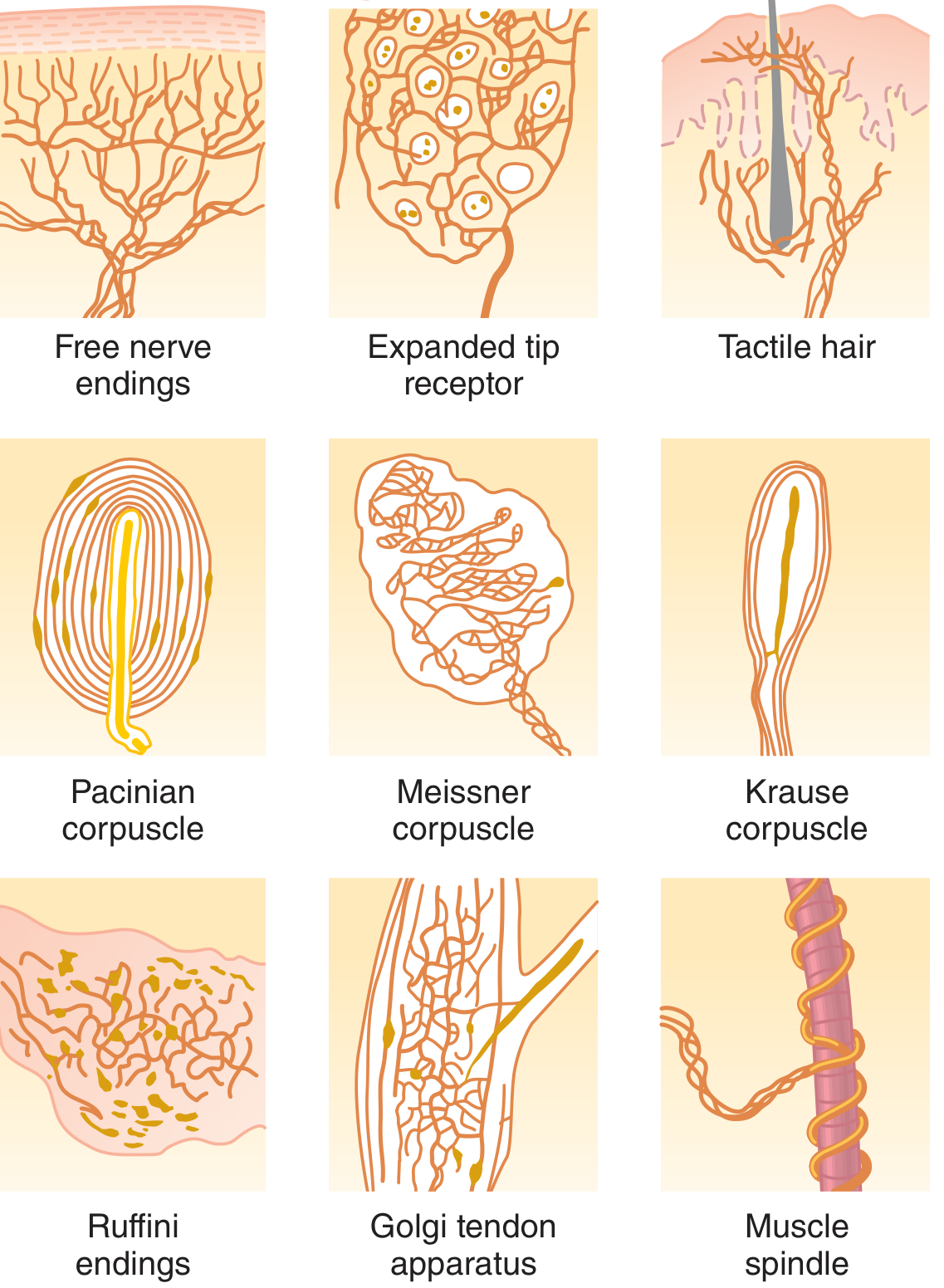
Key Functional Properties of Receptors
1. Differential Sensitivity ("Adequate Stimulus")
Each receptor is highly sensitive to one specific type of stimulus and nearly nonresponsive to others. Examples:
- Rods and cones respond to light but not to heat or pressure
- Osmoreceptors in the hypothalamus detect fluid osmolality but not sound
- Pain receptors are silent to normal touch but fire with tissue damage
This principle is called "labeled line coding" - each sensory fiber type leads to a specific area of the CNS, and perception depends on where the signal terminates, not what stimulus triggered it.
2. Receptor Potential Generation
Receptors generate potentials via four mechanisms:
- Mechanical deformation - stretches membrane, opens stretch-sensitive ion channels
- Chemical application - opens ion channels or activates second messenger systems
- Temperature change - alters temperature-sensing ion channels and metabotropic receptors
- Electromagnetic radiation - (e.g., light) directly/indirectly changes membrane characteristics
The maximum receptor potential amplitude is ~100 mV at extremely high stimulus intensity.
3. Adaptation of Receptors
Receptors adapt (reduce their firing rate) to a constant stimulus over time.
| Type | Examples | Function |
|---|---|---|
| Rapidly adapting ("phasic receptors") | Pacinian corpuscle, hair receptors | Detect change or rate of stimulus - fire only when stimulus changes; also called "rate receptors" or "movement receptors" |
| Slowly adapting ("tonic receptors") | Muscle spindles, Golgi tendon organs, pain receptors, baroreceptors, vestibular macula | Detect continuous/sustained stimulus strength; keep brain informed of ongoing body status |
The Pacinian corpuscle adapts within hundredths of a second (to "extinction"); baroreceptors may take ~2 days. Pain receptors and chemoreceptors never fully adapt.
- Predictive function of rate receptors: The semicircular canals and joint rate receptors let the CNS predict body position a few seconds ahead - essential for running, balance, and coordinated movement.
4. Membrane Receptor Functions (Signal Transduction)
Beyond sensory receptors, Guyton & Hall describes membrane protein receptors that bind signaling molecules:
- Ion channel-linked receptors (ionotropic) - e.g., nicotinic acetylcholine receptors at the neuromuscular junction and autonomic ganglia; directly gate ion flow when ligand binds
- G-protein coupled receptors (metabotropic) - e.g., muscarinic acetylcholine receptors on parasympathetic effectors; activate intracellular second messenger cascades (e.g., cAMP via adenylate cyclase)
- Enzyme-linked receptors - e.g., growth hormone receptor; binding activates intracellular enzyme activity directly
Autonomic Receptor Functions (Chapter 61)
Cholinergic Receptors
| Receptor | Location | Mechanism | Function |
|---|---|---|---|
| Muscarinic | Postganglionic parasympathetic effectors | G-protein coupled | Smooth muscle contraction, gland secretion, cardiac slowing |
| Nicotinic | Autonomic ganglia, neuromuscular junctions | Ligand-gated ion channel | Fast synaptic transmission, skeletal muscle contraction |
Adrenergic Receptors (Table 61.1)
| Receptor | Key Effects |
|---|---|
| Alpha-1 | Vasoconstriction, iris dilation, intestinal relaxation, bladder sphincter contraction |
| Alpha-2 | Inhibits norepinephrine release (presynaptic), decreases GI motility |
| Beta-1 | Increased heart rate, increased contractility, lipolysis |
| Beta-2 | Vasodilation, bronchodilation, glycogenolysis, uterine relaxation |
| Beta-3 | Lipolysis in fat tissue |
- Norepinephrine - excites mainly alpha receptors, lesser effect on beta
- Epinephrine - excites both alpha and beta receptors approximately equally
Summary Table: Receptor Classification by Stimulus Type
| Class | Stimulus | Example Receptors |
|---|---|---|
| Mechanoreceptors | Touch, pressure, vibration, stretch | Pacinian, Meissner, Ruffini, Muscle spindle |
| Thermoreceptors | Temperature | Cold and warm receptors |
| Nociceptors | Tissue damage | Free nerve endings |
| Electromagnetic | Light | Rods, Cones |
| Chemoreceptors | Chemical concentrations | Carotid body, taste buds, olfactory receptors |
All content sourced from: Guyton and Hall Textbook of Medical Physiology (ISBN 9780443111013), Chapter 47 (Sensory Receptors), Chapter 61 (Autonomic Nervous System).
This is a shared conversation. Sign in to Orris to start your own chat.