
Typhoid fever and Paratyphold A.B. (Cinical and laboratory diagnostics. Complications. Treatment) tell me each and everything about these disease
typhoid fever paratyphoid clinical features diagnosis treatment complications 2024
typhoid fever rose spots rash Salmonella typhi
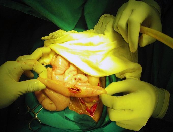
This clinical photograph shows an intraoperative view of an exploratory laparotomy, demonstrating a bowel perforation in the context of typhoid fever (Salmonella Typhi). The image focuses on a loop of small intestine held by a surgeon's gloved hands, revealing a discrete, longitudinal perforation with hyperemic and slightly everted edges. The surrounding serosa exhibits generalized inflammation and congestion. In the background, other segments of the intestine appear edematous and pale-pink, with visible white-yellowish peritoneal nodules and purulent exudate suggestive of concurrent miliary peritoneal tuberculosis and secondary peritonitis. Surgical instruments, suction tubing, and green sterile drapes define the surgical field. This image serves as an educational example of the severe gastrointestinal complications of typhoid fever, specifically intestinal perforation, and illustrates the challenging clinical scenario of co-infection with tuberculosis in an acute surgical setting.
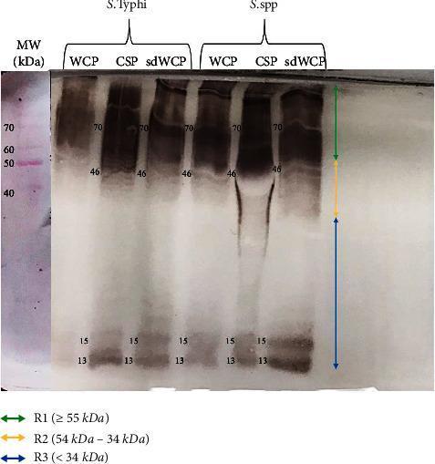
A diagnostic Western blot (WB) analysis image showing the immunoreactivity of antigens derived from Salmonella typhi and Salmonella spp. The image displays six experimental lanes representing three different protein extraction methods—Whole Cell Protein (WCP), Cell Surface Protein (CSP), and sodium deoxycholate-extracted WCP (sdWCP)—for each bacterial species. A molecular weight (MW) marker lane on the far left, stained with Ponceau S, shows reference bands from 40 kDa to 70 kDa. The WB profile is categorized into three vertical regions: R1 (≥ 55 kDa), R2 (54–34 kDa), and R3 (< 34 kDa). Prominent, high-intensity immunoreactive bands are observed in both species at approximately 70 kDa and 46 kDa, with a noticeable smear in the CSP lanes. Low molecular weight bands are consistently identified at 13 kDa and 15 kDa across all preparations. This visual provides comparative data on the antigenic profiles and antibody isotype reactivity against somatic and flagellar antigens, relevant for typhoid fever serodiagnosis research.
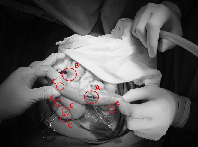
This monochrome clinical photograph captures an intraoperative view of the abdominal cavity during a surgical procedure for bowel perforation. The image illustrates two distinct infectious pathologies concurrently affecting the intestines and peritoneum. Label 'A' identifies a typhoid intestinal perforation, appearing as an irregular, longitudinal breach in the seromuscular layer of the small bowel, typical of advanced Salmonella typhi infection. Label 'B' shows a non-perforated typhoid lesion, characterized by localized inflammation and thickening without a full-thickness defect. Surrounding these intestinal segments, multiple small, discrete, whitish nodules are indicated by label 'C', representing miliary peritoneal tuberculosis. These nodules are scattered across the serosal surfaces and mesentery, demonstrating a grain-like distribution characteristic of disseminated mycobacterial infection. The image serves as a high-level educational example of acute surgical abdomen complications resulting from co-infection of typhoid fever and miliary tuberculosis, highlighting diagnostic landmarks such as transmural perforation and widespread peritoneal seeding.
Typhoid Fever & Paratyphoid A, B, C - Complete Guide
Sources: Harrison's Principles of Internal Medicine 22E (2025), Tintinalli's Emergency Medicine, Park's Textbook of Preventive and Social Medicine, CDC Guidelines
1. DEFINITION & CAUSATIVE ORGANISMS
| Disease | Organism | Notes |
|---|---|---|
| Typhoid fever | Salmonella enterica serovar Typhi (S. Typhi) | More severe |
| Paratyphoid A | S. enterica serovar Paratyphi A | Milder, gastrointestinal-predominant |
| Paratyphoid B | S. enterica serovar Paratyphi B | Less common |
| Paratyphoid C | S. enterica serovar Paratyphi C | Rare |
- Man is the ONLY reservoir - no animal reservoir exists for any of these organisms (unlike non-typhoidal Salmonella)
- Gram-negative, facultative anaerobic, motile bacilli
- Killed by drying, pasteurization, and common disinfectants
- S. Paratyphi A thought to cause milder disease, but a large prospective study (Kathmandu, Nepal, n=669) found typhoid and paratyphoid clinically indistinguishable
2. EPIDEMIOLOGY
- Global burden: 9.2-21 million cases of typhoid and ~5 million cases of paratyphoid per year; 110,000-280,000 deaths/year
- Highest burden: Indian subcontinent (India, Pakistan, Bangladesh, Nepal), Eastern Mediterranean, sub-Saharan Africa, Southeast Asia, some Pacific regions
- Incidence can exceed 1,000 cases per 100,000 children in some urban areas
- High correlation with mixing of drinking water and human sewage
- In endemic regions: more common in poor urban neighborhoods, children, and adolescents
- Peak age: 5-19 years; incidence falls after age 20 (immunity acquired from subclinical infection)
- Males more commonly infected; carrier rate higher in females
- Contaminated drinking water or ice
- Food from street vendors
- Raw fruits/vegetables grown in sewage-fertilized fields
- Ill household contacts
- Poor hand hygiene
- Prior H. pylori infection (reduced gastric acidity)
- International travel (78% of US cases are travel-associated, mainly from Indian subcontinent)
3. TRANSMISSION & PATHOGENESIS
- Ingestion of organism - gastric acid is a major barrier (reduced gastric acid = higher susceptibility)
- Penetration of small intestinal mucosa via M cells overlying Peyer's patches
- Uptake by macrophages - survive and replicate intracellularly (this is why cell-mediated immunity is the primary defense)
- Spread to mesenteric lymph nodes → thoracic duct → bloodstream = primary bacteremia (end of incubation)
- Seeding of reticuloendothelial system (liver, spleen, bone marrow) and gallbladder
- Secondary bacteremia - produces clinical illness
- In the third week - Peyer's patches become necrotic and ulcerate, risking perforation and hemorrhage
- Organisms can persist in gallbladder for years (chronic carrier state)
4. CLINICAL FEATURES (WEEK BY WEEK)
Week 1 - Onset
- Step-ladder fever rising to 38.8-40.5°C (101.8-104.9°F)
- Headache (80%), chills (35-45%), malaise
- Cough (30%), myalgia (20%), arthralgia
- Relative bradycardia (pulse slow relative to the height of fever - "Faget's sign") - present in <50% of cases
- Anorexia (55%), nausea (18-24%), vomiting (18%)
- Constipation more common than diarrhea in early stages
- Coated tongue (51-56%)
Week 2 - Full Illness
- High continuous fever, now well established
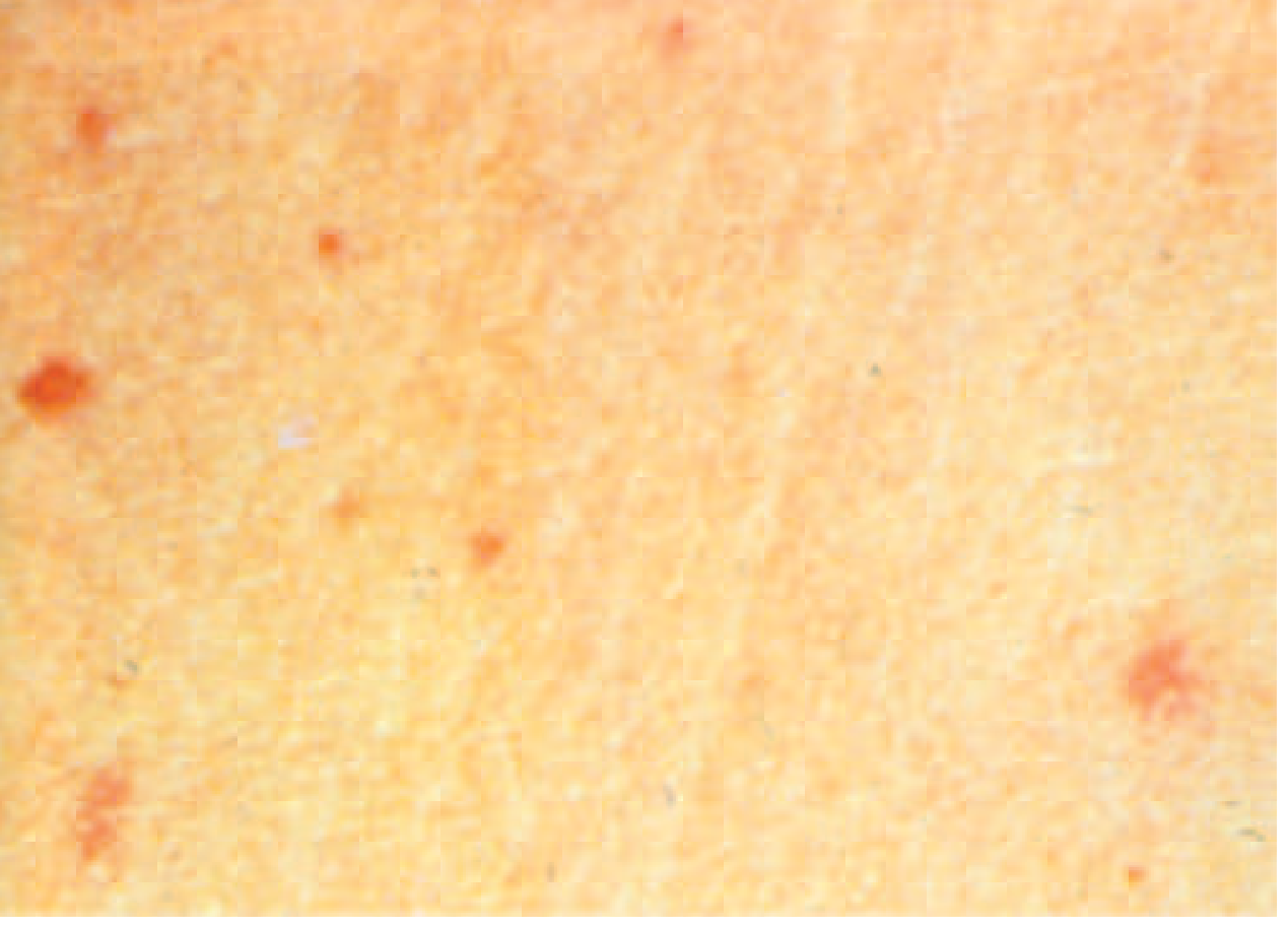
- "Rose spots" - faint, salmon-colored, blanching, maculopapular rash on the trunk and chest
- Appear in ~30% of patients at end of week 1 to beginning of week 2
- 2-4 mm, blanch on pressure, fade without scarring
- Seen best in fair-skinned individuals
- Represent Salmonella-infected macrophages in the skin
- Splenomegaly and hepatomegaly (5-6%)
- Abdominal distension, diarrhea (22-28%) or constipation (13-16%)
- Epistaxis in some patients
Week 3-4 - Complications or Recovery
- Severe: intestinal hemorrhage or perforation
- Neurological manifestations possible
- Without treatment: fever persists for up to 4 weeks
- Milder course with shorter duration
- More prominent gastrointestinal symptoms from the start
- Lower complication and fatality rates
5. LABORATORY DIAGNOSTICS
Blood Tests
| Test | Findings |
|---|---|
| CBC | Leukopenia (WBC often 3,000-5,000/mm³), mild anemia, thrombocytopenia |
| Liver enzymes | Mildly elevated ALT/AST (hepatitis pattern in some) |
| ESR | Elevated |
| CRP | Elevated |
- Leukocytosis can occur in children and with intestinal perforation (should raise suspicion for complication)
Microbiological Cultures (Gold Standard)
| Specimen | Optimal Timing | Sensitivity |
|---|---|---|
| Blood culture | Week 1-2 | 40-80% (most sensitive in 1st week) |
| Bone marrow culture | Any time | 90-95% - most sensitive overall (positive even after antibiotics started) |
| Stool culture | Week 2-3 | 30-50% |
| Urine culture | Week 2-3 | 25-30% |
| Bile (duodenal drainage) | Carrier detection | High yield in chronic carriers |
- Blood cultures are the most practical first-line test
- Bone marrow culture is the definitive test - organism can be recovered even after antibiotic treatment has begun
- Stool cultures become positive in weeks 2-3 as organisms shed via bile into the gut
Serological Tests
- Detects agglutinating antibodies against O (somatic) and H (flagellar) antigens of S. Typhi
- O antibodies - appear in week 1, rise in weeks 2-3, fall quickly after recovery - indicates active infection
- H antibodies - appear later, persist for months/years - may reflect past infection or vaccination
- Diagnostic criteria: 4-fold rise in paired sera (taken 2 weeks apart) OR single titre ≥1:160 (O) in a non-endemic area
- Limitations: Low sensitivity and specificity; false positives in other Salmonella infections, malaria, liver disease, and in vaccinated individuals; results vary by lab
- Not reliable for diagnosis in endemic areas where baseline titres may be elevated
- For paratyphoid: similar test using Paratyphi A (AO, AH), B (BO, BH) antigens
- IgM and IgG ELISA against a specific 50-kDa outer membrane protein of S. Typhi
- Typhidot-M (detects IgM only) is more specific - eliminates IgG that persists from previous infection
- Results available in 3-4 hours
- Better than Widal in endemic areas
- Detects IgM antibodies to O9 antigen of S. Typhi
- Rapid test, good specificity
- Does not cross-react with vaccination
- High sensitivity, identifies organisms even after antibiotic initiation
- Not universally available
Key Diagnostic Approach Summary
- Culture blood in week 1 (best yield)
- Culture stool + urine from week 2
- Bone marrow culture if blood culture negative and high clinical suspicion
- Widal test - supportive evidence only (not definitive in endemic areas)
- Rapid tests (Typhidot, Tubex) for resource-limited settings
6. DIFFERENTIAL DIAGNOSIS
- Malaria - most important; always exclude first in travelers
- Brucellosis
- Leptospirosis
- Typhus / Rickettsiosis
- Dengue fever
- Sepsis from other causes
- Viral hepatitis
- Tuberculosis (in chronic fever)
7. COMPLICATIONS (10-15% of hospitalized patients)
Gastrointestinal
| Complication | Details |
|---|---|
| Intestinal hemorrhage | Erosion of Peyer's patches into blood vessels; presents with melena or bright rectal bleeding in week 3; occurs in 1-5% |
| Intestinal perforation | Most feared; ileal perforation near ileocecal valve; presents as acute abdomen; 1-3% incidence; mortality 10-32% even with surgery |
| Hepatitis | Mild jaundice and elevated transaminases in up to 40% |
| Cholecystitis | Acute or chronic; gallbladder is the reservoir for carriers |
Cardiovascular
- Myocarditis - ECG changes, arrhythmias
- Mycotic aneurysm (rare)
Hematological
- DIC (Disseminated Intravascular Coagulopathy)
- Severe anemia (from GI blood loss + hemolysis)
- Thrombocytopenia
Neurological ("Typhoid Encephalopathy")
- Altered consciousness, confusion, delirium
- Meningismus, seizures
- Psychosis
- Ataxia, cranial nerve palsies
- Deafness (sensorineural - a specific sequela)
- Occurs in 1-10% of severe cases
Respiratory
- Pneumonia (bronchopneumonia)
- Pleural effusion
Renal
- Glomerulonephritis, renal failure (rare)
Other
- Septic arthritis
- Osteomyelitis (especially in sickle cell disease)
- Pancreatitis
8. RELAPSE
- Occurs in ~10% of inadequately treated patients (and some adequately treated)
- Typically 1-3 weeks after clinical recovery (CDC) / "2-3 weeks after recovery" (MSF 2024)
- Relapse illness is usually milder than the primary attack
- Usually NOT due to antibiotic resistance
- Re-treatment with the same antibiotic is usually effective
- Second relapse possible (rare)
9. CARRIER STATE
- Chronic carrier: Excretion of S. Typhi in stool or urine for >1 year after infection
- Affects 1-4% of typhoid fever cases (Park: 2-5%)
- Organisms persist in the gallbladder and biliary tract
- More common in women, elderly, and those with pre-existing gallbladder disease
- The famous historical case: "Typhoid Mary" (Mary Mallon) - caused >1,300 cases over her lifetime
- Faecal carriers more frequent than urinary carriers
- Chronic urinary carrier state often associated with urinary tract abnormalities
- Vi antibody detected in ~80% of chronic carriers (useful screening test)
10. TREATMENT
General Principles
- Antibiotics shorten clinical course, reduce complication risk, and lower mortality
- Treatment decisions complicated by high rates of antimicrobial resistance
- Susceptibility testing should guide treatment whenever possible
Antibiotic Resistance Profile
| Drug Category | Resistance Situation |
|---|---|
| Chloramphenicol, Ampicillin, TMP-SMX | MDR strains widely prevalent since 1980s; unreliable |
| Fluoroquinolones | Decreased susceptibility to ciprofloxacin (DSC) widespread on Indian subcontinent (66% of US travel-acquired cases from India); full resistance emerging |
| Ceftriaxone / 3rd gen cephalosporins | Generally effective; resistance emerging |
| Azithromycin | Effective for uncomplicated typhoid; resistance emerging in some areas |
| XDR (Extensively Drug Resistant) S. Typhi | Pakistan outbreak 2016-2019; resistant to all 1st-line + fluoroquinolones + 3rd-gen cephalosporins; only azithromycin and carbapenems active |
Antibiotic Regimens
| Drug | Dose | Duration |
|---|---|---|
| Azithromycin | 500 mg OD (or 1g on day 1 then 500 mg) | 5-7 days |
| Cefixime | 15-20 mg/kg/day in 2 divided doses | 7-14 days |
| Ciprofloxacin (if susceptible) | 500 mg BD | 5-7 days |
| Drug | Dose | Duration |
|---|---|---|
| Ceftriaxone | 2 g IV OD (adults); 60 mg/kg/day in children | 10-14 days |
| Azithromycin IV (if available) | 500 mg OD | 7-10 days |
| Meropenem | For XDR typhoid; 1 g IV TDS | 14 days |
- Dexamethasone 3 mg/kg IV loading dose, then 1 mg/kg every 6 hours for 48 hours (for typhoid shock or encephalopathy)
- Reduces mortality in severe disease with neurological involvement
Supportive Treatment
- IV rehydration
- Antipyretics (paracetamol; avoid NSAIDs due to GI bleeding risk)
- Blood transfusion if significant GI blood loss
- Nutritional support
- Close monitoring for complications (abdominal exam, stool examination)
Treatment of Complications
| Complication | Management |
|---|---|
| Intestinal hemorrhage | IV fluids, blood transfusion, conservative management initially; surgery if massive bleeding |
| Intestinal perforation | Emergency surgery (primary repair or resection), IV antibiotics covering gram-negatives and anaerobes |
| Typhoid encephalopathy | Dexamethasone + antibiotics |
| DIC | Fresh frozen plasma, platelets, heparin in some cases |
Treatment of Chronic Carriers
- Ampicillin or Amoxicillin 4-6 g/day + Probenecid 2 g/day for 6 weeks (concentrated in bile; eradicates carrier state in ~70%)
- Ciprofloxacin 750 mg BD for 4 weeks (preferred if susceptible)
- Cholecystectomy with concurrent antibiotic therapy - reserved for carriers with gallstones when medical treatment fails
- Chloramphenicol is ineffective at clearing the carrier state
11. PARATYPHOID A, B, C - SPECIFIC NOTES
| Feature | Typhoid | Paratyphoid A | Paratyphoid B | Paratyphoid C |
|---|---|---|---|---|
| Organism | S. Typhi | S. Paratyphi A | S. Paratyphi B | S. Paratyphi C (rare) |
| Severity | More severe | Milder (but not always) | Milder | Variable |
| Predominant symptoms | Systemic > GI | GI-predominant | GI-predominant | Variable |
| Carrier rate | 1-4% | Lower | Lower | - |
| CFR | ~1% (treated) | ~0.5% (roughly half) | ~0.5% | - |
| Rose spots | Yes (~30%) | Can occur | Can occur | - |
| Treatment | Same as above | Same as above | Same as above | Same |
| Widal | O/H agglutinins | AO, AH agglutinins | BO, BH agglutinins | CO, CH |
- Paratyphoid fever accounts for ~5 million cases/year globally
- S. Paratyphi A is the most common paratyphoid
- Paratyphoid is slightly more common in older adults (median age 29 vs 23 for typhoid in US data)
- Complications occur less frequently but are otherwise the same in type
- The two diseases cannot be distinguished on clinical grounds - only culture separates them
12. PROGNOSIS & MORTALITY
| Scenario | Mortality |
|---|---|
| Pre-antibiotic era typhoid | >10-30% |
| Treated typhoid (modern) | <1% |
| With intestinal perforation | 10-32% |
| Paratyphoid (treated) | ~0.5% - lower than typhoid |
| MDR/XDR typhoid untreated | Higher |
- With early diagnosis and appropriate treatment, mortality is ~1%
- Without treatment: disease lasts ~4 weeks; historical mortality 10-30%
13. PREVENTION & CONTROL
Vaccination
| Vaccine | Type | Route | Efficacy | Duration |
|---|---|---|---|---|
| Ty21a (Vivotif) | Live attenuated oral | Oral (4 capsules) | 50-80% | 3-7 years |
| Vi polysaccharide (ViPS) | Injectable subunit | IM | 55-72% | 2-3 years |
| Typhoid conjugate vaccine (TCV, Typbar-TCV) | Vi conjugated to tetanus toxoid | IM single dose | >80% | Longer; protective in <2 yrs |
- No vaccine covers paratyphoid - vaccines only protect against S. Typhi
- WHO recommends TCV for routine use in children >6 months in endemic countries
- Vaccines are not 100% effective - food and water precautions remain essential
Sanitation & Control
- Safe water supply and sewage treatment are the most important measures
- The "weakest link" in transmission is sanitation (most amenable to control)
- Food handling hygiene, hand washing
- Identification and treatment of chronic carriers
- Isolation of cases until 3 bacteriologically negative stool and urine samples obtained
- Disinfection: 5% cresol for 2 hours (stools and urine); 2% chlorine for soiled linen
Summary Table - At a Glance
| Feature | Detail |
|---|---|
| Causative agent | S. Typhi (typhoid); S. Paratyphi A, B, C (paratyphoid) |
| Only reservoir | Humans (cases + carriers) |
| Incubation | 5-21 days (mean 10-14 days) |
| Classic fever | Step-ladder, continuous, 38.8-40.5°C |
| Pathognomonic sign | Rose spots on trunk (30% of cases) |
| Relative bradycardia | <50% of cases |
| Gold standard diagnosis | Blood culture (week 1); bone marrow culture (any time, most sensitive) |
| Widal test | Supportive only; unreliable in endemic areas |
| Complications | Perforation, hemorrhage, encephalopathy, DIC, myocarditis |
| Complication timing | Week 3+ |
| Drug of choice | Azithromycin (uncomplicated, oral); Ceftriaxone (severe, IV) |
| Dexamethasone | Add for shock/encephalopathy |
| Chronic carrier Rx | Ciprofloxacin 750 mg BD x 4 weeks |
| Mortality (treated) | <1% (typhoid); ~0.5% (paratyphoid) |
Typhoid fever and Paratyphold A.B. (Cinical and laboratory diagnostics. Complications. Treatment) tell me in details
🦠 Typhoid Fever & Paratyphoid A, B — Complete Clinical Guide
Based on Harrison's Principles of Internal Medicine 22E (2025), Tintinalli's Emergency Medicine, Park's Textbook of Preventive & Social Medicine, CDC Guidelines 2024
1. DEFINITION & CAUSATIVE ORGANISMS
| Disease | Causative Organism | Severity |
|---|---|---|
| Typhoid fever | Salmonella enterica serovar Typhi | More severe |
| Paratyphoid A | S. enterica serovar Paratyphi A | Milder (GI-predominant) |
| Paratyphoid B | S. enterica serovar Paratyphi B | Milder |
| Paratyphoid C | S. enterica serovar Paratyphi C | Rare, variable |
- Gram-negative, facultative anaerobic, motile bacilli
- Man is the ONLY reservoir - no animal reservoir
- Killed by drying, pasteurization, and common disinfectants
- Survive for weeks in water and ice
- Virulence factors include the Vi (virulence) capsular polysaccharide antigen, which inhibits complement activation and phagocytosis
2. EPIDEMIOLOGY
- Global burden: 9.2-21 million typhoid cases + ~5 million paratyphoid cases annually; 110,000-280,000 deaths/year
- Highest incidence: Indian subcontinent (India, Pakistan, Bangladesh, Nepal), Eastern Mediterranean, sub-Saharan Africa, Southeast Asia
- Can exceed 1,000 cases per 100,000 children in some urban slum areas
- Peak age: 5-19 years (incidence falls after 20 due to acquired immunity)
- Males more commonly infected; carrier rate higher in females
- Correlates strongly with mixing of drinking water and sewage
- Contaminated water or ice
- Street food vendors
- Raw fruits/vegetables grown in sewage-fertilized soil
- Ill household contacts
- Poor hand hygiene, no toilet access
- Prior H. pylori infection (reduced gastric acid = easier bacterial colonization)
- International travel (78% of US cases are travel-associated, mainly Indian subcontinent)
3. TRANSMISSION & PATHOGENESIS
Route of Transmission — the "5 Fs"
- Primary sources: feces and urine of cases or chronic carriers
- Secondary sources: contaminated water, food, flies, fingers
Pathogenesis (Step by Step)
Ingestion (10³–10⁹ organisms)
        ↓
Gastric acid barrier (1st defense — reduced acidity increases susceptibility)
        ↓
Penetration of small bowel mucosa via M cells overlying Peyer's patches
        ↓
Uptake by macrophages → survive intracellularly (cell-mediated immunity is primary defense)
        ↓
Mesenteric lymph nodes → thoracic duct → PRIMARY BACTEREMIA (silent, end of incubation)
        ↓
Seeding of reticuloendothelial system: liver, spleen, bone marrow, gallbladder
        ↓
Replication in macrophages → SECONDARY BACTEREMIA → clinical illness begins
        ↓
Week 3-4: Peyer's patches enlarge, necrose, ulcerate
        ↓
Risk of intestinal perforation and hemorrhage
- Organisms persist in the gallbladder (chronic carrier state)
- Intracellular survival explains why bacteremia can recur and why treatment requires bacteriostatic agents with intracellular penetration
4. CLINICAL FEATURES — WEEK BY WEEK
WEEK 1 — Prodrome / Onset
| Symptom | Frequency |
|---|---|
| Fever (step-ladder rise, 38.8-40.5°C) | >75% |
| Headache | 80% |
| Chills | 35-45% |
| Malaise, lethargy | Very common |
| Anorexia | 55% |
| Dry cough | 30% |
| Myalgia | 20% |
| Constipation (early) | 13-16% |
| Nausea, vomiting | 18-24% |
| Arthralgia | 2-4% |
- Fever character: Begins low, rises progressively in a step-ladder pattern, becomes continuous by end of week 1
- Relative bradycardia (Faget's sign): Pulse unusually slow for the height of fever — present in <50% of cases; when present, strongly suggestive
- Coated tongue: 51-56%
WEEK 2 — Full Illness
- Fever now sustained, high (40-40.5°C)
- "Rose spots" — pathognomonic rash
- Faint, salmon-colored, blanching maculopapular rash
- Located primarily on trunk and chest
- 2-4 mm diameter, fade without scarring
- Appear in ~30% of patients, more visible in fair-skinned individuals
- Represent Salmonella-infected macrophages in the skin
- Resolve spontaneously in 2-5 days
- Hepatosplenomegaly (5-6%)
- Abdominal distension and tenderness (4-5%)
- Diarrhea (22-28%) — "pea-soup" diarrhea; or persistent constipation
- Epistaxis in some patients
- Toxemic facies — patient appears acutely unwell
WEEK 3-4 — Complications or Resolution
- If untreated: fever persists for up to 4 weeks
- Maximum risk of intestinal perforation and hemorrhage
- "Typhoid state" — profound toxemia, muttering delirium, picking at bedclothes
- Neurological features may emerge
- With treatment: defervescence begins within 3-5 days; clinical improvement in 4-5 days
Paratyphoid A & B - Specific Features
- Clinically identical to typhoid (a large prospective study of 669 consecutive cases in Kathmandu found them indistinguishable)
- Paratyphoid A traditionally considered milder with more GI-predominant symptoms
- Rose spots can occur
- Complications occur less frequently
- Cannot be distinguished from typhoid by clinical examination alone — only culture differentiates
5. LABORATORY DIAGNOSTICS
A. Routine Blood Tests
| Test | Finding in Typhoid | Significance |
|---|---|---|
| WBC count | Leukopenia (2,000-5,000/mm³) | Characteristic; leukocytosis suggests perforation or superinfection |
| Differential | Relative lymphocytosis, eosinopenia | |
| Hemoglobin | Mild normocytic anemia | From hemolysis + bone marrow suppression |
| Platelets | Mild thrombocytopenia | |
| LFTs (ALT/AST) | Mildly elevated (2-3× normal) | Hepatic involvement in 40% |
| Bilirubin | Mildly elevated in some | |
| Serum electrolytes | Hyponatremia common | |
| ESR/CRP | Elevated | Nonspecific |
B. Microbiological Cultures (Gold Standard)
| Culture Type | Best Timing | Sensitivity | Notes |
|---|---|---|---|
| Blood culture | Week 1 (first 7-10 days) | 40-80% | Most practical; 3 samples preferred; 10 mL per bottle; positivity falls after antibiotics |
| Bone marrow culture | Any time | 90-95% | MOST SENSITIVE - positive even after antibiotics; use when blood culture negative |
| Stool culture | Week 2-3 | 30-50% | Positive as organisms shed via bile into gut |
| Urine culture | Week 2-3 | 25-30% | Low yield |
| Bile (duodenal drainage) | Carrier detection | High | Used to detect chronic carriers |
| Rose spot biopsy | Week 2 | ~60% | Rarely done |
- Never delay blood culture - it is the most important and practical test
- 3 blood cultures in 24 hours before antibiotics significantly increases yield
- Bone marrow culture is the gold standard - should be used when:
- Blood culture is negative but clinical suspicion is high
- Patient has already received antibiotics
- Carrier state investigation
- Cultures must be incubated for 7-10 days before reporting as negative
C. Serological Tests
1. Widal Test (Tube Agglutination Test)
| Antigen | Antibody Type | Significance |
|---|---|---|
| O (somatic) | IgM | Appears week 1; rises weeks 2-3; falls quickly after recovery → indicates ACTIVE infection |
| H (flagellar) | IgG | Appears later; persists months-years → may reflect past infection or vaccination |
- AO, AH - Paratyphi A agglutinins
- BO, BH - Paratyphi B agglutinins
- Significant titer: Single titer ≥1:160 (O antigen) in a non-endemic area
- Definitive diagnosis: 4-fold rise in paired sera taken 10-14 days apart
- In endemic areas, baseline titers may be elevated — single titer is unreliable
- False positives: malaria, other Salmonella infections, liver disease, autoimmune conditions, prior vaccination
- False negatives: early disease (before antibody production), immunocompromised patients, early antibiotic treatment
- Does not differentiate acute from past infection reliably in endemic areas
- Not recommended as sole diagnostic test by WHO
2. Typhidot Test
- Detects IgM and IgG against a specific 50 kDa outer membrane protein of S. Typhi
- Typhidot-M (detects IgM only) is more specific — eliminates cross-reactivity from past infection IgG
- Results in 3-4 hours
- Better performance in endemic areas than Widal test
3. TUBEX Test
- Detects IgM against O9 antigen of S. Typhi
- Rapid colorimetric test
- Good specificity; does NOT cross-react with typhoid vaccination
- Semi-quantitative
4. Blood Culture PCR / Molecular Tests
- High sensitivity; can identify organisms even after antibiotic initiation
- Not widely available in endemic regions
- Multiplex PCR panels can differentiate S. Typhi from S. Paratyphi
5. Vi Antibody Test
- Vi agglutinins present in ~80% of chronic carriers
- Used to screen for chronic carrier state
- Not useful for acute diagnosis
D. Summary: Diagnostic Approach by Week
| Week of illness | Best test |
|---|---|
| Week 1 | Blood culture × 3 |
| Week 2-3 | Blood culture + Stool and urine culture + Widal (paired sera) |
| Any time (culture negative) | Bone marrow culture |
| Chronic carrier screening | Vi antibody + stool/urine culture + duodenal drainage |
6. COMPLICATIONS
A. Gastrointestinal Complications
1. Intestinal Perforation (Most Feared)
- Incidence: 1-3%
- Usually in the terminal ileum, near the ileocecal valve (at Peyer's patch ulcers)
- Presents with: sudden worsening of abdominal pain, rigid abdomen ("board-like"), absent bowel sounds, rebound tenderness
- X-ray/CT: free air under the diaphragm
- Mortality: 10-32% even with surgery
- Management: emergency laparotomy (primary repair or resection + anastomosis), broad-spectrum IV antibiotics
2. Intestinal Hemorrhage
- Incidence: 1-5%
- From erosion of a blood vessel at the base of an ulcerated Peyer's patch
- Presents with: melena or bright red rectal bleeding, sudden drop in temperature and fever, tachycardia
- Management: IV fluids, blood transfusion, conservative initially; surgery if massive or uncontrolled
3. Hepatitis
- Mild jaundice and elevated transaminases in up to 40% of patients
- Usually self-limiting
4. Cholecystitis
- Acute or chronic
- Gallbladder is the site of persistence in chronic carriers
- May require cholecystectomy in refractory carriers
B. Neurological Complications ("Typhoid Encephalopathy") — 1-10%
- Altered consciousness, confusion, stupor, coma
- Muttering delirium ("typhoid state")
- Meningismus (meningeal irritation without meningitis)
- Seizures
- Sensorineural deafness (a specific recognized sequela)
- Ataxia, cranial nerve palsies
- Psychosis, behavioral changes
C. Cardiovascular Complications
| Complication | Details |
|---|---|
| Myocarditis | ECG changes (ST abnormalities, arrhythmias), elevated troponin; 1-5% |
| Mycotic aneurysm | Rare; arterial wall infection |
| Peripheral circulatory failure / Typhoid shock | In severe cases |
D. Hematological Complications
| Complication | Notes |
|---|---|
| DIC (Disseminated Intravascular Coagulopathy) | Coagulopathy, bleeding from multiple sites |
| Severe anemia | From GI blood loss + hemolysis + bone marrow suppression |
| Thrombocytopenia | Common; severe in some |
| Hemophagocytic syndrome | Rare but life-threatening |
E. Respiratory Complications
- Pneumonia / Bronchopneumonia — from bacteremia seeding the lungs, or superinfection
- Pleural effusion (uncommon)
F. Renal Complications
- Glomerulonephritis (immune complex deposition)
- Acute tubular necrosis / Acute kidney injury
- Urinary tract infection
G. Other Complications
| Complication | Notes |
|---|---|
| Septic arthritis | Especially in children; large joints |
| Osteomyelitis | Risk increases with hemoglobinopathies (sickle cell disease) |
| Pancreatitis | Rare |
| Orchitis | Rare |
H. Relapse
- Occurs in ~10% of inadequately treated patients
- Onset 1-3 weeks after apparent clinical recovery
- Usually milder than primary illness
- Generally NOT due to antibiotic resistance
- Re-treatment with the same antibiotic is effective
- Second relapse possible but rare
7. TREATMENT
A. Antibiotic Resistance Landscape (Critical Context)
| Drug Class | Resistance Status |
|---|---|
| Chloramphenicol, Ampicillin, TMP-SMX | MDR strains widespread since 1980s — no longer reliable as first-line |
| Fluoroquinolones (Ciprofloxacin) | Decreased susceptibility (DSC) prevalent on Indian subcontinent (66% of US travel cases from India); full resistance growing |
| 3rd-gen Cephalosporins (Ceftriaxone) | Still largely effective; resistance emerging |
| Azithromycin | Effective for uncomplicated; resistance emerging in some regions |
| XDR (Extensively Drug-Resistant) S. Typhi | Emerged in Pakistan 2016-2019; resistant to ALL 1st-line drugs + fluoroquinolones + 3rd-gen cephalosporins — only azithromycin and carbapenems active |
- Always perform antimicrobial susceptibility testing when culture is obtained
- A thorough travel history guides empiric choices while awaiting results
B. Treatment Regimens
Uncomplicated Typhoid/Paratyphoid (Oral, Outpatient)
| Drug | Adult Dose | Duration | Notes |
|---|---|---|---|
| Azithromycin (preferred) | 500-1000 mg OD | 5-7 days | Best for uncomplicated; good intracellular penetration; use for DSC strains |
| Cefixime | 20 mg/kg/day in 2 doses (max 400 mg/day) | 7-14 days | Oral 3rd-gen cephalosporin; good for MDR strains |
| Ciprofloxacin (only if fully susceptible) | 500 mg BD | 5-7 days | Avoid in DSC/resistant strains; do NOT use empirically from Indian subcontinent |
| Ofloxacin (if susceptible) | 400 mg BD | 7 days | Alternative fluoroquinolone |
Severe / Complicated Typhoid (IV, Inpatient)
| Drug | Adult Dose | Duration | Notes |
|---|---|---|---|
| Ceftriaxone (drug of choice) | 2 g IV OD (60 mg/kg/day in children) | 10-14 days | Standard of care for severe disease |
| Azithromycin IV | 500 mg IV OD | 7-10 days | Transition to oral when tolerating |
| Meropenem | 1 g IV TDS | 14 days | For XDR typhoid only; last resort |
Adjuvant Dexamethasone (For Severe Toxicity)
- Dexamethasone 3 mg/kg IV loading dose, then 1 mg/kg every 6 hours for 48 hours
- Shown to reduce mortality in severe disease with neurological involvement
- Do not use for uncomplicated disease (may prolong carrier state and increase relapse risk)
C. Supportive Treatment
| Measure | Details |
|---|---|
| IV rehydration | Essential for febrile, anorexic, or diarrheal patients |
| Antipyretics | Paracetamol preferred; avoid NSAIDs (increase GI bleeding risk) |
| Nutrition | Soft, easily digestible diet; high-calorie; avoid spicy or hard food |
| Rest | Complete bed rest during fever |
| Blood transfusion | For GI hemorrhage or severe anemia |
| Monitoring | Daily abdominal exam, stool inspection, vital signs, LFTs, CBC |
D. Treatment of Complications
| Complication | Management |
|---|---|
| Intestinal hemorrhage | Bed rest, IV fluids, blood transfusion; NPO; conservative initially; surgery if massive |
| Intestinal perforation | Emergency laparotomy (primary repair or resection); IV antibiotics + metronidazole; ICU |
| Typhoid encephalopathy | Dexamethasone (3 mg/kg load + 1 mg/kg q6h × 48h) + IV ceftriaxone |
| DIC | FFP, platelets, packed red cells; treat underlying infection aggressively |
| Myocarditis | Bed rest, cardiac monitoring; corticosteroids in severe cases |
E. Treatment of Chronic Carriers
| Regimen | Dose | Duration | Efficacy |
|---|---|---|---|
| Ciprofloxacin (preferred) | 750 mg BD | 4 weeks | ~80% clearance |
| Ampicillin + Probenecid | Ampicillin 4-6 g/day + Probenecid 2 g/day | 6 weeks | ~70% clearance |
| Amoxicillin + Probenecid | Similar to ampicillin | 6 weeks | ~70% |
| Cholecystectomy | Surgery | - | Reserved for carriers with gallstones unresponsive to medical therapy |
- Chloramphenicol is ineffective for clearing the carrier state
- Follow-up stool/urine cultures at 3-4 months and 12 months post-treatment
8. PARATYPHOID A & B — SPECIFIC COMPARISON
| Feature | Typhoid | Paratyphoid A | Paratyphoid B |
|---|---|---|---|
| Organism | S. Typhi | S. Paratyphi A | S. Paratyphi B |
| Global cases/year | 9.2-21 million | Most of ~5 million | Less common than A |
| Severity | More severe | Milder | Milder |
| Predominant symptoms | Systemic > GI | GI-predominant | GI-predominant |
| Rose spots | ~30% | Can occur | Can occur |
| Relative bradycardia | Present | May be present | May be present |
| Complications | 10-15% | Less frequent | Less frequent |
| Carrier rate | 1-4% | Lower | Lower |
| CFR (treated) | <1% | ~0.5% | ~0.5% |
| Widal antigens | O, H | AO, AH | BO, BH |
| Vaccine protection | Yes (Ty21a, Vi, TCV) | No | No |
| Treatment | Same | Identical | Identical |
9. CHRONIC CARRIER STATE
- Definition: Excretion of S. Typhi in stool or urine for >1 year after infection
- Affects 1-4% of typhoid fever cases
- Organisms persist in gallbladder and biliary tract (bile concentrations remain high)
- Higher risk: women, elderly, pre-existing gallbladder disease
- Faecal carriers more common than urinary carriers
- Chronic urinary carrier state associated with urinary tract abnormalities
- Vi antibody positive in ~80% of chronic carriers (screening test)
- Famous example: "Typhoid Mary" (Mary Mallon) - caused >1,300 cases over her lifetime while remaining asymptomatic
10. PROGNOSIS
| Scenario | Mortality |
|---|---|
| Typhoid fever — untreated (historical) | 10-30% |
| Typhoid fever — treated | <1% |
| With intestinal perforation | 10-32% |
| With encephalopathy (untreated) | High |
| Paratyphoid fever — treated | ~0.5% (roughly half of typhoid) |
| MDR/XDR typhoid without appropriate treatment | Significantly higher |
11. PREVENTION
Vaccines
| Vaccine | Type | Route | Efficacy | Duration | Covers |
|---|---|---|---|---|---|
| Ty21a (Vivotif) | Live attenuated | Oral × 4 capsules | 50-80% | 3-7 years | S. Typhi only |
| Vi polysaccharide (ViPS) | Injectable subunit | IM single dose | 55-72% | 2-3 years | S. Typhi only |
| Typhoid Conjugate Vaccine (TCV / Typbar-TCV) | Vi-tetanus conjugate | IM single dose | >80% | Longer-lasting; works in <2 yrs | S. Typhi only |
- No vaccine protects against paratyphoid - vaccines only cover S. Typhi
- WHO recommends TCV for children >6 months in endemic countries
- Vaccines are NOT 100% effective — food/water precautions are always necessary
Sanitation (Most Important)
- Safe drinking water and sewage treatment — the most impactful public health measure
- Hand hygiene (before eating, after toilet use)
- Safe food handling
- Pasteurization of dairy products
Control of Cases & Carriers
- Case isolation until 3 consecutive negative stool and urine cultures
- Notification to public health authorities (mandatory in most countries)
- Disinfection of stools and urine with 5% cresol for 2 hours
- Soiled linen: 2% chlorine solution or steam sterilization
- Carrier identification and treatment
- Exclusion of carriers from food handling and patient care
QUICK REFERENCE SUMMARY TABLE
| Feature | Detail |
|---|---|
| Causative agents | S. Typhi (typhoid); S. Paratyphi A, B (paratyphoid) |
| Only reservoir | Humans (cases + chronic carriers) |
| Incubation period | 5-21 days (mean 10-14 days) |
| Fever type | Step-ladder, continuous, 38.8-40.5°C |
| Pathognomonic sign | Rose spots on trunk (30%) |
| Relative bradycardia | <50% of cases |
| Gold standard diagnosis | Blood culture (week 1); Bone marrow culture (any time) |
| Most sensitive culture | Bone marrow (90-95%) |
| Widal test | Supportive only; unreliable in endemic areas |
| Leukopenia | Characteristic; leukocytosis suggests perforation |
| Typical complication timing | Week 3+ |
| Most feared complication | Intestinal perforation |
| Drug of choice (uncomplicated) | Azithromycin (oral) or Cefixime |
| Drug of choice (severe) | Ceftriaxone IV |
| Add dexamethasone for | Shock, encephalopathy |
| XDR typhoid treatment | Meropenem ± Azithromycin |
| Chronic carrier treatment | Ciprofloxacin 750 mg BD × 4 weeks |
| Mortality (treated) | <1% typhoid; ~0.5% paratyphoid |
| Relapse rate | ~10% (1-3 weeks after recovery) |
| Carrier rate | 1-4% (typhoid) |
Typhoid fever and Paratyphold A.B. (Cinical and laboratory diagnostics. Complications. Treatment) tell me in details
Typhoid Fever & Paratyphoid A, B — Complete Detailed Guide
Sources: Harrison's Principles of Internal Medicine 22E (2025), Tintinalli's Emergency Medicine, Park's Textbook of Preventive & Social Medicine, CDC Clinical Guidance 2024
1. DEFINITION & CAUSATIVE ORGANISMS
| Disease | Causative Organism | Relative Severity |
|---|---|---|
| Typhoid fever | Salmonella enterica serovar Typhi | More severe |
| Paratyphoid A | S. enterica serovar Paratyphi A | Milder, GI-predominant |
| Paratyphoid B | S. enterica serovar Paratyphi B | Milder |
| Paratyphoid C | S. enterica serovar Paratyphi C | Rare, variable |
- Gram-negative, non-spore-forming, facultative anaerobic, motile bacilli
- Man is the ONLY reservoir - no animal reservoir (unlike non-typhoidal Salmonella)
- Killed by drying, pasteurization, and common disinfectants
- Survive weeks in water and ice
- Key virulence factor: Vi (virulence) capsular polysaccharide - inhibits complement and phagocytosis
- The disease was called "typhoid" for its clinical similarity to typhus; the term "enteric fever" was proposed in 1869 based on the anatomic site of infection
2. EPIDEMIOLOGY
- Global burden: 9.2-21 million typhoid cases + ~5 million paratyphoid cases per year; 110,000-280,000 deaths/year
- Highest incidence: Indian subcontinent (India, Pakistan, Bangladesh, Nepal), Eastern Mediterranean, sub-Saharan Africa, Southeast Asia
- Can exceed 1,000 cases per 100,000 children in some urban areas
- Peak age group: 5-19 years (incidence falls after age 20 due to acquired immunity from repeated subclinical exposure)
- Males more commonly infected (greater exposure); carrier rate higher in females
- Strong correlation with mixing of drinking water and human sewage
- Contaminated drinking water or ice
- Street food vendors
- Raw fruits/vegetables grown in sewage-fertilized soil
- Ill household contacts
- Poor hand hygiene and lack of toilet access
- Prior H. pylori infection (chronically reduced gastric acid lowers the infective dose needed)
- International travel (78% of US cases are travel-associated, predominantly from India, Pakistan, Bangladesh)
3. TRANSMISSION
Route - the "5 Fs"
Feces/Urine (of cases or carriers)
      ↓
Food — Fingers — Fomites — Flies — Fluids (Water)
      ↓
New host (fecal-oral route)
- Primary sources: feces and urine of cases or chronic carriers
- Secondary sources: contaminated water, food, flies, dirty fingers
- Transmission is dose-dependent - severity varies with the size of the inoculum
- No evidence that organisms are excreted in sputum or milk
4. PATHOGENESIS (Step by Step)
STEP 1: Ingestion of organisms
        Gastric acid is the first barrier — reduced acidity increases susceptibility

STEP 2: Organisms reach small bowel mucosa
        Penetrate via M cells overlying Peyer's patches

STEP 3: Uptake by submucosal macrophages
        Survive and replicate INTRACELLULARLY
        (This is why cell-mediated immunity — not antibody — is the primary defense)

STEP 4: Spread to mesenteric lymph nodes → thoracic duct
        → PRIMARY (SILENT) BACTEREMIA — marks end of incubation period

STEP 5: Seeding of reticuloendothelial system
        Liver, spleen, bone marrow, GALLBLADDER

STEP 6: Organisms replicate in macrophages of these organs
        → SECONDARY BACTEREMIA → Clinical illness begins

STEP 7: Week 3-4 — Peyer's patches enlarge, become necrotic, ULCERATE
        → Risk of intestinal perforation and hemorrhage

STEP 8: Organisms may persist in gallbladder indefinitely
        → CHRONIC CARRIER STATE
5. CLINICAL FEATURES — WEEK BY WEEK
WEEK 1 — Onset / Prodrome
| Symptom | Frequency |
|---|---|
| Fever (progressive step-ladder rise to 38.8-40.5°C) | >75% |
| Headache | 80% |
| Chills | 35-45% |
| Anorexia | 55% |
| Dry cough | 30% |
| Malaise, lethargy | Very common |
| Myalgia | 20% |
| Nausea / Vomiting | 18-24% |
| Constipation (early stage) | 13-16% |
| Arthralgia | 2-4% |
- Pulse is abnormally slow relative to the height of fever
- Present in <50% of cases
- When present, is strongly suggestive of enteric fever
- May be absent in elderly or those with cardiac disease
WEEK 2 — Full Illness / Peak
- Fever now sustained and high (40-40.5°C), truly continuous
- "Rose Spots" - the pathognomonic rash:
- Faint, salmon-colored, blanching, maculopapular spots
- Located on trunk and chest
- 2-4 mm in diameter; fade without scarring in 2-5 days
- Present in approximately 30% of patients
- More visible in fair-skinned individuals
- Represent Salmonella-infected macrophages in the skin
- Can occur in both typhoid and paratyphoid
- Hepatosplenomegaly (5-6%); abdominal distension
- Diarrhea (22-28%) - classically described as "pea-soup diarrhea" - OR constipation (13-16%)
- Abdominal tenderness (4-5%)
- Epistaxis in some patients
- Toxemic facies - patient appears acutely unwell, drowsy, expressionless
WEEK 3-4 — Complications or Defervescence
- Maximum risk of intestinal perforation and hemorrhage (Peyer's patches at peak ulceration)
- "Typhoid state" in severe untreated cases: profound toxemia, muttering delirium, picking at bedclothes, incontinence
- Neurological features can emerge (see complications)
- Without treatment: fever persists for up to 4 weeks
- With treatment: fever begins to resolve within 3-5 days of starting appropriate antibiotics; clinical improvement within 4-5 days
Paratyphoid A & B - Specific Points
- A prospective study of 669 consecutive cases in Kathmandu, Nepal found typhoid and paratyphoid clinically indistinguishable
- Paratyphoid A traditionally considered milder with more GI-predominant (gastroenteritis-like) onset
- Rose spots and relative bradycardia can occur in paratyphoid
- Complications occur less frequently and mortality is roughly half that of typhoid
- Cannot be distinguished from typhoid clinically - only culture differentiates them
6. LABORATORY DIAGNOSTICS
A. Routine Blood Tests
| Test | Typical Finding | Clinical Significance |
|---|---|---|
| WBC count | Leukopenia (2,000-5,000/mm³) | Characteristic of typhoid; leukocytosis suggests perforation, superinfection, or early disease in children |
| Differential | Relative lymphocytosis; eosinopenia | Eosinopenia strongly supportive |
| Hemoglobin | Mild normocytic anemia | Hemolysis + bone marrow suppression |
| Platelets | Mild-moderate thrombocytopenia | |
| ALT/AST | Mildly elevated (2-3× normal) | Hepatic involvement in ~40% of cases |
| Bilirubin | Mildly elevated in some | |
| Serum sodium | Hyponatremia (common) | |
| ESR, CRP | Elevated | Nonspecific markers of inflammation |
| Prothrombin time | May be prolonged in severe hepatitis or DIC |
B. Microbiological Cultures (Gold Standard)
| Culture Type | Optimal Timing | Sensitivity | Key Points |
|---|---|---|---|
| Blood culture | Week 1 (days 1-10) | 40-80% | Most practical first-line test; take 3 sets of 10 mL each before antibiotics; sensitivity falls after antibiotics started |
| Bone marrow culture | Any time | 90-95% | The most sensitive test overall; positive even after antibiotics have been given; use when blood culture is negative or patient already on antibiotics |
| Stool culture | Week 2-3 | 30-50% | Becomes positive as organisms shed via bile into gut; repeat cultures increase yield |
| Urine culture | Week 2-3 | 25-30% | Low yield; useful as supplementary test |
| Bile / Duodenal drainage | Carrier detection | High in carriers | Detects organisms in the biliary tract of chronic carriers |
| Rose spot biopsy culture | Week 2 | ~60% | Rarely performed in practice |
- Take blood cultures before starting antibiotics - sensitivity drops dramatically thereafter
- Take 3 blood cultures within 24 hours to maximize yield
- Bone marrow culture is the definitive test when blood culture is negative - organisms are recoverable even 5-7 days after antibiotics have been started
- Cultures must be incubated for 7-10 days before reporting as negative
- Organism identification confirms the diagnosis AND allows susceptibility testing to guide treatment
C. Serological Tests
1. Widal Test (Tube Agglutination Test)
| Antigen | Antibody | Timing | Significance |
|---|---|---|---|
| O (Somatic/LPS) | IgM | Rises week 1-2; falls quickly after recovery | Indicates ACTIVE or recent infection |
| H (Flagellar) | IgG | Rises later; persists months to years | Reflects past infection or prior vaccination — less useful for acute diagnosis |
- Paratyphi A: AO and AH agglutinins
- Paratyphi B: BO and BH agglutinins
- Definitive criterion: 4-fold rise in titer in paired sera taken 10-14 days apart
- Presumptive criterion (non-endemic areas): Single O titer ≥ 1:160
- In endemic areas, cut-off titers must be higher (local baseline titers are elevated)
| Problem | Explanation |
|---|---|
| False positives | Malaria, other Salmonella infections, liver disease, autoimmune disease, prior vaccination |
| False negatives | Early disease (before antibody production), immunocompromised patients, early antibiotic treatment |
| Cannot distinguish acute from past infection in endemic areas | Background titers are elevated; single titer is unreliable |
| Does not cover paratyphoid unless specific antigens included | Many labs only test O and H for Typhi |
2. Typhidot Test
- Detects IgM and IgG against a specific 50 kDa outer membrane protein of S. Typhi
- Typhidot-M (modified) detects IgM only - more specific for acute infection; eliminates IgG that persists from past infection
- Results available in 3-4 hours
- Better performance than Widal in endemic areas
- Sensitivity ~70%, specificity ~80%
3. TUBEX Test
- Detects IgM antibodies against O9 antigen of S. Typhi
- Semi-quantitative colorimetric test
- Good specificity; does NOT cross-react with typhoid vaccination
- Results in 2-3 hours
- Useful for rapid diagnosis in resource-limited settings
4. Molecular Methods (PCR)
- Blood culture PCR / multiplex PCR panels
- Can identify S. Typhi vs S. Paratyphi A/B with high sensitivity
- Can detect organisms even after antibiotics have been started
- Not yet widely available in endemic regions but increasingly used in reference labs
5. Vi Antibody Test
- Detects antibody against the Vi capsular antigen of S. Typhi
- Vi agglutinins present in ~80% of chronic carriers
- Used to screen for chronic carrier state, not for acute diagnosis
- Elevated Vi titer in a patient without active fever = suspect chronic carrier
D. Diagnostic Algorithm by Week
| Week of Illness | Priority Tests |
|---|---|
| Week 1 | Blood culture × 3 sets (before antibiotics); CBC, LFTs |
| Week 2-3 | Blood culture + stool culture + urine culture; Widal (paired sera if week 1 sample available) |
| Any time (culture-negative) | Bone marrow culture |
| Suspected chronic carrier | Vi antibody test; stool/urine culture × 3; duodenal drainage |
E. Differential Diagnosis
| Disease | Key Differentiating Features |
|---|---|
| Malaria | Most important; blood film is mandatory in returned travelers; responds to antimalarials |
| Brucellosis | Animal exposure history; undulant fever; joint pain |
| Leptospirosis | Water/animal exposure; jaundice; conjunctival suffusion; Weil's disease |
| Typhus / Rickettsiosis | Rash + eschar; insect bite history |
| Dengue | Rapid onset; thrombocytopenia; myalgia/bone pain; dengue serology |
| Tuberculosis | Chronic fever; cough; chest X-ray; Mantoux |
| Visceral leishmaniasis | Prolonged fever; massive splenomegaly; from endemic area |
| Infectious endocarditis | Heart murmur; multiple blood cultures; echocardiogram |
7. COMPLICATIONS
A. Gastrointestinal Complications
Intestinal Perforation (Most Feared)
- Incidence: 1-3% of hospitalized cases
- Site: Terminal ileum, 20-30 cm proximal to ileocecal valve (site of Peyer's patches)
- Pathology: full-thickness necrosis of ulcerated Peyer's patch with bacterial spillage into peritoneum
- Clinical presentation:
- Sudden worsening of abdominal pain
- Abdomen becomes "board-like" (rigidity)
- Absent bowel sounds
- Rebound tenderness, guarding
- Paradoxically, fever may temporarily drop
- Signs of peritoneal sepsis / septic shock
- Investigations: Erect chest X-ray or abdominal X-ray shows free air under the diaphragm (pneumoperitoneum); CT abdomen is more sensitive
- Management:
- Emergency laparotomy (primary repair of small perforations, or resection + anastomosis for large/multiple perforations)
- IV broad-spectrum antibiotics covering Gram-negatives AND anaerobes (e.g., ceftriaxone + metronidazole)
- ICU care, IV fluids, vasopressors if in shock
- Mortality: 10-32% even with prompt surgery
Intestinal Hemorrhage
- Incidence: 1-5%
- Mechanism: erosion of a mesenteric blood vessel at the base of an ulcerated Peyer's patch
- Clinical presentation: Melena (dark tarry stools) or bright red rectal bleeding; sudden fall in body temperature and fever; tachycardia; hypotension in massive hemorrhage
- Management:
- IV fluids, blood transfusion (keep Hb >8 g/dL)
- NPO, IV antibiotics continued
- Conservative management initially (most stop spontaneously)
- Surgery (bowel resection) for massive or uncontrolled hemorrhage
- Endoscopy not typically feasible in acute typhoid
Hepatitis
- Elevated transaminases in up to 40%; jaundice in some
- Usually self-limiting; severe hepatic failure is rare
Cholecystitis
- Acute or chronic acalculous cholecystitis
- The gallbladder is the persistent reservoir in chronic carriers
- May require cholecystectomy in refractory carriers with gallstones
B. Neurological Complications ("Typhoid Encephalopathy") — 1-10%
| Feature | Details |
|---|---|
| Altered consciousness | Confusion, stupor, coma |
| Muttering delirium | "Typhoid state" — patient mutters, picks at bedclothes, incontinent |
| Meningismus | Neck stiffness without true meningitis (CSF usually normal) |
| Seizures | Generalized or focal |
| Cranial nerve palsies | Rare |
| Ataxia | Cerebellar signs |
| Psychosis | Behavioral disturbances post-illness |
| Sensorineural deafness | A recognized specific sequela; may be permanent |
C. Cardiovascular Complications
| Complication | Details |
|---|---|
| Myocarditis | 1-5%; ECG changes (ST abnormalities, arrhythmias); elevated troponin; can cause sudden death |
| Typhoid shock / circulatory failure | Severe sepsis-induced; high mortality |
| Mycotic aneurysm | Rare; arterial wall seeding by bacteremia |
| Phlebitis / thrombophlebitis | IV line-related or bacteremic seeding |
D. Hematological Complications
| Complication | Notes |
|---|---|
| DIC | Coagulopathy, bleeding from multiple sites; elevated D-dimer, low fibrinogen |
| Severe anemia | GI blood loss + hemolysis + bone marrow suppression |
| Thrombocytopenia | Common; severe in some (<50,000/mm³) |
| Hemophagocytic lymphohistiocytosis (HLH) | Rare but life-threatening; marked cytopenia, hyperferritinemia |
E. Respiratory Complications
- Pneumonia (bronchopneumonia): from hematogenous seeding or aspiration; occurs in ~1-3%
- Pleural effusion: uncommon
- Adult respiratory distress syndrome (ARDS): in severe sepsis
F. Renal Complications
| Complication | Notes |
|---|---|
| Glomerulonephritis | Immune-complex deposition; proteinuria, hematuria |
| Acute tubular necrosis / AKI | From hypotension or direct toxin effect |
| Urinary tract infection | Organisms shed in urine in week 2-3 |
G. Other Complications
| Complication | Notes |
|---|---|
| Septic arthritis | Large joints; especially in children; Salmonella osteomyelitis in sickle cell disease |
| Osteomyelitis | Highest risk in sickle cell disease and other hemoglobinopathies |
| Pancreatitis | Rare; abdominal pain + elevated amylase/lipase |
| Orchitis / epididymitis | Rare |
| Splenic abscess / rupture | Very rare; sudden worsening + left-sided pain |
H. Relapse
- Occurs in approximately 10% of inadequately treated patients (and some fully treated ones)
- Onset: 1-3 weeks after apparent clinical recovery
- Usually milder than the primary illness
- Generally NOT due to antibiotic resistance - organisms remain sensitive
- Re-treatment with the same antibiotic is effective
- Second relapse is possible but rare
- Thought to occur due to persisting intracellular organisms reactivating from the reticuloendothelial system
8. TREATMENT
A. Antimicrobial Resistance — Critical Context for Treatment Decisions
| Drug Category | Resistance Status |
|---|---|
| Chloramphenicol + Ampicillin + TMP-SMX | MDR strains widespread since 1980s; plasmid-mediated; no longer reliable as first-line drugs |
| Fluoroquinolones (Ciprofloxacin) | Decreased susceptibility to ciprofloxacin (DSC) prevalent across Indian subcontinent (66% of US travel-associated cases from India are DSC); full resistance growing — do NOT use empirically for travelers from South Asia |
| 3rd-gen cephalosporins (Ceftriaxone, Cefixime) | Generally still effective; resistance emerging in some regions |
| Azithromycin | Effective for uncomplicated typhoid; resistance emerging in parts of South Asia |
| XDR (Extensively Drug-Resistant) S. Typhi | Major outbreak Pakistan 2016-2019; resistant to ALL 1st-line drugs + fluoroquinolones + 3rd-gen cephalosporins; only azithromycin and carbapenems remain active |
B. Antibiotic Treatment Regimens
Uncomplicated Typhoid/Paratyphoid — Oral, Outpatient
| Drug | Adult Dose | Duration | Notes |
|---|---|---|---|
| Azithromycin (preferred oral agent) | 500-1000 mg once daily | 5-7 days | Excellent intracellular penetration; drug of choice for DSC strains; effective for uncomplicated disease |
| Cefixime | 400 mg BD (or 20 mg/kg/day in 2 doses in children) | 7-14 days | Good for MDR strains; oral 3rd-gen cephalosporin |
| Ciprofloxacin (only if fully susceptible on testing) | 500 mg BD | 5-7 days | Do NOT use empirically for travelers from Indian subcontinent; confirm susceptibility first |
| Ofloxacin (only if susceptible) | 400 mg BD | 7 days | Alternative fluoroquinolone |
Severe / Complicated Typhoid — IV, Inpatient
| Drug | Adult Dose | Duration | Notes |
|---|---|---|---|
| Ceftriaxone (drug of choice for severe disease) | 2 g IV once daily (60 mg/kg/day in children) | 10-14 days | Standard of care for hospitalized patients; highly effective |
| Azithromycin IV | 500 mg IV once daily | 7-10 days | Transition to oral when patient can tolerate |
| Meropenem (for XDR typhoid only) | 1 g IV three times daily | 14 days | Last resort; for extensively drug-resistant strains confirmed on testing |
Adjuvant Dexamethasone — For Severe Toxicity
- Typhoid shock (circulatory failure)
- Altered consciousness / encephalopathy
- Severe delirium
- Loading dose: 3 mg/kg IV over 30 minutes
- Maintenance: 1 mg/kg IV every 6 hours for 48 hours (8 doses total)
- Shown to significantly reduce mortality in severe disease with neurological involvement
- Do NOT use for uncomplicated disease - may prolong shedding and increase relapse risk
C. Supportive Treatment
| Measure | Recommendation |
|---|---|
| IV rehydration | Essential for all febrile, anorexic, or diarrheal patients — correct electrolytes |
| Antipyretics | Paracetamol (Acetaminophen) is preferred; avoid NSAIDs and aspirin — increase GI bleeding risk |
| Nutrition | Soft, easily digestible, high-calorie diet; avoid spicy, hard, or fibrous food (reduces mechanical trauma to ulcerated bowel) |
| Strict bed rest | During the febrile phase; reduces risk of hemorrhage and perforation |
| Blood transfusion | For GI hemorrhage or hemoglobin <7-8 g/dL |
| Stool softeners | To avoid straining which can precipitate perforation |
| Monitoring | Daily abdominal examination, stool inspection for blood, vital signs, CBC, LFTs, renal function |
D. Treatment of Specific Complications
| Complication | Management |
|---|---|
| Intestinal perforation | Emergency laparotomy; primary repair or resection; IV ceftriaxone + metronidazole; ICU |
| Intestinal hemorrhage | NPO; IV fluids; blood transfusion; conservative management; surgery if massive |
| Typhoid encephalopathy | Dexamethasone (3 mg/kg load + 1 mg/kg q6h × 48h) + IV ceftriaxone |
| DIC | Fresh frozen plasma, platelets, packed red cells; treat primary infection aggressively |
| Myocarditis | Bed rest; cardiac monitoring; corticosteroids in severe cases |
| AKI | IV fluids; dialysis if refractory |
E. Treatment of Chronic Carriers
| Regimen | Dose | Duration | Efficacy |
|---|---|---|---|
| Ciprofloxacin (preferred) | 750 mg BD | 4 weeks | ~80% eradication |
| Ampicillin + Probenecid | Ampicillin 4-6 g/day + Probenecid 2 g/day | 6 weeks | ~70% eradication |
| Amoxicillin + Probenecid | Similar to ampicillin | 6 weeks | ~70% eradication |
| Cholecystectomy + antibiotic | Surgery | - | For carriers with cholelithiasis unresponsive to medical therapy |
- Chloramphenicol is ineffective at clearing the carrier state
- Probenecid increases biliary concentration of beta-lactams
- Follow-up stool/urine cultures at 3-4 months and 12 months post-treatment
9. PARATYPHOID A & B — COMPARISON TABLE
| Feature | Typhoid Fever | Paratyphoid A | Paratyphoid B |
|---|---|---|---|
| Organism | S. Typhi | S. Paratyphi A | S. Paratyphi B |
| Global annual cases | 9.2-21 million | Most of ~5 million paratyphoid | Less common than A |
| Severity | More severe | Generally milder | Milder |
| Predominant presentation | Systemic > GI | GI-predominant (gastroenteritis-like onset) | GI-predominant |
| Clinical distinction from typhoid | — | Impossible on clinical grounds | Impossible on clinical grounds |
| Rose spots | ~30% | Can occur | Can occur |
| Relative bradycardia | <50% | May be present | May be present |
| Complications | 10-15% | Less frequent | Less frequent |
| Chronic carrier rate | 1-4% | Lower | Lower |
| Case-fatality rate (treated) | <1% | ~0.5% (roughly half) | ~0.5% |
| Widal agglutinins | O (1:160+), H | AO, AH | BO, BH |
| Vaccine protection | Yes (Ty21a, Vi, TCV) | No vaccine available | No vaccine available |
| Treatment | Same regimen | Identical | Identical |
| Diagnosis | Blood culture | Blood culture (identifies S. Paratyphi A) | Blood culture (identifies S. Paratyphi B) |
10. CHRONIC CARRIER STATE
- Definition: Excretion of S. Typhi in stool or urine for more than 1 year after the primary infection
- Affects 1-4% of typhoid fever cases (Park's quotes 2-5%)
- Organisms persist in the gallbladder and biliary tract where bile concentration maintains them
- Higher risk: women, elderly, individuals with pre-existing gallbladder pathology
- Faecal carriers are more common than urinary carriers
- Chronic urinary carrier state is often associated with structural urinary tract abnormality
- Vi antibody positive in ~80% of chronic carriers (best screening test)
- Convalescent carriers excrete bacilli for 6-8 weeks; only ~4% still excrete at 3 months; ~3% at 1 year
- Famous historical case: "Typhoid Mary" (Mary Mallon) — an asymptomatic chronic cook-carrier responsible for more than 1,300 typhoid cases during her lifetime
11. PROGNOSIS
| Scenario | Mortality |
|---|---|
| Typhoid — pre-antibiotic era | 10-30% |
| Typhoid fever — treated appropriately | <1% |
| Paratyphoid fever — treated | ~0.5% (roughly half of typhoid) |
| Typhoid with intestinal perforation | 10-32% (even with surgery) |
| Typhoid with encephalopathy — untreated | High |
| MDR/XDR typhoid without appropriate drug | Significantly higher |
12. PREVENTION
Vaccination
| Vaccine | Type | Route | Efficacy | Duration | Age | Covers |
|---|---|---|---|---|---|---|
| Ty21a (Vivotif) | Live attenuated | Oral × 4 capsules (alternate days) | 50-80% | 3-7 years | >6 years | S. Typhi only |
| Vi polysaccharide (ViPS) | Purified capsular antigen | IM single dose | 55-72% | 2-3 years | >2 years | S. Typhi only |
| Typhoid Conjugate Vaccine (TCV / Typbar-TCV) | Vi conjugated to tetanus toxoid | IM single dose | >80% | Longer-lasting | >6 months | S. Typhi only |
- No vaccine protects against paratyphoid A or B
- WHO recommends TCV for routine childhood immunization in typhoid-endemic countries
- Vaccines are NOT 100% effective - food and water precautions remain essential for all travelers
Sanitation (Single Most Important Measure)
- Safe water supply and sewage treatment - "the weakest link in transmission is sanitation" (Park's)
- Hand hygiene before eating and after toilet use
- Safe food handling and cooking practices
- Pasteurization of dairy products
- Elimination of flies from food preparation areas
Control of Cases and Carriers
- Case isolation until 3 consecutive bacteriologically negative stool and urine samples (on separate days)
- Notification to public health authorities (typhoid is a notifiable disease)
- Disinfection of stools and urine: 5% cresol for 2 hours
- Soiled linen: 2% chlorine solution or steam sterilization
- Exclusion of carriers from food handling, catering, healthcare, and childcare
- Follow-up cultures at 3-4 months and 12 months post-treatment for all cases
QUICK REFERENCE CARD
| Feature | Key Fact |
|---|---|
| Causative agents | S. Typhi (typhoid); S. Paratyphi A, B (paratyphoid) |
| Only reservoir | Humans — cases and chronic carriers |
| Route | Fecal-oral (water, food, flies, fingers) |
| Incubation | 5-21 days (mean 10-14 days) |
| Fever type | Step-ladder rising to continuous, 38.8-40.5°C |
| Pathognomonic sign | Rose spots on trunk (~30%) |
| Relative bradycardia | <50% of cases (Faget's sign) |
| WBC | Leukopenia characteristic; leukocytosis = complication |
| Best Week 1 test | Blood culture × 3 sets |
| Most sensitive test | Bone marrow culture (90-95%; works after antibiotics) |
| Widal test value | Supportive only; unreliable in endemic areas; paired sera needed |
| Complication timing | Week 3 onward |
| Most feared complication | Intestinal perforation (ileum) |
| Other key complications | Hemorrhage, encephalopathy, myocarditis, DIC |
| Drug of choice (uncomplicated) | Azithromycin oral OR Cefixime oral |
| Drug of choice (severe) | Ceftriaxone IV |
| Add for encephalopathy/shock | Dexamethasone 3 mg/kg load then 1 mg/kg q6h × 48h |
| XDR typhoid | Meropenem IV |
| Avoid | Aspirin, NSAIDs, straining, raw food during illness |
| Chronic carrier treatment | Ciprofloxacin 750 mg BD × 4 weeks |
| Relapse | ~10%, 1-3 weeks after recovery, usually milder |
| Mortality (treated) | <1% typhoid; ~0.5% paratyphoid |
| Vaccine covers paratyphoid? | No - vaccines cover S. Typhi only |