
shoulder joint injection A-z
shoulder joint injection technique glenohumeral anatomy
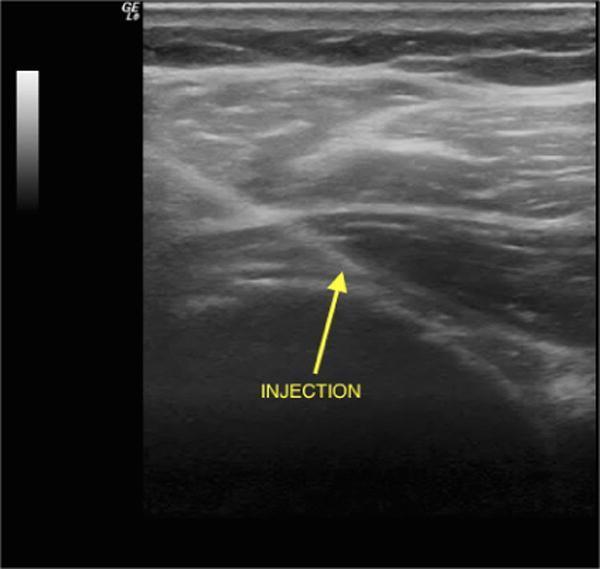
This diagnostic ultrasound image demonstrates an ultrasound-guided intra-articular injection of the glenohumeral joint using a posterior, lateral-to-medial approach. The grayscale B-mode image shows the musculoskeletal anatomy of the shoulder, including the superficial soft tissue and underlying musculature with characteristic striated, heterogenous echogenicity. A hyperechoic, linear needle trajectory is visible, oriented at an oblique angle as it penetrates the deep fascia and approaches the hypoechoic joint space. An annotation arrow labeled 'INJECTION' points to the needle tip positioned at the target site between the humeral head and the glenoid rim. The image illustrates a common orthopedic procedural technique for managing adhesive capsulitis or osteoarthritis, emphasizing the use of real-time imaging to ensure accurate needle placement and optimal delivery of therapeutic agents such as corticosteroids or hyaluronic acid into the joint capsule.
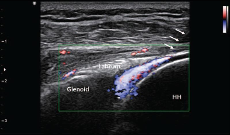
A musculoskeletal ultrasound image in a posterior view demonstrates an ultrasound-guided intra-articular injection of the shoulder joint. The B-mode image shows the glenohumeral joint anatomy, with the glenoid on the left, the humerus head (HH) on the right, and the triangular, hyperechoic posterior labrum situated between them. White arrows indicate the trajectory of the needle approaching the joint space from a posterior-lateral direction. A green rectangular color Doppler box is superimposed over the joint, displaying prominent red and blue signals that confirm the dynamic flow of the injected substance into the articular cavity. This technique is used in clinical practice to ensure precise delivery of therapeutic agents, such as triamcinolone acetonide (TA) or polydeoxyribonucleotide (PDRN), for conditions like hemiplegic shoulder pain (HSP).
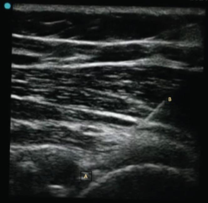
This diagnostic ultrasound image in a transverse view demonstrates an ultrasound-guided interventional procedure on the glenohumeral joint (A) using a posterior approach. The image shows the musculoskeletal anatomy of the shoulder, including the superficial subcutaneous tissue and muscular layers (deltoid and infraspinatus) characterized by heterogeneous echogenicity with hyperechoic fascial planes and hypoechoic muscle bundles. An injecting needle (B) is clearly visible as a distinct, linear, hyperechoic structure traversing the soft tissue planes toward the posterior aspect of the glenohumeral joint capsule. The educational focus of this image is to illustrate the precise needle trajectory required for hydrodilatation or intra-articular injection. The visualization confirms the needle's position relative to the humeral head and glenoid margin, highlighting the clinical significance of real-time ultrasound guidance in ensuring accurate delivery of therapeutics into the joint space while avoiding neurovascular structures.
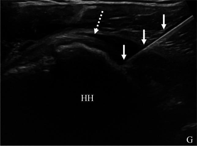
Diagnostic Image: Ultrasound (US) showing a glenohumeral joint corticosteroid injection via a posterior approach in a 46-year-old patient. The ultrasound frame displays a longitudinal view of the posterior shoulder anatomy including the humeral head (HH) and the glenoid (G). The bony surfaces appear as hypoechoic lines with deep acoustic shadowing. A more hyperechoic, fibrillar structure representing the infraspinatus tendon is indicated by a dashed arrow. An echogenic needle (solid arrows) is visualized in-plane using a medial-to-lateral trajectory. The needle tip is correctly positioned deep to the infraspinatus tendon, resting on the articular surface of the humeral head within the joint space. This clinical photograph demonstrates the real-time visualization required for accurate intra-articular needle placement to treat chronic shoulder pain or rotator cuff pathology while avoiding extra-articular injection.
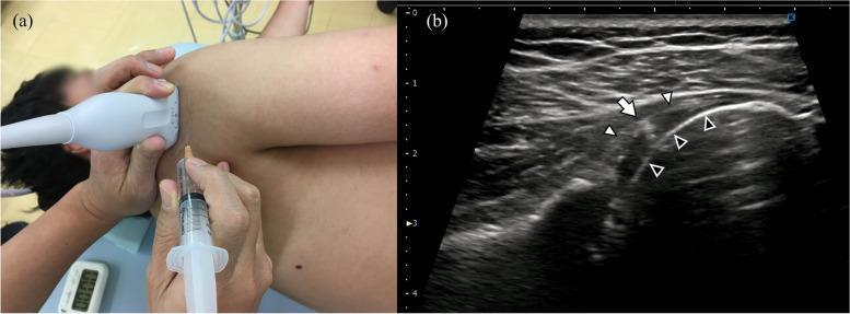
This composite image illustrates an ultrasound-guided glenohumeral joint injection procedure. Panel (a) is a clinical photograph showing the procedural setup: a patient in a lateral recumbent position with a clinician holding a linear ultrasound transducer against the posterior shoulder. A syringe and needle are being inserted out-of-plane relative to the transducer for joint access. Panel (b) is a corresponding ultrasound image in a transverse plane showing the musculoskeletal anatomy. The white arrowheads identify the hyperechoic glenohumeral joint capsule. The black arrowheads mark the humeral head, which exhibits a curvilinear hyperechoic cortex. A white arrow points to the needle tip, appearing as a focal hyperechoic reflective point positioned deep to the capsule within the joint space. This educational material demonstrates the precision of needle placement for intra-articular steroid or anesthetic delivery in orthopedics and physical medicine.
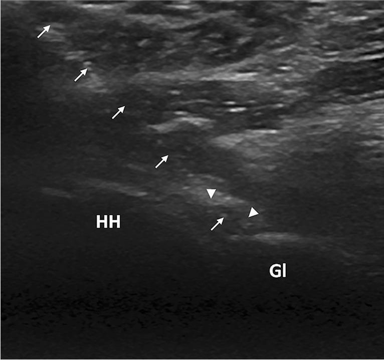
This diagnostic ultrasound image demonstrates a real-time, US-guided glenohumeral joint hydrodistension procedure using a posterior approach. The image shows the relevant musculoskeletal anatomy, including the humeral head (labeled HH) and the glenoid (labeled Gl). A needle is visualized as a hyperechoic linear structure, marked by white arrows, following a lateral-to-medial trajectory. The needle tip is correctly positioned within the glenohumeral joint space. Specifically, the tip terminates within the posterior joint recess, which is shown as a slightly distended, hypoechoic region indicated by white arrowheads. This visual illustrates the precise placement required for intra-articular injection in conditions like adhesive capsulitis (frozen shoulder). The imaging captures the interface between the bony articular surfaces and the joint capsule, highlighting the 'in-plane' needle technique used to monitor the depth and direction of the needle relative to the glenoid labrum and humeral cartilage.
Shoulder Joint Injection: A-Z Guide
Anatomy Relevant to Injection
- Glenohumeral (GH) joint - the true ball-and-socket joint between the humeral head and glenoid
- Subacromial bursa (SAB) - the most commonly injected shoulder structure; lies between the deltoid and the supraspinatus
- Acromioclavicular (AC) joint - small synovial-lined joint at the lateral clavicle
Indications
| Target | Indications |
|---|---|
| Glenohumeral joint | Osteoarthritis (OA), rheumatoid/inflammatory arthritis, frozen shoulder (adhesive capsulitis), joint aspiration for effusion |
| Subacromial bursa | Rotator cuff syndrome, subacromial impingement, calcific tendinitis, subacromial bursitis |
| AC joint | AC joint arthritis, post-traumatic AC pain |
| Bicipital tendon sheath | Bicipital tendinitis |
Contraindications
- Prosthetic joint (requires orthopedic surgeon in sterile environment)
- Active skin infection/cellulitis overlying the injection site
- Psoriasis or eczema at the injection site
- Systemic infection or bacteremia
- Known allergy to any drug to be injected
-
Coagulopathy or significant thrombocytopenia; raised INR outside therapeutic range (note: direct oral anticoagulants appear safe for joint aspiration/injection)
-
Poorly controlled diabetes mellitus (transient hyperglycemia risk)
-
Joint injection within 3 months of planned joint replacement (some orthopedic preferences)
-
Immunocompromised state (increased septic arthritis risk)
-
Firestein & Kelley's Textbook of Rheumatology, 2-Vol. Set, p. 1041
Equipment & Materials
- 21- to 23-gauge needle, 5-mL syringe
- Corticosteroid: triamcinolone acetonide, triamcinolone hexacetonide, or methylprednisolone 20-40 mg (± 1 mL 1% lidocaine)
- 22-gauge, 1- to 1½-inch needle
- 5-7 mL 1% lidocaine + 30-40 mg methylprednisolone acetate or equivalent
- 20-gauge, 1½-inch needle
- 5-7 mL 1% lidocaine + 20-40 mg methylprednisolone acetate
- 25-gauge needle, 2-mL syringe (insulin syringe can be used)
- Pfenninger & Fowler's Procedures for Primary Care, 3rd ed., p. 1338
- Firestein & Kelley's Textbook of Rheumatology, 2-Vol. Set, p. 1041
Approaches - Glenohumeral Joint
Posterior Approach (Preferred)
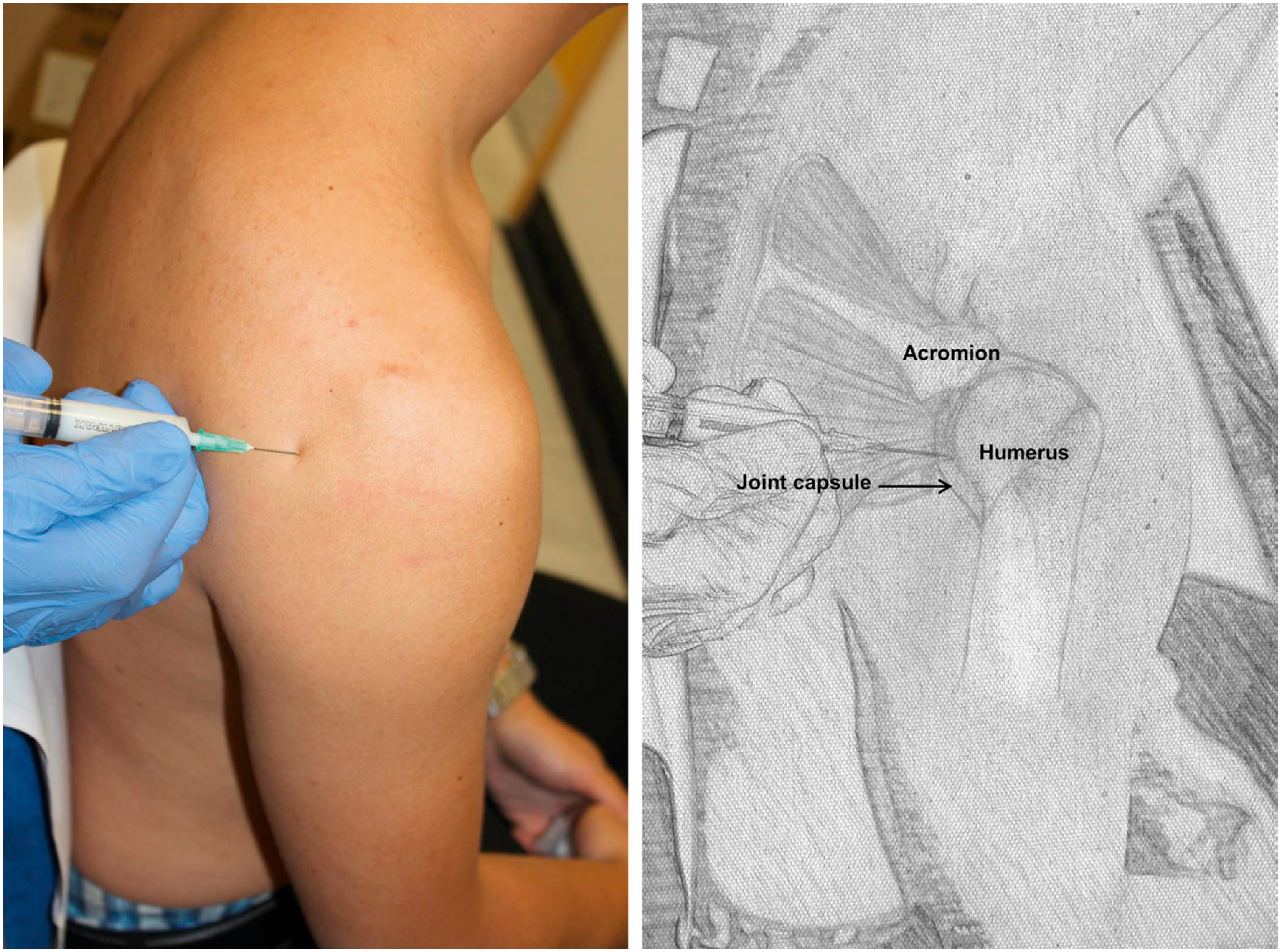
- Seat the patient with their back toward you
- Palpate the joint space 2-3 cm inferior and medial to the acromial angle
- Internal/external rotation of the shoulder helps confirm the joint line
- Advance the needle in an anterior direction toward the coracoid process
- Aim toward the index finger placed on the coracoid (index finger anterior, thumb on inferoposterior acromion)
- Insert 2-3 cm deep
- Aspirate before injecting; ensure there is little or no resistance to plunger
Anterior Approach
- Externally rotate the shoulder - this opens the joint space
- Identify the coracoid process
- Insert needle 1 cm inferior and 1 cm lateral to the coracoid process
- Direct perpendicular or slightly laterally into the joint
- Properly placed needle should not contact bone
- Pfenninger & Fowler's Procedures for Primary Care, 3rd ed., p. 1343 (Fig. 192-17)
- Firestein & Kelley's Textbook of Rheumatology, 2-Vol. Set, p. 1041
Subacromial Bursa Injection
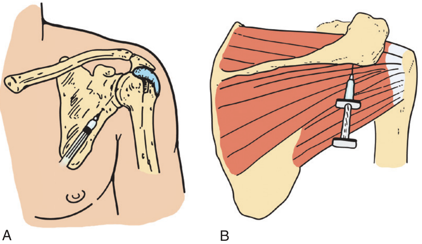
- Palpate the superior surface of the shoulder progressing laterally until the slight drop-off of the lateral edge of the acromion is felt
- The palpable soft spot above the humeral head marks the bursa
- Direct the needle perpendicular to the surface, through the deltoid into the bursa
- The needle should be free-floating (not in muscle or tendon - no resistance)
- The supraspinatus tendon is directly medial to the bursa (a gritty sensation on deeper entry suggests calcific tendinitis)
- Inject within the bursa, not the tendon
- Pfenninger & Fowler's Procedures for Primary Care, 3rd ed., p. 1342 (Fig. 192-13)
Ultrasound Guidance
- The joint space is narrow (e.g., AC joint with osteophytes)
- Body habitus makes landmarks difficult
- Previous "blind" injections failed
- Greater diagnostic confidence in injection placement is needed
- Use a multifrequency transducer; identify target structure
- Cleanse skin; inject local anesthetic parallel to the transducer
- Keep needle constantly in view as it advances; inject small amounts of anesthetic ahead of needle tip
- Confirm placement using power Doppler - visible flow in target = correct placement
- Hold needle with forceps, swap to therapeutic syringe, inject
- Pfenninger & Fowler's Procedures for Primary Care, 3rd ed., pp. 1337-1339
Corticosteroid Choices
| Agent | Relative Potency | Duration | Notes |
|---|---|---|---|
| Triamcinolone acetonide | Intermediate | 3-4 weeks | Lower postinjection flare risk |
| Triamcinolone hexacetonide | High | 4-6 weeks | Longest duration; best for intra-articular |
| Methylprednisolone acetate | Intermediate | 3-4 weeks | Higher postinjection flare risk; do not inject close to skin surface |
| Betamethasone | High | 3-4 weeks | More soluble; shorter acting |
Complications
Local
- Postinjection flare - crystal-induced synovitis in ~2-5% of cases; pain and swelling begin a few hours post-injection, lasting up to 3 days. Management: ice, analgesics. If symptoms exceed 48 hours or are associated with fever, rule out infection.
- Septic arthritis - rare; incidence increased in elderly and immunocompromised patients
- Tendon rupture - do NOT inject against resistance; never inject directly into the Achilles tendon
- Skin/fat atrophy and depigmentation - occurs with superficial corticosteroid leakage; evident 6 weeks to 3 months post-injection; usually reversible within 6 months
- Hemarthrosis / bleeding - use smaller gauge needles in anticoagulated patients
- Nerve damage - avoid injection without knowledge of neurovascular landmarks
- Osteonecrosis - rare with repeated frequent injections
- Cartilage damage - with multiple frequent injections
- Intra-articular calcification - rare
- Steroid "chalk" deposits
Systemic
- Transient hyperglycemia - important warning for diabetic patients; typically 24-48 hours
- Facial flushing / palpitations - within 24 hours; due to systemic steroid absorption
- Adrenal suppression - rare with single injections
Technique to Minimize Atrophy Risk
- Roberts & Hedges' Clinical Procedures in Emergency Medicine, pp. 1228-1229
- Firestein & Kelley's Textbook of Rheumatology, 2-Vol. Set, p. 1041
Frequency / Dosing Limits
Post-Injection Care
- Apply a sterile dressing post-injection
- Advise relative rest for 24-48 hours (reduces postinjection flare risk)
- Warn the patient about the possibility of a short-term pain flare in the first 12-24 hours
- Return for review if fever, increasing warmth, redness, or severe pain develops beyond 48 hours (to rule out septic arthritis)
- Diabetic patients: monitor blood glucose for 24-48 hours
Summary: Quick Reference Card
| Parameter | Glenohumeral | Subacromial Bursa | AC Joint |
|---|---|---|---|
| Needle | 21-23 G | 22 G, 1-1.5" | 25 G |
| Volume | 2-5 mL | 5-7 mL | 1-2 mL |
| Steroid dose | 20-40 mg | 30-40 mg | 10-20 mg |
| Preferred approach | Posterior | Lateral | Superior/direct |
| Landmark | 2-3 cm inf/medial to acromial angle | Lateral acromion edge | AC joint line |
| Free-floating | Yes | Yes (bursa) | Firm endpoint |
- Firestein & Kelley's Textbook of Rheumatology, 2-Vol. Set - Chapter 54, pp. 1041-1042
- Pfenninger & Fowler's Procedures for Primary Care, 3rd ed. - Chapter 192, pp. 1337-1343
- Roberts & Hedges' Clinical Procedures in Emergency Medicine - pp. 1228-1229
- Textbook of Family Medicine, 9th ed. - Chapter 32
metformin and vitamin b12
metformin vitamin B12 deficiency
metformin vitamin B12 deficiency mechanism supplementation
PMID: 42144864
Metformin and Vitamin B12
Background
Mechanism
- Harrison's Principles of Internal Medicine, 22nd ed., p. 829
- Goldman-Cecil Medicine, p. 985
Magnitude of Effect
- Long-term metformin use is associated with 20-30% lower blood levels of vitamin B12 compared to non-users.
- Overt deficiency (serum B12 below the laboratory lower limit) occurs in a meaningful proportion of long-term users.
- In elderly patients on metformin, elevated methylmalonic acid (MMA) - a functional marker of cellular B12 deficiency - is present in up to 40% of the oldest old.
- Risk increases with higher dose and longer duration of metformin therapy.
-
Pooled B12 deficiency rate: 1.4% in RCTs vs. 18.7% in observational studies (the wide gap reflecting heterogeneity and the importance of real-world use patterns vs. trial conditions)
-
B12 levels declined after 6 and 12 months of use, though not always reaching statistical significance
-
Goodman & Gilman's Pharmacological Basis of Therapeutics, p. 2884
-
Goldman-Cecil Medicine, p. 1734
Clinical Consequences
1. Megaloblastic Anemia
- B12 is required for DNA synthesis; deficiency causes large, poorly functional red blood cells (macrocytic/megaloblastic anemia).
- However, anemia may be absent or masked, especially if folate status is adequate.
2. Peripheral Neuropathy
-
Cobalamin is essential for myelin maintenance. Deficiency causes subacute combined degeneration - peripheral neuropathy and/or spinal cord (dorsal column + lateral corticospinal tract) degeneration.
-
This is the most serious risk: neurological damage can develop and progress even in the absence of anemia, and can become irreversible if not caught and treated promptly.
-
Metformin users already have T2DM - itself a cause of peripheral neuropathy - making it difficult to distinguish diabetic neuropathy from B12-deficiency neuropathy clinically.
-
Goldman-Cecil explicitly notes: "long-term use of metformin has been associated with vitamin B12 deficiency and worsening of symptoms of neuropathy"
-
Goldman-Cecil Medicine, p. 989
-
Goldman-Cecil Medicine, Chapter 150 (Cobalamin Deficiency)
3. Elevated Homocysteine
- B12 is a cofactor for homocysteine remethylation to methionine. Deficiency raises plasma homocysteine, a risk factor for cardiovascular and thrombotic disease.
4. Neuropsychiatric Symptoms
- Even B12 levels in the low-normal range (200-350 pg/mL) can be associated with neuropsychiatric complications, particularly in older adults, even without overt megaloblastic anemia.
Screening & Diagnosis
| Test | Notes |
|---|---|
| Serum B12 | First-line; levels 200-350 pg/mL are "low-normal" but may still cause neurological symptoms |
| Methylmalonic acid (MMA) | Elevated MMA = functional (cellular) B12 deficiency; more sensitive and specific; preferred when serum B12 is borderline |
| Homocysteine | Elevated in B12 AND folate deficiency; less specific than MMA |
- All patients on long-term metformin (>4 years)
- Elderly patients on metformin (more vulnerable)
- Any metformin user presenting with new or worsening neuropathy, unexplained anemia, or fatigue
- Frequency: annually (as recommended by both The Clozapine Handbook and Goldman-Cecil for patients on metformin)
Treatment & Supplementation
Oral B12 Supplementation
- At least 500-1000 µg/day is needed to correct elevated MMA in most patients with metformin-related B12 malabsorption.
- Goldman-Cecil recommends at least 1000 µg/day for individuals at risk of malabsorption (including metformin users), as passive diffusion (which bypasses the intrinsic factor/calcium-dependent pathway) requires high oral doses to be effective.
- Note: Standard multivitamin preparations (~6 µg B12) are inadequate for correcting deficiency.
When to Escalate to Parenteral (IM) B12
- Severe deficiency with neurological involvement
- Confirmed malabsorption where oral high-dose therapy has failed to normalize MMA
Calcium Supplementation
- Some evidence suggests calcium supplementation (1.2 g/day) can reverse metformin-induced B12 malabsorption, consistent with the calcium-dependent mechanism. However, this is not universally adopted in guidelines.
Key Drug Interactions / Compounding Factors
- Proton pump inhibitors (PPIs) - reduce gastric acid needed for releasing protein-bound B12 from food
- H2 blockers
- Other drugs reducing B12 absorption: colchicine, neomycin, para-aminosalicylate, slow-release potassium chloride, anticonvulsants
Clinical Pitfalls
- Masking by folate: High-dose folic acid can partially correct megaloblastic anemia but will not stop neurological progression in B12 deficiency - irreversible damage can occur.
- Low-normal B12: Don't dismiss a level of 200-350 pg/mL in a symptomatic metformin user - check MMA.
- Neuropathy attribution: New or worsening neuropathy in a diabetic patient on metformin should always prompt a check of B12 status, not just tighter glycemic control.
- Pediatric/adolescent users: The 2026 systematic review [PMID: 42144864] confirms B12 levels fall in younger patients too, though deficiency rates in RCTs are lower than in observational data.
Summary: Practical Approach
Start metformin
        ↓
Baseline B12 level (± MMA if borderline)
        ↓
Annual monitoring: serum B12 (± MMA if symptoms or low-normal B12)
        ↓
B12 < 300 pg/mL or elevated MMA → supplement with 1000 µg/day oral B12
        ↓
Neurological symptoms → check MMA, consider IM B12 if severe
- Goldman-Cecil Medicine, International Ed. - Ch. 199 (Metformin section, p. 989; B12 deficiency pp. 1728-1734)
- Goodman & Gilman's Pharmacological Basis of Therapeutics - Biguanides, p. 2884
- Harrison's Principles of Internal Medicine, 22nd ed. - Table 104-3, p. 829
- Fuster & Hurst's The Heart, 15th ed. - Table 7-5, p. 313
- The Clozapine Handbook - Metformin monitoring guidance
- Tahir EA et al. (2026). Metformin & B12 in children/adolescents - systematic review. Endocrinol Diabetes Metab. PMID: 42144864