
Breech/Transverse lie/FACE/BROW presentation theory ,clinical and treatment and management
Reading File
Searching sources...
This is a broad obstetrics topic. Let me run parallel searches across all four malpresentations.
Finding Sources
Finding Sources
Finding Sources
Finding Sources
Finding Sources
Finding Sources
Excellent - found key sections. Now let me read the primary sources in parallel.
Reading File
Finding Sources
Now let me read the key sections - Deflection Abnormalities (face/brow), transverse lie, and Rosen's malpresentation:
Reading File
Reading File
Reading File
Reading File
Reading File
Reading File
Reading File
Reading File
I now have comprehensive data from Creasy & Resnik (Maternal-Fetal Medicine) and Rosen's Emergency Medicine. Let me also fetch the breech vaginal delivery technique and ECV details.
Reading File
I now have all the key information needed. Let me compile the comprehensive answer.
Loading Image
Loading Image
Loading Image
I now have all the information needed. Let me compile the comprehensive, structured answer.
Malpresentations in Obstetrics: Breech, Transverse Lie, Face & Brow
Sources: Creasy & Resnik's Maternal-Fetal Medicine; Rosen's Emergency Medicine
RELATIVE INCIDENCE (Rosen's EM)
| Malpresentation | Incidence |
|---|---|
| Breech | 1/25 live births (~3-4%) |
| Shoulder (Transverse lie) | 1/300 live births |
| Face presentation | 1/550 live births |
| Brow presentation | 1/1400 live births |
I. BREECH PRESENTATION
Theory / Definition
Breech presentation = fetal buttocks or lower extremities present at the pelvic inlet. Incidence decreases with advancing gestation: 24% at 18-22 weeks → 8% at 28-30 weeks → 7% at 34 weeks → 2.8% at 38-40 weeks.
Types:
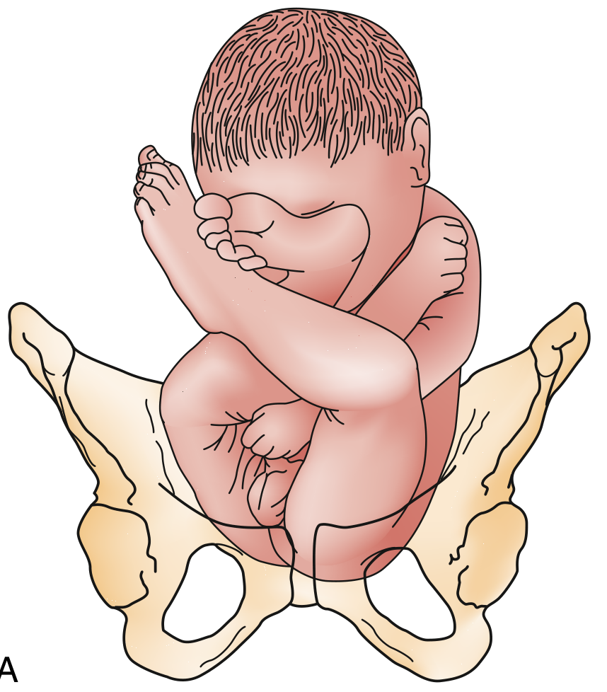
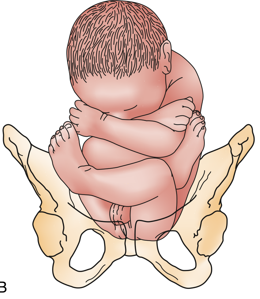
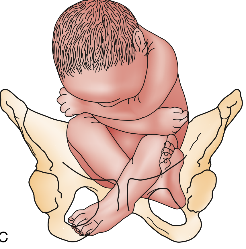
Reference point for position: Sacrum (e.g., sacrum anterior, sacrum posterior)
Etiology / Predisposing Factors
- Fundal-cornual placental implantation (occurs in 7% of pregnancies) - the fetus finds breech position more comfortable
- Prematurity
- Multiparity
- Fetal abnormalities / neuromuscular disorders (reduced fetal mobility)
- Polyhydramnios
- Uterine anomalies (bicornuate, fibroids)
- Prior breech presentation
- Multiple gestation
Note: Children born in breech scored less well on motor skills at age 5 regardless of delivery mode - suggesting some breech fetuses remain breech because of diminished neuromuscular ability.
Risks / Complications
- Umbilical cord prolapse - especially footling breech (presenting part does not fill the cervical os)
- Entrapment of the after-coming head by incompletely dilated cervix - particularly in preterm infants <1500 g
- Trauma from head extension or nuchal arm position
- Brachial plexus injury
- Asphyxia
- Significant variable decelerations in labor (umbilical cord in lower segment during late 1st and 2nd stage)
Diagnosis / Clinical Features
- Leopold maneuvers (before labor): hard, round, ballotable head in fundus; soft, irregular breech at the inlet; FHR heard at or above umbilicus
- Vaginal examination: softer, irregular presenting part; anus felt between two bony prominences (vs. vertex where you feel fontanelles/sutures in a complete bony circle)
- Key distinguishing tip: "the face and skull have a complete circle of bone, whereas the anus is flanked by bone on only two sides"
- If fontanel is not identified on vaginal exam, suspect breech
- Ultrasound (gold standard): confirms type, gestational age, fetal weight, arm/neck position, hyperextended neck (contraindication to vaginal delivery)
Management
A. Antepartum - External Cephalic Version (ECV)
When: Offered at 36-37 weeks with intact membranes, before labor onset
Contraindications to ECV:
- Uterine anomalies
- Third-trimester bleeding / placenta previa
- Multiple gestation
- Oligohydramnios
- Evidence of uteroplacental insufficiency
- Nuchal cord on USS
- Previous cesarean section or significant uterine surgery
- Obvious CPD
Procedure:
- Confirm reactive NST
- Administer tocolytic (terbutaline 0.25 mg SC) to relax uterus
- Attempt forward roll (or backward roll if unsuccessful)
- Monitor FHR intermittently with Doppler during procedure
- Fetal bradycardia occurs in ~20% but almost always resolves when manipulation stops
- Post-procedure FHR monitoring for 1 hour
- Rh-negative mothers: give 300 mcg Rh immune globulin (feto-maternal transfusion risk 6-28%)
Outcome: Reduces cesarean delivery risk by ~50% overall at 36 weeks
B. Mode of Delivery - Key ACOG Position
ACOG recommends: If ECV fails (or is not possible), planned cesarean delivery for term breech.
Criteria for a trial of vaginal breech delivery (selected cases only, with skilled obstetrician):
- Frank or complete breech (NOT footling)
- Estimated fetal weight 2000-3800 g
- Normal gynecoid pelvis with adequate measurements (AP inlet ≥11 cm, transverse inlet ≥12 cm, interspinous ≥9 cm)
- Flexed fetal head (hyperextended neck = contraindication)
Zatchuni Prognostic Index (Diagnostic Index for vaginal breech feasibility - scored on parity, gestational age, EFW, previous breech, cervical dilation, station): Score ≤3 = cesarean preferred.
C. Vaginal Breech Delivery Technique
- Allow buttocks/legs time to dilate cervix completely
- Do NOT pull on hips - this brings shoulders before the cervix is fully dilated, trapping the head
- Support but do not traction the presenting part
- Mauriceau maneuver for the after-coming head: insert finger into fetal mouth to flex the neck and flex chin onto chest, avoiding neck extension (associated with cord injuries)
- Only attempt once fetal elbows and chin have entered the pelvic inlet
- Support fetal pelvis to avoid abdominal injury
- Generous episiotomy may be needed
- Piper forceps can be applied to the after-coming head
- Fetal monitoring throughout (variable decels common); keep membranes intact as long as possible for hydraulic cord protection
D. Cesarean Section in Breech
- For most term breeches
- Uterine incision: low transverse usually adequate; low vertical or classical incision for transverse/back-down shoulder; Durfee high transverse incision is an option
II. TRANSVERSE LIE (Shoulder Presentation)
Theory / Definition
The fetal long axis is perpendicular to the maternal long axis. The shoulder is the presenting part. Incidence ~1/300 deliveries.
Shoulder presentation reference point: Acromion/scapula
Etiology
- Prematurity (38% of cases)
- High parity (87% had ≥3 prior deliveries)
- Premature rupture of membranes (30%)
- Placenta previa (10%)
- Uterine abnormalities
- Polyhydramnios
Risks
- Cord prolapse - potentially catastrophic (patient often arrives with asphyxiated/dead fetus)
- High perinatal mortality (3.9%-24%) - largely due to prevalence of low-birth-weight infants
- If labor progresses untreated with transverse lie: "neglected shoulder presentation" with uterine rupture risk
Diagnosis
- Palpation/Leopold: No fetal poles at fundus or inlet; fetal head felt laterally on one side, breech on the other
- Abdomen appears unusually wide transversely
- Patient may notice the abnormal position
- Ultrasound confirms diagnosis
Management
Decision framework based on gestational age + membrane status:
| Clinical Scenario | Management |
|---|---|
| <viable threshold (<600g) in labor | No intervention; may deliver vaginally (conduplicato corpore) without maternal harm |
| Viable fetus, membranes intact, no labor, <36 wks | Expectant - monitor for spontaneous version; avoid ECV before 36-37 wks (risk of cord entanglement, placental trauma) |
| Viable fetus, membranes intact, ≥36-37 wks, no labor | External version + induction at 38 wks (after excluding CPD and placenta previa); admit to hospital |
| Viable fetus, membranes RUPTURED or in active labor | Cesarean delivery mandatory |
| Placenta previa present | Cesarean delivery mandatory |
| CPD suspected | Cesarean delivery mandatory |
"Unstable lie" protocol (Edwards & Nicholson): Admit all patients ≥37 weeks with unstable lie. Exclude CPD/previa, attempt ECV, induce labor at ≥38 weeks. This policy reduced cord prolapse from 10/50 to 1/102 cases and eliminated perinatal deaths.
Cesarean uterine incision for transverse lie:
- Low transverse incision: usually adequate for "back-up" transverse lie
- Low vertical or classical incision: for "back-down" shoulder presentation
- Durfee high transverse incision: alternative option
III. FACE PRESENTATION
Theory / Definition
Maximum deflection of the cephalic presentation - the occiput extends to contact the fetal back, and the face (chin/mentum) becomes the presenting part.
Engaging diameter: Submento-bregmatic (~9.5 cm) - approximately 0.8 cm larger than vertex presentation.
Reference point for position: Mentum (chin) - e.g., Mentum Anterior (MA), Mentum Transverse (MT), Mentum Posterior (MP)
Frequency: ~1/500 deliveries (though likely higher if all presentations assessed early in labor)
Etiology
- CPD (cephalopelvic disproportion)
- Increased parity
- Prematurity
- Premature rupture of membranes
- Anencephaly - ALMOST ALWAYS results in face presentation (absent cranial vault allows maximum extension)
- Fetal anomalies otherwise do NOT account for most deflection problems
Clinical Features / Diagnosis
- Vaginal exam: Orbital ridges, nose, mouth, and chin felt - a complete bony ring around a soft central orifice (vs. anus in breech = bony on only 2 sides)
- Labor may be prolonged (Friedman found face presentation does NOT significantly affect labor course in nulliparas or multiparas - contrary to clinical impression)
- 50% of deflection diagnoses not made until 2nd stage of labor
- Diagnosis confirmed on ultrasound
Management
Guiding principle: "If a face presentation is progressing, leave it alone."
- Mentum anterior (MA): Usually delivers vaginally - mechanism is flexion of the head as it passes under the symphysis (chin leads)
- Mentum transverse (MT): Frequently rotates spontaneously to MA → vaginal delivery
- 70%-90% of face presentations result in spontaneous delivery
- Mentum posterior (MP, persistent): Cannot deliver vaginally at term (chin cannot flex past sacrum - the occiput would need to enter the pelvis which is too large) → Cesarean section mandatory
- Labor arrest with face presentation → Cesarean section
- Cord prolapse with face presentation → Cesarean section
- No attempts at manual conversion of face presentation
- Oxytocin augmentation may be used if labor is progressing and no CPD
IV. BROW PRESENTATION
Theory / Definition
Partial deflection of the cephalic presentation - intermediate between vertex and face. The frontal bone (between anterior fontanelle and supraorbital ridges) is the presenting part.
Engaging diameter: Mento-occipital (~13-14 cm) - the LARGEST diameter of the fetal head, approximately 1.5 cm larger than vertex. This is why brow presentation causes the most severe dystocia among head presentations.
Reference point: Frontum (forehead)
Frequency: ~1/1400 deliveries
Etiology
Same as face presentation:
- CPD
- Increased parity
- Prematurity
- PROM
- Fetal anomalies (rare)
Clinical Features / Diagnosis
- Vaginal exam: Anterior fontanelle + orbital ridges palpable; neither the occiput posteriorly nor the chin anteriorly can be felt
- If lambdoid sutures and posterior fontanelle cannot be identified centrally in the pelvis → suspect deflection problem
- Brow presentation associated with abnormalities of descent and longer second stage (unlike face presentation)
- Friedman: 10.9% of brow presentations had clinical + radiographic CPD vs. 2.7% of vertex controls
- Ultrasound confirms diagnosis
Management
Key principle: Most brow presentations convert spontaneously.
Decision pathway:
- Labor progressing + converting to face or vertex → manage expectantly for vaginal delivery
- Labor arrested + CPD suspected → Cesarean delivery
- Labor arrested + poor contractions + NO CPD → carefully monitored oxytocin augmentation ± radiographic pelvimetry to exclude CPD
- Brow fails to convert + fetus is term → Cesarean delivery (mento-occipital diameter too large to negotiate pelvis)
- For preterm/small infants: brow presentation is almost always unstable and converts spontaneously to occiput or face
Summary rule: "Successful descent in brow presentation at term depends on conversion to face or vertex." If this conversion does not occur → cesarean.
SUMMARY TABLE: Key Differentiating Points
| Feature | Breech | Transverse Lie | Face | Brow |
|---|---|---|---|---|
| Incidence | 3-4% | 1/300 | 1/550 | 1/1400 |
| Presenting part | Buttocks/feet | Shoulder | Chin | Forehead |
| Reference point | Sacrum | Acromion | Mentum | Frontum |
| Engaging diameter | Bitrocanteric | N/A | Submentobregmatic | Mento-occipital (largest) |
| Most common cause | Prematurity, fundal placenta | High parity, prematurity | CPD, anencephaly | CPD, prematurity |
| Key danger | Cord prolapse, head entrapment | Cord prolapse, neglected shoulder | MP cannot deliver | Largest engaging diameter |
| Spontaneous resolution | Rare after 36 wks | Variable | 70-90% deliver vaginally | >50% convert spontaneously |
| Primary management | ECV → CS if fails | CS if membranes ruptured/labor | Expectant if MA/MT; CS if MP | Expectant if converting; CS if not |
| Vaginal delivery possible? | Selected cases with criteria | Only in non-viable/very preterm | Yes, if MA or MT converts | Only if converts to vertex/face |
CARDINAL RULES TO REMEMBER
- Breech: Never pull - only support. Mauriceau maneuver for after-coming head.
- Transverse lie + ruptured membranes = cesarean. Admit all unstable lie patients ≥37 weeks.
- Face presentation: Mentum anterior = go; Mentum posterior = cesarean.
- Brow presentation: The largest engaging diameter. Almost always needs to convert or → cesarean at term.
- Cord prolapse complicates all malpresentations and is an absolute indication for emergency cesarean.
- ECV (contraindications: placenta previa, ruptured membranes, oligohydramnios, previous uterine surgery, multiple gestation, cord around neck, CPD).
- Creasy & Resnik's Maternal-Fetal Medicine, pp. 953-958 | Rosen's Emergency Medicine, pp. 3415-3418
This is a shared conversation. Sign in to Orris to start your own chat.