
Massive transfusion
"massive transfusion" protocol hemorrhage
massive transfusion protocol blood products ratio
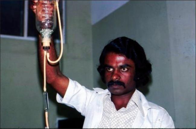
A clinical photograph of a historical blood transfusion setup from the early 1970s. The image displays a clear glass transfusion bottle held vertically by a medical professional. The interior of the bottle contains numerous small, round glass beads, which functioned as an early filtration system to trap clots or debris during the administration of blood products. Thick, translucent rubber tubing is connected to the base of the bottle, leading to an in-line vertical glass drip chamber. This chamber allowed for the visual monitoring of flow rates and the trapping of air bubbles before the blood reached the patient. The photograph illustrates the evolution of transfusion medicine and the transition from reusable glass and rubber equipment to modern, sterile, disposable plastic systems. This content is relevant for medical history, hematology, and the study of global health resource development in South Asia.

Summary : This image is a logo for a medical specialty or department related to blood transfusion. logo: TRANSFUSION MEDICINE Visible Elements : • The logo contains the text "TRANSFUSION MEDICINE" in uppercase white letters on a dark blue rectangular background. • To the left of the text, there are several stylized red blood cells depicted in a scattered arrangement, suggesting the theme of blood or hematology. • The background is white, and the overall design is horizontal. Dimensions & Placement : • The text occupies the right two-thirds of the logo, while the red blood cell graphics are on the left. • The logo is rectangular and oriented horizontally. Analysis : • The use of red blood cells visually reinforces the focus on transfusion medicine, which deals with blood products and their clinical use. • The dark blue background for the text provides strong contrast, making the department or specialty name highly legible.
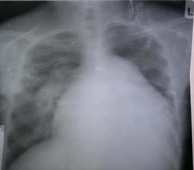
This diagnostic image is a posteroanterior (PA) view chest X-ray demonstrating significant cardiothoracic abnormalities. The most prominent feature is massive cardiomegaly, with a cardiothoracic ratio (CTR) estimated at 0.8, indicating the cardiac silhouette occupies 80% of the thoracic width. A notable bulge in the pulmonary conus is visible along the left heart border, suggestive of pulmonary hypertension or pulmonary artery dilation. The lung fields exhibit pulmonary plethora, characterized by increased vascular markings and congestion extending to the peripheral zones, typical of a left-to-right shunt (such as an atrial septal defect). The combination of these findings—cardiomegaly, a prominent pulmonary segment, and plethoric lung fields—is clinically significant for chronic volume overload and increased pulmonary blood flow, frequently seen in congenital heart disease with Eisenmenger physiology or advanced valvular disease. The image serves as an educational example of cardiac remodeling and pulmonary vascular redistribution in the context of high-flow cardiac shunts.
Massive Transfusion
Definition
- >3 units of pRBCs within 1 hour, or
- 4 blood components within 30 minutes, or
- Active bleeding >150 mL/min
- Replacement of 50% of total blood volume within 3 hours
Causes / Indications
- Trauma (most common - both military and civilian)
- Ruptured aortic aneurysm
- Gastrointestinal hemorrhage (especially with chronic liver disease)
- Obstetric emergencies (postpartum hemorrhage, placenta previa/accreta)
- Liver transplantation and other major surgical procedures
The Lethal Triad
| Component | Mechanism |
|---|---|
| Hypothermia | Radiant heat loss, cool IV fluids/blood products, reduced metabolism from tissue hypoxia, anesthesia |
| Acidosis | Tissue hypoperfusion, large-volume crystalloid, citrate from banked blood if liver overwhelmed |
| Coagulopathy | Dilution, consumption of clotting factors/platelets, DIC, platelet dysfunction |
Massive Transfusion Protocol (MTP)
Why a Protocol?
Blood Product Ratios
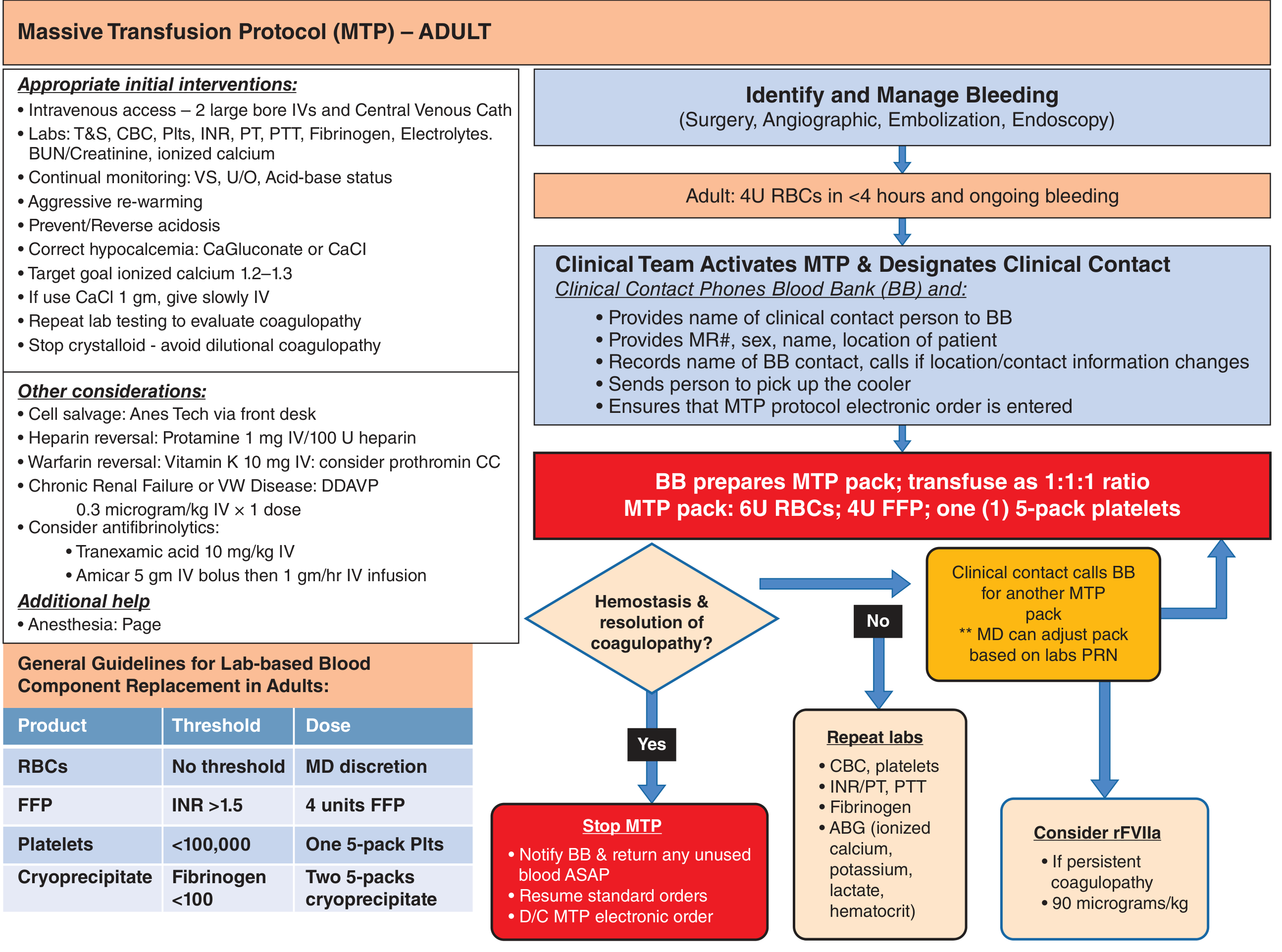
MTP Pack (University of Michigan Level I Trauma Center as an example):
- 6 units pRBCs
- 4 units FFP
- 1 five-pack platelets
Damage Control Resuscitation (DCR)
| DCR Component | Details |
|---|---|
| Permissive hypotension | Goal: palpable radial pulse (unless head injury); avoid over-resuscitation |
| Reduced crystalloid use | Avoid dilutional coagulopathy; stop crystalloid when initiating MTP |
| Early hemorrhage control | Surgery, angioembolization, endoscopy |
| 1:1:1 blood product ratios | pRBCs : FFP : platelets |
| Early coagulopathy correction | Factor VIIa, PCC, tranexamic acid (TXA) |
| Hypertonic saline | Used at some centers |
Lab-Based vs. Protocol-Based Approach
Key Adjuncts
Tranexamic Acid (TXA)
- Antifibrinolytic; reduces mortality in trauma hemorrhage
- Give as early as possible after injury
- Dose: 10 mg/kg IV (or 1 g IV over 10 min, then 1 g over 8 hrs per CRASH-2)
- Also effective in postpartum hemorrhage and elective/emergency cesarean
Calcium
- pRBCs and FFP contain citrate (preservative), which chelates calcium
- Results in life-threatening hypocalcemia with rapid, large-volume transfusion
- Most MTPs include calcium supplementation
- Calcium chloride is preferred over gluconate (gluconate requires hepatic metabolism)
- Target ionized calcium ≥ 0.9 mmol/L (ideal: 1.2-1.3 mmol/L)
Whole Blood
- Used at some centers for initial trauma resuscitation
- Supplies all blood elements in appropriate ratio without coordination of multiple products
- Limitation: no universally compatible whole blood (low-titer O whole blood used with volume limits of 1-2 units if ABO type unknown)
Liquid Plasma
- Plasma from whole-blood donation kept at 2-6°C (never frozen); available without thawing
- Suitable for trauma but limited to short storage (<5-7 days due to labile factor degradation)
Type A Plasma
- Used when AB plasma supply is insufficient; compatible with 84% of the population (A + O types)
rFVIIa (Recombinant Factor VIIa)
- Consider for persistent coagulopathy unresponsive to standard MTP
- Dose: ~90 mcg/kg
Prothrombin Complex Concentrate (PCC)
- Used for warfarin reversal (Vitamin K + consider PCC) or persistent coagulopathy
Complications of Massive Transfusion
| Complication | Notes |
|---|---|
| Hypothermia | Use blood warmers, warmed IV fluids, warming blankets/lights |
| Hypocalcemia | Citrate chelation; monitor ionized Ca²⁺, supplement with CaCl₂ |
| Hypomagnesemia | Monitor and replace |
| Hyper- or hypokalemia | Old blood releases K⁺; monitor frequently |
| Dilutional coagulopathy | Prevented by balanced resuscitation |
| Metabolic acidosis | From hypoperfusion + citrate overload; optimize O₂ delivery; NaHCO₃ not routinely recommended |
| Metabolic alkalosis | Citrate metabolism to bicarbonate in liver |
| Transfusion-related acute lung injury (TRALI) | Donor antibody-mediated; most serious pulmonary complication |
| TACO (transfusion-associated circulatory overload) | Volume overload |
| Hypotension (non-hemolytic) | Bradykinin-mediated |
| Cross-matching confusion | After ~1 blood volume transfused, new specimens contain so much donor blood that cross-matching becomes unreliable; collect early specimens |
Practical Tips
- Obtain blood specimens early in the course of MT; after ~1 blood volume of transfused products, specimens will contain predominantly donor blood and confuse cross-matching
- Stop crystalloid once MTP is activated to prevent dilutional coagulopathy
- Warm all blood and IV fluids
- Identify the source of bleeding in parallel (surgery, angioembolization, endoscopy)
- Use type O, Rh-negative pRBCs if blood type unknown and female of childbearing potential; type AB FFP (or type A if AB unavailable) as universal plasma
- The platelet threshold in MT context is < 100,000/mm³ in multisystem trauma or intracranial hemorrhage (vs. < 50,000 in most other active bleeding contexts)
Recent Evidence (2024-2026)
- A 2024 EAST systematic review and meta-analysis (PMID: 38531812) on whole blood resuscitation in injured patients requiring transfusion supports its use with practice management guidelines.
- A 2025 Cochrane review (PMID: 39911088) examined transfusion strategies for postpartum hemorrhage management.
- A 2026 systematic review and meta-analysis (PMID: 42228445) evaluated machine learning models for predicting MT needs in trauma, showing promise for early triage decisions.