
Somites
Reading File
Somites are blocks of paraxial mesoderm that form during embryonic development and give rise to most of the musculoskeletal structures of the body.
Searching sources...
Finding Sources
Finding Sources
Reading File
Reading File
Finding Sources
Finding Sources
Reading File
Reading File
Loading Image
Loading Image
Here is a comprehensive overview of somites, drawn from the textbooks The Developing Human (Moore & Persaud), Langman's Medical Embryology, Fischer's Mastery of Surgery, and Creasy & Resnik's Maternal-Fetal Medicine.
Somites
Definition and Origin
Somites (Greek: soma, body) are paired, cuboidal blocks of paraxial mesoderm that flank the developing neural tube on either side. They are derived from cells of the primitive node and form via condensation and segmentation of the thick longitudinal columns of paraxial mesoderm that lie adjacent to the notochord.
Formation (Somitogenesis)
- Paraxial mesoderm begins to segment around the end of the third week (day ~20).
- Segmentation proceeds in a strict craniocaudal direction, starting in the future occipital region.
- The first pair appears just caudal to the otic placode.
- Between approximately day 26-32, 38-39 pairs form (the "somite period").
- By the end of week 5, 42-44 pairs are present in total.
Distribution of somite pairs:
| Region | Pairs |
|---|---|
| Occipital | 4 |
| Cervical | 8 |
| Thoracic | 12 |
| Lumbar | 5 |
| Sacral | 5 |
| Coccygeal | 10-12 |
The first occipital and the most caudal 5-7 coccygeal somites disappear; the rest persist as structural precursors.
Clinical use: Because somites are so prominent and countable during weeks 4-5, their number is used as one of the standard criteria for estimating embryonic age.
Molecular Clock of Somitogenesis
Somite formation is not random - it is driven by a segmentation clock: periodic expression of C-Hairy and Notch pathway genes results in one new pair of somites forming approximately every 4 hours. Key molecular players include:
- WNT, FGF, NOTCH signaling pathways - regulate periodic segmentation
- HOX genes - control craniocaudal patterning of each somite
- FoxC1 / FoxC2 forkhead transcription factors - precede somite formation
- Delta-Notch signaling - regulates the craniocaudal segmental pattern
- Tbx6 (T-box gene family) - plays an important role in somitogenesis
(The Developing Human, p. 213; Creasy & Resnik)
Differentiation of the Somite
Each somite differentiates into two main regions:
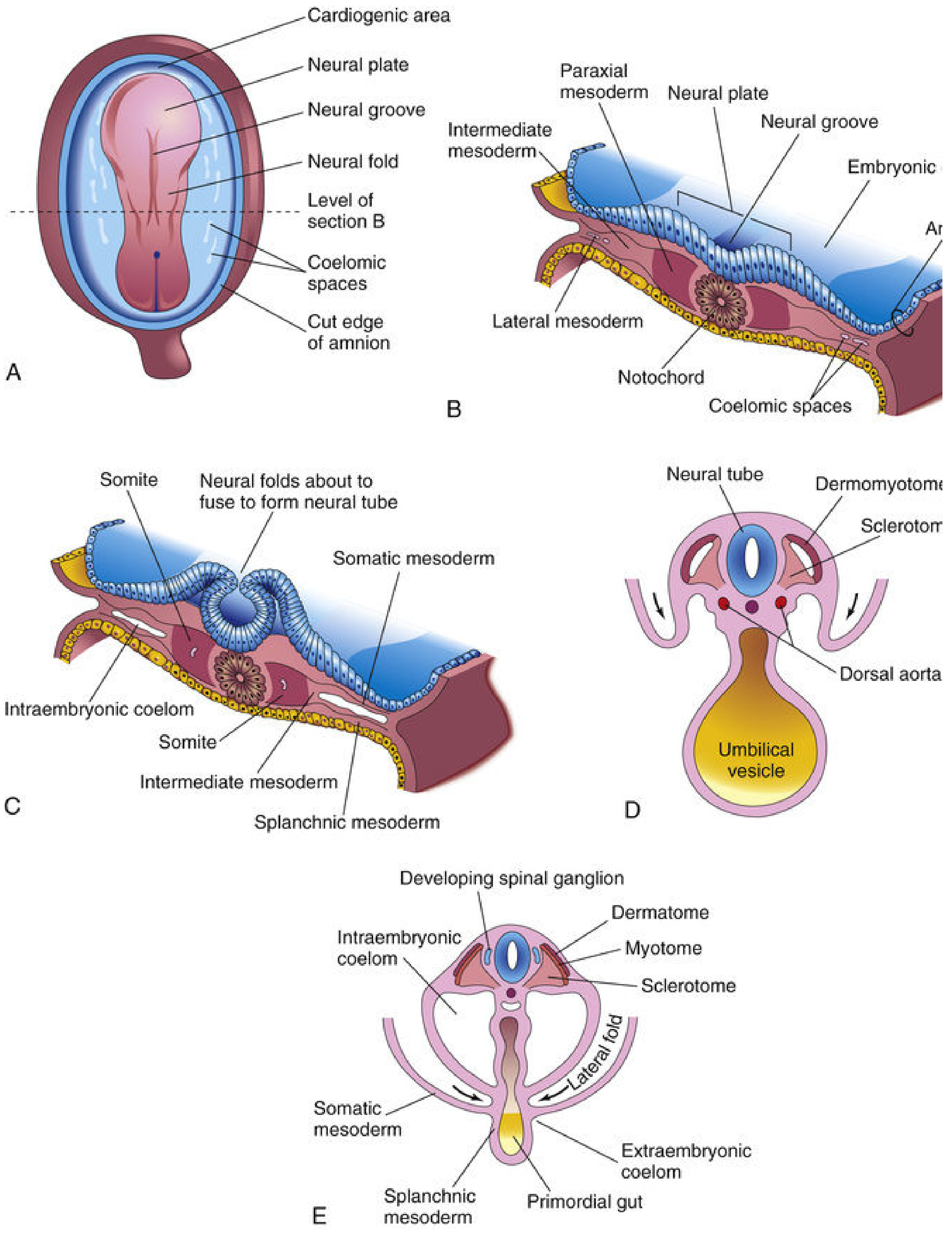
FIG. 14.1 - Formation and early differentiation of somites (The Developing Human)
1. Sclerotome (ventromedial)
- Cells lose their epithelial character and migrate around the neural tube and notochord
- Give rise to the vertebrae and ribs (axial skeleton)
- Form loosely woven mesenchyme at the end of week 4, which later undergoes endochondral ossification
- Bony rib components = primaxial sclerotome cells; cartilaginous rib components that attach to the sternum = sclerotome cells that cross the lateral somitic frontier (abaxial cells)
2. Dermomyotome (dorsolateral)
This region further splits into:
- Myotome - myoblast precursors that form the musculature of the body wall and back
- Dorsomedial (DML) cells → primaxial muscles (back muscles; innervated by dorsal rami)
- Ventrolateral (VLL) cells → can cross the "lateral somitic frontier" into lateral plate mesoderm → form abaxial muscles (body wall, limb girdle)
- Dermatome - forms dermis (fibroblasts) of the back and trunk
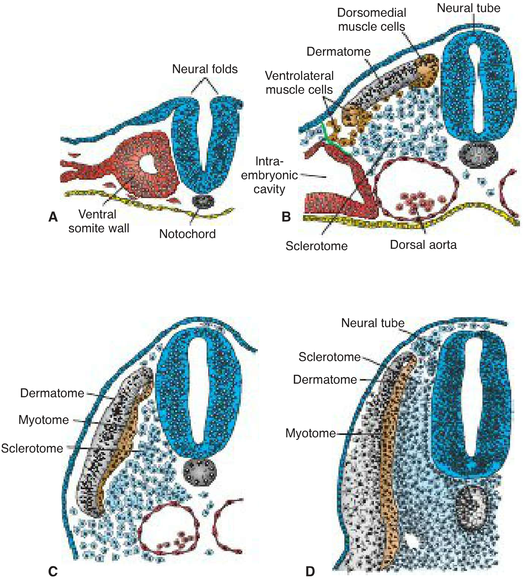
FIGURE 11.1 - Stages of somite differentiation (Langman's Medical Embryology)
Summary of Somite Derivatives
| Somite Region | Derivative |
|---|---|
| Sclerotome | Vertebrae, ribs (axial skeleton) |
| Myotome | Skeletal muscles of back and body wall |
| Dermatome | Dermis of back and trunk |
Additional note on innervation: Each myotome receives its innervation from the spinal nerve derived from the same segmental level - the basis of segmental (dermatomal/myotomal) innervation seen clinically in radiculopathy.
Clinical Significance
- Abnormalities in somite number or patterning underlie vertebral segmentation defects (e.g., Klippel-Feil syndrome, hemivertebrae)
- Disruption of the Notch/Delta or Tbx6 pathways causes congenital scoliosis due to vertebral malformations
- The segmental organization of somites is the embryological basis for the dermatome maps used in neurological examination
(The Developing Human, Fischer's Mastery of Surgery, Langman's)
This is a shared conversation. Sign in to Orris to start your own chat.