
Iugr
- Causes: placental insufficiency, maternal factors (hypertension, malnutrition, smoking), fetal factors (chromosomal anomalies, infections), uterine anomalies
- Classified as symmetric (early onset, global) or asymmetric (late onset, head-sparing)
- Diagnosed by ultrasound: EFW, AC, Doppler studies (umbilical artery, MCA, ductus venosus)
- Management depends on gestational age and Doppler findings
"fetal growth restriction" OR "intrauterine growth restriction"
IUGR intrauterine growth restriction Doppler ultrasound fetal
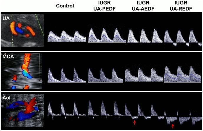
This diagnostic ultrasound comparison chart illustrates fetal Doppler spectral waveforms in the assessment of intrauterine growth restriction (IUGR). The image displays a matrix of flow velocity waveforms across three fetal vessels: the umbilical artery (UA), middle cerebral artery (MCA), and aortic isthmus (AoI). Four clinical states are compared: a normal Control fetus and three progressive stages of IUGR: Present End-Diastolic Flow (PEDF), Absent End-Diastolic Flow (AEDF), and Reversed End-Diastolic Flow (REDF). Key diagnostic features include: 1) UA progression from high-diastolic flow in the control to complete loss (AEDF) and reversal (REDF) of flow during diastole, indicating increased placental resistance. 2) MCA changes showing the 'brain-sparing effect,' where diastolic flow increases as IUGR severity progresses. 3) AoI waveforms demonstrating increasingly pathological retrograde flow during late systole and diastole, highlighted by red arrows in the AEDF and REDF cases. This visual resource is critical for teaching fetal hemodynamics, placental insufficiency, and the criteria for identifying severe fetal distress in obstetric radiology.
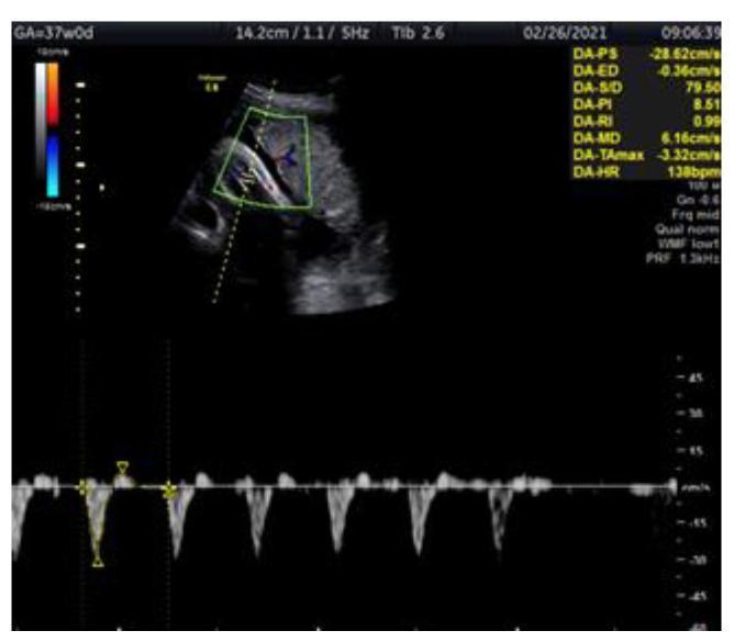
A diagnostic ultrasound image showing a color Doppler assessment of the fetal tibial artery in a 37-week fetus diagnosed with intrauterine growth restriction (IUGR). The split-screen display consists of a top panel showing a cross-sectional grayscale view of the fetal leg with a color Doppler box overlaid on the tibial artery, where red and blue hues indicate blood flow direction. A yellow dashed line marks the sampling gate. The bottom panel displays the corresponding spectral Doppler waveform. The waveform shows a high-resistance pulsatile pattern characterized by sharp systolic peaks and a notable pathological finding: reverse flow (diastolic flow dipping below the baseline). On-screen quantitative data indicates a Pulsatility Index (PI) of 8.51, which is above the 95th percentile for gestational age, a resistive index (RI) of 0.99, and a heart rate of 138 bpm. This visual evidence highlights the role of peripheral artery Doppler in monitoring fetal well-being and identifying hemodynamic deterioration.
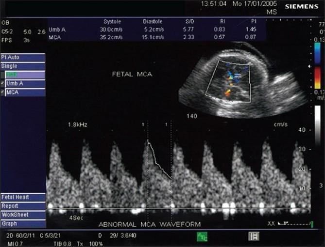
Diagnostic ultrasound image demonstrating fetal middle cerebral artery (MCA) Doppler velocimetry in a case of intrauterine growth restriction (IUGR). The image includes a 2D grayscale fetal head cross-section in the upper right with a color Doppler gate positioned over the Circle of Willis to identify the MCA. The lower portion displays a spectral Doppler waveform labeled 'ABNORMAL MCA WAVEFORM.' The waveform shows a low-resistance pattern characterized by high end-diastolic flow (15.1 cm/s) relative to peak systolic velocity (35.2 cm/s). On-screen calculations indicate a Pulsatility Index (PI) of 0.87 and a Resistive Index (RI) of 0.57. For comparison, umbilical artery (Umb A) data is also listed (PI 1.45), suggesting a reversed cerebro-umbilical (C/U) ratio. This visual finding is characteristic of the 'brain-sparing effect,' where cerebral vasodilation occurs as a compensatory mechanism for chronic fetal hypoxia and placental insufficiency.
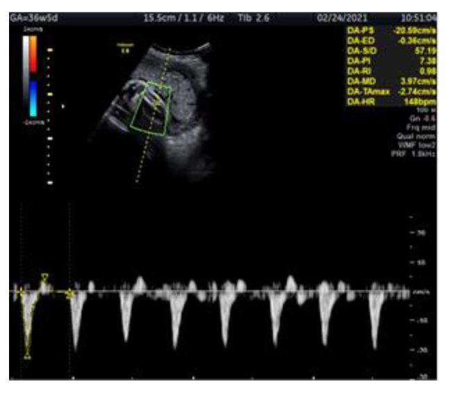
This diagnostic image is a Doppler ultrasound showing a longitudinal assessment of the fetal tibial artery in a 36-week and 5-day gestation fetus with intrauterine growth restriction (IUGR). The top portion displays a grayscale B-mode image of the fetal lower extremity with a superimposed color Doppler box highlighting blood flow within the tibial artery; red and blue hues indicate the direction of flow relative to the transducer. The bottom portion features a pulse-wave Doppler spectral waveform showing highly pulsatile flow with a high-resistance pattern. Numerical hemodynamic indices are displayed on the right, including a peak systolic velocity (PS) of -20.58 cm/s and a pathologically elevated pulsatility index (PI) of 7.38, which is above the 95th percentile for gestational age. The clinical significance of this finding in an IUGR context is its role as a potential early indicator of fetal hemodynamic redistribution or deteriorating fetal condition when conventional arterial markers may still appear normal.
Intrauterine Growth Restriction (IUGR) / Fetal Growth Restriction (FGR)
Definition
- Creasy & Resnik's Maternal-Fetal Medicine, p. 168
Types
| Type | Characteristic | Cause |
|---|---|---|
| Asymmetric (head-sparing) | AC disproportionately smaller; head relatively preserved | Placental/maternal causes; late onset |
| Symmetric | All parameters equally small; brain NOT spared | Early onset; intrinsic fetal causes (infections, chromosomal) |
- Textbook of Family Medicine 9e, p. 496
Pathophysiology
- Reduced umbilical blood flow per kg of fetal weight (because fewer terminal villi = smaller umbilical capillary bed) - umbilical artery pulsatility index is above normal
- Reduced vasosyncytial membranes - greater fraction of oxygen must diffuse across thick oxygen-consuming segments of the placental barrier, so placental oxygen permeability falls
- Reduced glucose permeability - transplacental glucose diffusion is impaired
- Creasy & Resnik's Maternal-Fetal Medicine, p. 296
Etiology
Maternal Factors (most common)
- Vascular disease: preeclampsia, chronic hypertension (most important)
- Hypercoagulable states (acquired or inherited)
- Malnutrition, prolonged hypoglycemia
- Substance use: cigarette smoking, alcohol, narcotics
- Drugs: phenytoin and other teratogens
- Connective tissue disorders, diabetes with vascular involvement
Fetal Factors
- Chromosomal disorders (trisomy 18, 13, 21)
- Congenital anomalies
- Congenital infections (TORCH: Toxoplasma, Rubella, CMV, Herpes/HIV)
- Metabolic disorders
Placental Factors
- Placenta previa (low implantation)
- Placental abruption
- Placental infarction, chronic villitis, hemorrhagic endovasculitis
- Placental mosaicism
- Partial abruptions, hematomas
Note: The etiology of FGR is unknown in approximately 60% of cases.
- Robbins & Kumar Basic Pathology, p. 137; Creasy & Resnik's MFM, p. 168
Diagnosis
Clinical
- Fundal height measurement: fundal height in cm between 18-40 weeks used for screening; a single measurement at 32-34 weeks has ~85% sensitivity, 96% specificity
- Fundal height lagging by ≥3 cm below expected = indication for USS
Ultrasound
- Abdominal circumference (AC) - first parameter to fall behind (soft tissue = glycogen in liver)
- EFW calculated from AC, BPD, HC, FL - plotted on growth curves
- Serial measurements every 2-4 weeks
- Overall US sensitivity for FGR: 80-90% depending on measurements used
- Amniotic fluid index (AFI) - oligohydramnios suggests chronic renal underperfusion
Doppler Studies (key for surveillance)
- Umbilical artery (UA) Doppler: most important initial test
- Elevated S/D ratio or PI → increased placental resistance
- Absent end-diastolic flow (AEDF) → severe placental disease
- Reversed end-diastolic flow (REDF) → imminent fetal compromise
- Middle cerebral artery (MCA): brain-sparing = low PI (high diastolic flow); abnormal when PI <5th percentile
- Cerebro-umbilical ratio (CPR): reversed ratio is a warning sign
- Ductus venosus: absent/reversed A-wave = pre-terminal, immediate delivery often indicated
- Aortic isthmus (AoI): retrograde flow in AEDF/REDF = severe compromise
Management
Antenatal Surveillance
- Umbilical artery Doppler is the most important first step in confirmed FGR
- Non-stress test (NST), biophysical profile (BPP) - for ongoing surveillance
- Contraction stress test if NST is non-reactive
Treatment
- Bed rest in lateral recumbent position - the main supportive measure; prevents IVC compression, maximizes venous return, cardiac output, and uteroplacental perfusion
- Treat underlying maternal conditions (hypertension, etc.)
- Corticosteroids (betamethasone) if preterm delivery is anticipated <34 weeks
- No proven pharmacologic therapy to increase fetal growth
Timing of Delivery
- After 34 weeks with idiopathic FGR: provider-initiated preterm birth is indicated to avoid stillbirth (prematurity complications wane significantly after 34 weeks)
- Preeclampsia or other maternal indication: delivery timing driven by maternal status
- Abnormal antenatal testing or persistent oligohydramnios: delivery may be warranted before term
- Otherwise: deliver at 38-40 weeks depending on severity
- REDF on umbilical artery / absent or reversed ductus venosus A-wave: immediate delivery regardless of gestational age
Neonatal Complications
| Complication | Notes |
|---|---|
| Hypothermia | Reduced fat stores |
| Hypoglycemia | Depleted glycogen reserves |
| Polycythemia / erythrocytosis | Chronic hypoxia stimulus |
| Low Apgar scores | Fetal distress |
| Meconium aspiration | Hypoxia-induced gut motility |
| Sepsis / infection | Immune compromise |
| Intrauterine fetal demise (IUFD) | Severe cases |
| Accelerated lung maturity | Stress-induced surfactant production |
Note: Anemia is NOT typically a complication of IUGR - polycythemia is more characteristic. Obesity is also not associated with IUGR.
Long-term Consequences (Barker Hypothesis / DOHaD)
-
Cerebral dysfunction, learning disabilities
-
Visual and hearing impairment
-
Increased risk of cardiovascular disease, type 2 diabetes, metabolic syndrome in adulthood (fetal programming)
-
Robbins & Kumar Basic Pathology, p. 137
Key Associations to Remember (Exam Points)
- Asymmetric IUGR - small liver/AC is hallmark (liver has most glycogen, loses it first)
- UA Doppler is the #1 surveillance tool after diagnosis
- AEDF/REDF on UA - most ominous Doppler finding, demands urgent delivery
- MCA brain-sparing = high diastolic flow = low PI = compensatory vasodilation
- Symmetric IUGR → think chromosomal/infection (TORCH)
- Asymmetric IUGR → think placental insufficiency/maternal hypertension
- FGR is a risk factor for stillbirth, PTB, and recurrence in subsequent pregnancies
- History of PTB is the highest risk factor for FGR recurrence