
Labelled diagram of submandibular gland , involving arteries and nerve supply too
labelled anatomical diagram submandibular gland arteries and nerve supply
"Submandibular Gland"[MeSH Terms] AND anatomy
submandibular gland anatomy arterial nerve supply anatomy
A clear educational labelled anatomical diagram of the right submandibular salivary gland, showing superficial and deep parts wrapping around the posterior border of the mylohyoid muscle, submandibular duct (Wharton's duct) running forward to the sublingual papilla, mandibular body, digastric muscle, lingual nerve looping below the duct, hypoglossal nerve below the duct, facial artery crossing the gland and giving the submental branch, lingual artery and its sublingual branch, submandibular ganglion suspended from the lingual nerve. Include a compact inset that traces parasympathetic secretomotor fibers: superior salivatory nucleus -> facial nerve -> chorda tympani -> lingual nerve -> submandibular ganglion -> gland; and sympathetic fibers: superior cervical ganglion -> periarterial plexus around facial/lingual arteries -> gland. Use accurate simple labels and arrows.
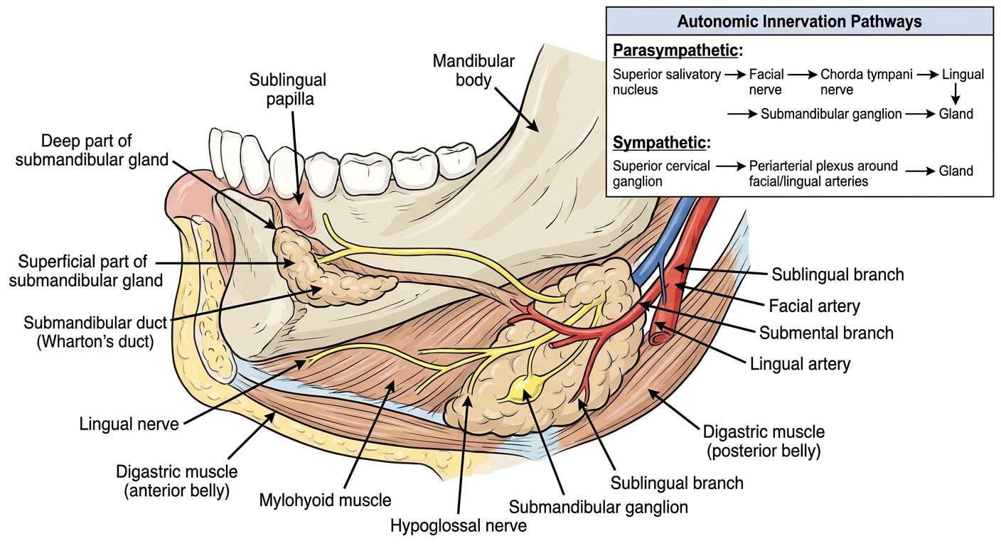
Submandibular gland: key labels
Parts and relations
- Superficial part: larger, lies below the mandible and superficial to the mylohyoid.
- Deep part: smaller, hooks around the posterior border of the mylohyoid into the floor of the mouth.
- Submandibular duct (Wharton's duct): arises from the deep part; runs forwards on hyoglossus, between the lingual nerve above and hypoglossal nerve below, then opens on the sublingual papilla beside the frenulum of the tongue.
- Lingual nerve: crosses lateral to the duct, then loops inferior to it and becomes medial to it anteriorly.
- Submandibular ganglion: hangs from the lingual nerve near the posterior border of mylohyoid.
Arterial supply
- Mainly by branches of the facial artery, particularly the submental artery.
- Additional supply from the lingual artery, including its sublingual branch.
- The facial artery is closely related to the posterior/superior part of the gland and may groove or traverse its capsule. NCBI anatomy reference
Nerve supply
| Type | Pathway | Main action |
|---|---|---|
| Parasympathetic | Superior salivatory nucleus → facial nerve (CN VII) → chorda tympani → joins lingual nerve (V3) → synapse in submandibular ganglion → postganglionic fibres to gland | Secretomotor, promotes salivation |
| Sympathetic | Superior cervical ganglion → periarterial plexus on facial and lingual arteries → gland | Vasomotor; modifies secretion |
| Sensory | Lingual nerve (V3) | General sensation |