I now have comprehensive material. Let me compile a thorough, well-organized answer on bilateral foot drop.
Bilateral Foot Drop
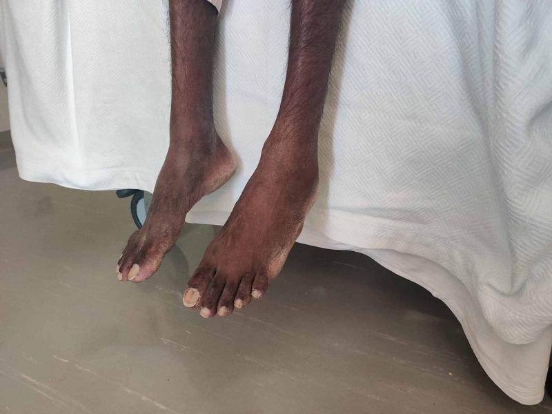
Foot drop is the inability to dorsiflex the foot due to weakness of the anterior tibialis and extensor muscles. When bilateral, the differential diagnosis is more restricted than for unilateral foot drop and strongly points toward systemic, spinal, or generalized neuromuscular disease rather than a single peripheral nerve lesion.
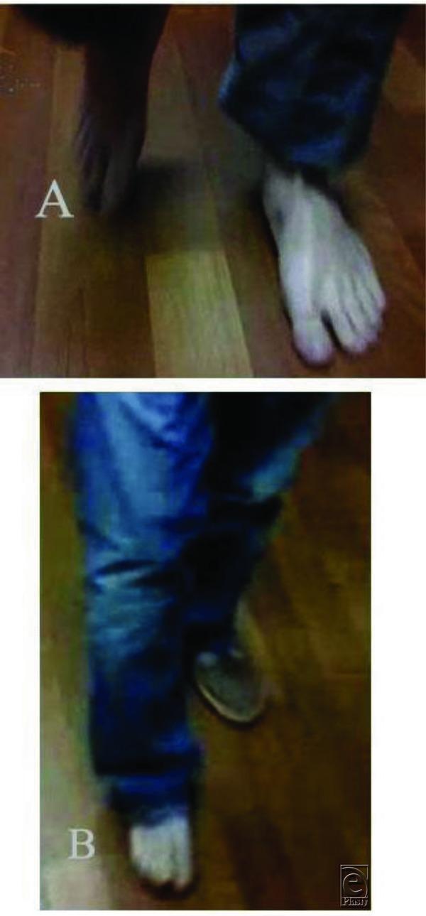
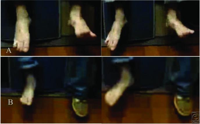
The classic gait is the steppage (equine) gait - the patient lifts the knee excessively high during the swing phase so the dangling foot clears the ground, then the sole slaps down at heel strike.
Anatomical Basis
Dorsiflexion is produced by:
- Tibialis anterior (deep peroneal nerve, L4-L5)
- Extensor digitorum longus and hallucis (deep peroneal nerve, L4-L5)
The pathway from cortex to foot: Motor cortex (parasagittal region) → corticospinal tract → L4-L5 anterior horn cells → lumbar roots → sciatic nerve → common peroneal nerve → deep peroneal nerve → tibialis anterior.
Causes of Bilateral Foot Drop
Organized by anatomical level (from central to peripheral):
1. Central / Upper Motor Neuron
| Cause | Key Features |
|---|
| Parasagittal meningioma or other mass (bilateral leg cortex) | Spastic, hyperreflexia, extensor plantar responses |
| High cervical myelopathy (rare) | Spastic paraparesis |
| Cerebral palsy (bilateral) | Long history, other UMN signs |
These produce spastic foot drop with increased tone and brisk reflexes - distinct from flaccid foot drop.
2. Spinal Cord
| Cause | Key Features |
|---|
| ALS / Motor neuron disease | Mixed UMN + LMN, no sensory loss, fasciculations |
| Progressive spinal muscular atrophy | Pure LMN, fasciculations, no sensory loss |
| Cervical/thoracic myelopathy | Spasticity below level, UMN signs |
3. Cauda Equina / Lumbosacral Roots
| Cause | Key Features |
|---|
| Cauda equina syndrome (central disc, tumour, abscess, haematoma) | Bilateral foot drop + saddle anaesthesia + sphincter disturbance - surgical emergency |
| Bilateral L4-L5 radiculopathy | Back pain, dermatomal sensory loss, reduced ankle jerks |
| Lumbosacral plexopathy | Diabetic, malignant infiltration, radiation |
| Spinal stenosis (neurogenic claudication) | Worse with walking, relieved by flexion |
4. Peripheral Nerve (Most Common for Bilateral)
| Cause | Key Features |
|---|
| Peripheral neuropathy (diabetic, alcoholic, toxic, nutritional) | Stocking-glove pattern, bilateral distal weakness, areflexia |
| Charcot-Marie-Tooth disease (CMT / hereditary motor-sensory neuropathy) | Gradual onset, pes cavus, inverted champagne bottle legs, family history |
| Vasculitis (mononeuritis multiplex → bilateral) | RA, PAN, SLE; asymmetric, systemic features |
| GBS (Guillain-Barré syndrome) | Ascending, areflexia, CSF albuminocytological dissociation |
| CIDP | Relapsing-remitting or progressive, elevated CSF protein |
| Bilateral common peroneal nerve compression | Weight loss ("slimmer's paralysis"), habitual leg crossing, prolonged squatting, childbirth (pushing palsy), bedridden patients |
| Leprosy | Endemic regions, thickened nerves, skin patches, sensory loss |
5. Anterior Horn Cell
| Cause | Key Features |
|---|
| ALS | Mixed UMN/LMN |
| Poliomyelitis / post-polio syndrome | Vaccine history, asymmetric, previous polio |
| Kennedy disease (SBMA) | X-linked, gynecomastia, bulbar involvement |
6. Neuromuscular Junction & Muscle
| Cause | Key Features |
|---|
| Myotonic dystrophy | Distal weakness, myotonia, cataracts, cardiomyopathy |
| Scapuloperoneal muscular dystrophy | Shoulder + peroneal distribution |
| Emery-Dreifuss / FSHD | FSH variant with bilateral foot drop known |
| Distal myopathies (Welander, Nonaka) | Rare, adult onset |
| Inflammatory myopathy | Usually proximal; distal rare |
Clinical Approach
History
- Onset: acute (vascular, compressive, GBS) vs. chronic (hereditary, neuropathy)
- Bilateral from start or sequential: simultaneous bilateral suggests systemic/central; staggered suggests mononeuritis multiplex or stepwise neuropathy
- Associated features: back pain (disc/cauda equina), bladder/bowel (cauda equina), burning/tingling (neuropathy), family history (CMT)
- Systemic history: diabetes, alcohol, chemotherapy (bortezomib), rheumatological disease, malignancy
Examination
- Tone: spastic (UMN) vs. flaccid (LMN/NMJ/myopathy)
- Reflexes: hyperreflexia (UMN), areflexia (neuropathy, severe LMN)
- Plantar responses: extensor = UMN
- Sensory pattern: dermatomal (root), stocking (neuropathy), saddle (cauda equina), absent (sensory ataxia component)
- Muscle wasting: pes cavus + inverted champagne bottle = CMT
- Sphincters: test for cauda equina
Investigations
| Test | Indication |
|---|
| MRI lumbar spine | Cauda equina, disc disease, spinal stenosis - urgent if bladder/bowel involved |
| MRI brain | Parasagittal lesion if UMN signs present |
| NCS/EMG | Localizes lesion: axonal vs. demyelinating; nerve vs. muscle vs. anterior horn |
| Blood: FBS/HbA1c, B12, folate, LFTs | Metabolic/nutritional neuropathy |
| Anti-ganglioside antibodies (anti-GM1) | GBS, multifocal motor neuropathy |
| CSF analysis | GBS (raised protein, normal cells), CIDP, malignancy |
| Genetic testing | CMT subtypes (PMP22 duplication most common) |
| Muscle biopsy | Myopathy |
| CK | Elevated in myopathy, ALS |
| Vasculitis screen (ANCA, ANA, RF) | Mononeuritis multiplex |
Red Flags Requiring Urgent Action
- Saddle anaesthesia + bilateral foot drop + sphincter disturbance = cauda equina syndrome - urgent MRI and surgical decompression
- Acute bilateral foot drop + ascending weakness + areflexia = possible GBS - admit, IVIG or plasmapheresis
- Foot drop with known malignancy = consider cord or plexus compression - urgent imaging
Management Principles
- Treat the underlying cause (glycaemic control in diabetes, immunotherapy in CIDP/GBS, surgery for cauda equina/tumour, B12 replacement)
- Ankle-foot orthosis (AFO) - the mainstay for any residual foot drop; prevents tripping and maintains positioning
- Physiotherapy - gait retraining, strengthening residual muscles, balance work
- Functional electrical stimulation (FES) - stimulates peroneal nerve during swing phase, improving gait quality
- Surgical options: tibialis posterior tendon transfer (converts a plantar-flexor to dorsiflexor, especially in CMT), peroneal nerve decompression (for fibular head compression), nerve repair/grafting
Key Teaching Points
- Bilateral foot drop almost never comes from isolated peroneal nerve damage - think systemically
- The most common cause overall is peripheral neuropathy (diabetic being most frequent)
- Hereditary cause (CMT) when insidious onset + pes cavus + family history + demyelinating pattern on NCS
- Always check for cauda equina signs - it is the most time-critical surgical emergency in this presentation
- NCS/EMG is the single most discriminating investigation for localizing the lesion level
Sources: Adams and Victor's Principles of Neurology 12th Ed; Bradley and Daroff's Neurology in Clinical Practice; Localization in Clinical Neurology 8th Ed; Harrison's Principles of Internal Medicine 22nd Ed (2025); Campbell's Operative Orthopaedics 15th Ed (2026)