

Diagnose these X-ray
left pleural effusion chest x-ray blunted costophrenic angle
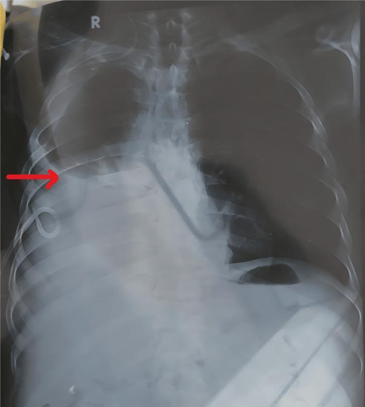
Anteroposterior (AP) chest X-ray demonstrating a moderate to large right-sided pleural effusion and a pigtail catheter in situ. The right hemithorax shows significant basal opacity with obscuration of the right costophrenic angle and hemidiaphragm. A curvilinear meniscus sign is visible at the upper margin of the fluid collection. A pigtail catheter is seen entering the right lateral chest wall, with its distal curled tip positioned superiorly within the pleural space. The left hemithorax displays a blunted costophrenic angle, suggestive of a concurrent mild left-sided pleural effusion. The cardiac silhouette is partially obscured, and the trachea remains midline. This diagnostic image illustrates pleural drainage management in a clinical setting, typically associated with conditions such as chronic liver disease or hepatohydrothorax, and highlights the radiological signs of bilateral pleural involvement and medical device placement.
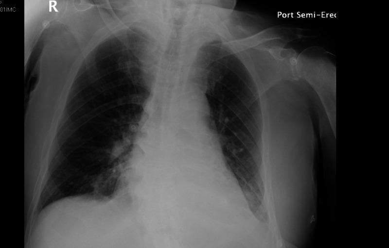
A portable semi-erect (Port Semi-Erect) frontal chest X-ray demonstrating multiple thoracic abnormalities. The cardiac silhouette is markedly enlarged, consistent with cardiomegaly. Evaluation of the pulmonary parenchyma reveals bilateral heterogeneous opacities, primarily concentrated in the bibasilar regions, suggesting airspace disease or infiltrates. In the pleural spaces, the right costophrenic angle remains relatively sharp, whereas the left costophrenic angle is blunted and obscured by an area of increased density along the left lateral chest wall, indicative of a small left pleural effusion. The pulmonary vasculature appears prominent, which, in the context of cardiomegaly, may represent pulmonary vascular congestion. These findings are characteristic of multifocal pneumonia, potentially superimposed on congestive heart failure. External artifacts, such as ECG leads, are visible on the chest wall.
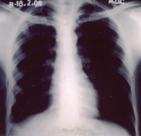
Posteroanterior (PA) view chest X-ray of a 24-year-old male, representing a follow-up assessment at 4 months post-treatment for pulmonary tuberculosis and bilateral pleural effusion. The diagnostic image shows significant resolution of previous pathology. The left lung field demonstrates clearing of the pleural space with a sharp costophrenic angle and normal aeration. In contrast, the right costophrenic angle remains blunted, a finding consistent with residual pleural thickening rather than active effusion. A subtle, resolving heterogeneous opacity is noted in the right upper zone, corresponding to the site of prior infection. The mediastinal contour and cardiac silhouette appear within normal limits. This image serves as a clinical example of radiological progression and sequelae following successful anti-tubercular therapy (ATT) in a case of sequential bilateral tuberculous pleurisy.
left lower lobe pneumonia consolidation chest x-ray
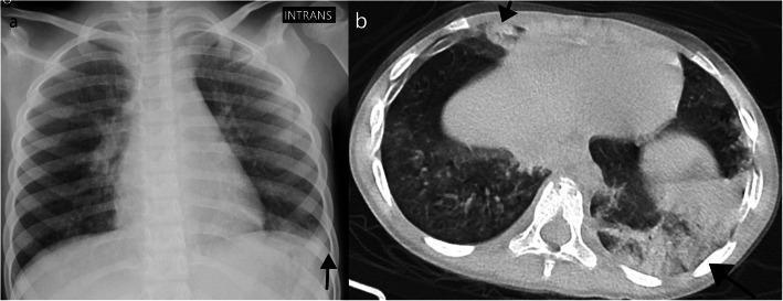
This composite diagnostic image features a pediatric chest X-ray (a) and an axial computed tomography (CT) scan (b) in a lung window, illustrating indeterminate pulmonary findings. The chest X-ray demonstrates a focal, unilateral opacity in the lower zone of the left lung, with relatively clear lung parenchyma elsewhere. The axial CT scan provides a more detailed view, revealing bilateral peripheral consolidations. The most prominent consolidation is located in the left lower lobe, with additional involvement noted in the middle lobe and the internal segment of the right lower lobe (indicated by arrows). These consolidations are subpleural in distribution and lack significant central or perihilar involvement. The case illustrates a ‘predominant consolidation’ pattern which, while maintaining a peripheral and lower lobe distribution, is considered indeterminate or atypical for standard COVID-19 presentations in pediatric patients. The images serve as an educational example of how CT can clarify findings that appear unilateral on plain radiography and help distinguish between typical, indeterminate, and atypical viral pneumonia patterns.
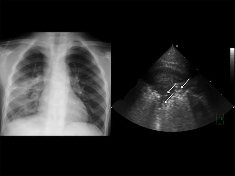
A multi-modal diagnostic image panel for pediatric pneumonia. (Left) An anteroposterior chest radiograph demonstrates a significant area of opacity in the right lower lobe, consistent with lung consolidation. The left lung remains relatively clear and air-filled. (Right) A corresponding transthoracic ultrasonography (TUS) of the right anterior lower lung showing a hepatized lung appearance (consolidation). The ultrasound features multiple punctate, hyperechoic (bright) spots indicated by white arrows, which represent static or dynamic air bronchograms—bubbles of air trapped within the consolidated, fluid-filled lung tissue. The clinical significance of this combination is to demonstrate the correlation between traditional radiography and point-of-care ultrasound in diagnosing community-acquired pneumonia (CAP) in children. Target educational concepts include identifying consolidation on X-ray and recognizing sonographic air bronchograms as a hallmark of pneumonia.
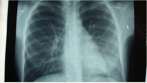
A posterior-anterior (PA) chest x-ray demonstrating localized pathology in the right lung. There is a notable area of increased opacity and consolidation in the right lower lobe, which partially obscures the right hemidiaphragm and suggests an infectious process such as pneumonia. In contrast, the left lung field appears relatively clear and well-aerated with normal vascular markings. The cardiac silhouette, mediastinal contours, and trachea appear within normal limits. The skeletal structures of the ribcage and clavicles are intact and clearly visible. This diagnostic image illustrates the typical radiographic presentation of community-acquired pneumonia, characterized by alveolar infiltration and consolidation in a specific anatomical lobe, used clinically to guide antibiotic treatment and monitor disease progression.
Chest X-Ray Analysis - Patient: BHALENDRA BORO
Important disclaimer: This is a photograph of a monitor displaying the X-ray (AP view), which introduces moiré artifacts, glare, and color distortion. This analysis is an educational aid only - formal radiologist interpretation of the original DICOM image is required for clinical decision-making.
Technical Details
- View: AP (Anteroposterior) - likely a portable/bedside film
- AP vs PA note: AP projections magnify the cardiac silhouette by ~20%, so heart size assessment must be interpreted cautiously
Systematic Findings
| Region | Finding |
|---|---|
| Trachea | Appears midline - no obvious deviation |
| Mediastinum | No gross widening appreciated |
| Heart | Appears mildly enlarged - but AP magnification effect cannot be excluded |
| Right lung | Relatively clear; right costophrenic angle is sharp and well-defined |
| Left lung | Dense, homogeneous opacity in the left lower zone |
| Left diaphragm | Completely obscured by the left lower zone opacity ("silhouette sign") |
| Left costophrenic angle | Obliterated - not visible |
| Right diaphragm | Well-defined and rounded |
Primary Abnormal Finding
- Completely obliterates the left hemidiaphragm (positive silhouette sign)
- Blunts/erases the left costophrenic angle
- Extends over the lower left hemithorax
Differential Diagnosis (in order of likelihood)
- Fluid in the pleural space accumulates at the base and erases the costophrenic angle
- Produces a concave meniscus at the upper border
- Common causes: heart failure, parapneumonic effusion, malignancy, TB, hypoalbuminemia
- Dense lobar or segmental opacity with a silhouette sign against the diaphragm
- Associated with fever, cough, productive sputum, raised WBC
- The opacity pattern is consistent with this diagnosis
- Very common - infection → pleural inflammation → secondary fluid collection
- This combination would explain both the density and the complete loss of the diaphragm border
- Collapse of lung tissue can produce a similar dense opacity
- Usually associated with shift of mediastinum/trachea toward the affected side
Recommended Next Steps
| Step | Purpose |
|---|---|
| Clinical correlation | Fever? Cough? Dyspnea? Pleuritic chest pain? Decreased breath sounds on the left? |
| Upright PA chest X-ray | Better image quality; true cardiothoracic ratio assessment |
| Lateral decubitus X-ray | Confirms freely mobile pleural effusion (fluid layers out) |
| Chest ultrasound | Gold standard for detecting and quantifying pleural effusion; guides drainage |
| CT chest | If diagnosis unclear; best for distinguishing effusion vs. consolidation vs. mass |
| Blood tests | FBC (WBC for infection), CRP, LDH, albumin, blood cultures |
| Pleural fluid analysis | If effusion confirmed and drained - Light's criteria to differentiate exudate vs. transudate |