

Dignosis
infected sebaceous cyst earlobe piercing
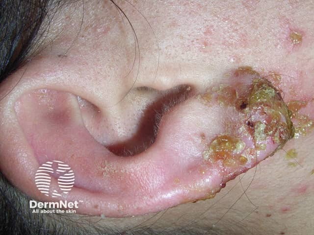
Imaging modality: Clinical photography. Technique: High‑resolution close‑up macro image of the left auricle. This dermatologic photograph documents an infected ear piercing with inflammatory skin changes around the earlobe. Anatomical site is the left external ear (pinna) with focal involvement of the earlobe and peripiercing tissue. Visual features include marked erythema and localized edema, yellow‑brown crusts and purulent exudate, superficial erosions, and flaking desquamation. The piercing tract is inflamed; surrounding skin is tender. There is no obvious deep cartilage involvement in this image, though deep infection cannot be excluded without clinical examination. The appearance is compatible with acute bacterial folliculitis or cellulitis secondary to piercing, with Staphylococcus aureus and Streptococcus species among common pathogens; allergic contact dermatitis to metal is also in consideration. Differential diagnosis includes perichondritis, cellulitis, dermatitis from jewelry, and foreign body reaction. Clinically relevant observations include disruption of skin integrity, risk of abscess formation, and potential progression to cartilaginous infection if untreated. Diagnostic significance lies in guiding immediate management: remove offending jewelry, perform gentle cleansing, consider topical antiseptics or oral antibiotics, assess for systemic symptoms, and refer if cartilage involvement is suspected. This image is educational for dermatology, wound care, and clinical triage. Useful for teaching and triage.
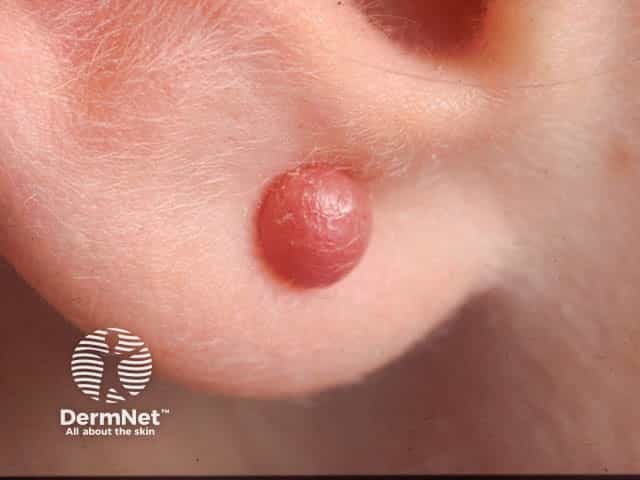
Imaging modality: Clinical photography; high-quality close-up macro view of the earlobe documenting a solitary keloid scar at a piercing site. Anatomical location: auricular lobule, periauricular skin, left or unspecified ear. Visual features: a firm, rounded, dome-shaped nodule with smooth, pink-red surface; surrounding skin shows mild dermal thickening without ulceration or purulent discharge; lesion is non-tender on palpation yet tethered within subcutaneous tissue by dense collagen; surface may appear shiny and slightly translucent. Pathophysiology: exuberant scar formation characterized by disorganized collagen deposition, scar tissue extending beyond original wound margins, with fibroblast proliferation and persistent inflammatory signaling; early matrix rich in type III collagen gradually replaced by type I fibers. Diagnostic relevance: classic piercing-related keloid; distinguishes from hypertrophic scar and granulomatous tissue; carries risk of recurrence after treatment if not adequately addressed. Clinical correlations: history of piercing trauma, possible itch or tenderness, cosmetic concern, psychological impact; imaging notes aid planning of therapy. Potential clinical applications: patient education, decision support for management strategies including intralesional corticosteroids, silicone therapy, laser ablation, cryotherapy, or surgical excision with adjuvant radiotherapy; monitor response and recurrence. Differential considerations: hypertrophic scar, epidermoid cyst, dermal granuloma, scar tissue. Clinical relevance: emphasizes cosmetic impact and informed, patient-centered management planning for outcomes.
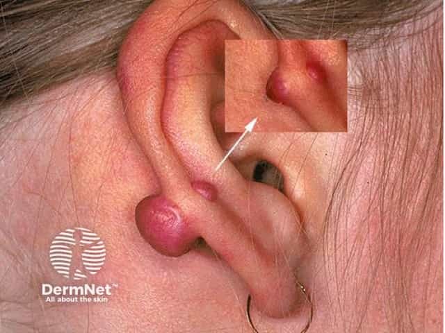
This is a clinical dermatology photograph of a left auricle (external ear) showing a solitary pink, pedunculated nodule arising from the earlobe with a smooth surface and firm consistency. A second small nodular lesion is faintly visible in the inset, near the superior helical region. The appearance is most compatible with piercing-related scar tissue such as a keloid or hypertrophic scar of the earlobe, though the differential diagnosis includes a pyogenic granuloma (lobular capillary hemangioma), epidermal inclusion cyst with surface erythema, or a benign adnexal neoplasm. The lesion demonstrates a well-circumscribed, rounded morphology with an elevated, nodular profile and a narrow stalk in some cases, suggesting an overgrowing scar rather than a fluid-filled cyst. The surrounding skin is mildly erythematous, but there is no ulceration or discharge evident in the photo. Clinically, history of piercing, trauma, and rapid growth would raise suspicion for keloid formation; a biopsy or excisional removal with histopathology would differentiate between scar tissue and vascular tumors. This image is useful for educational purposes in dermatology, medical training, and dermatoses differential diagnosis, and demonstrates typical appearance of ear-lobe nodules for teaching about wound healing disorders, scar augmentation, and management implications.
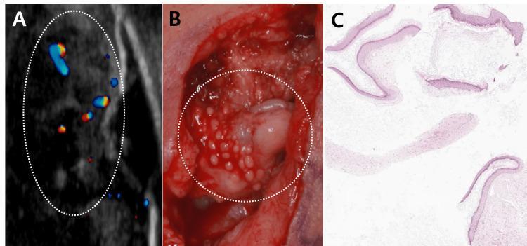
A three-panel figure demonstrating the multimodal diagnostic features of recurrent keloid-associated epidermoid cysts. Panel A shows a color Doppler ultrasonography of the earlobe, revealing multiple hypoechoic, heterogeneous lesions within a dotted oval area, with some internal vascularity signaled by blue and red flow indicators. Panel B is an intraoperative clinical photograph displaying a surgical field with multiple, small, whitish, round cyst-like structures embedded in dense, reddish scar tissue. Panel C presents a histopathological section (H&E stain, 40x magnification) showing multiple cyst walls lined with stratified squamous epithelium and containing lamellated keratin. This composite image illustrates the correlation between preoperative imaging, surgical findings, and definitive pathology for epidermoid cysts arising within keloid tissue, a rare clinical occurrence often triggered by trauma such as ear piercing.
Diagnosis: Infected Sebaceous (Epidermoid) Cyst - Posterior Earlobe
What the image shows
- Small (~3-5 mm), dome-shaped, well-circumscribed nodule
- Erythematous (pink-red) base with a central punctum (tiny pinpoint opening)
- Overlying skin is smooth, taut, and slightly shiny - indicating edema/early fluctuance
- No surrounding scaling or cellulitis (yet)
Differential Diagnoses
| Diagnosis | For | Against |
|---|---|---|
| Infected sebaceous cyst (most likely) | Central punctum, dome shape, posterior earlobe location, erythema | - |
| Epidermoid inclusion cyst (piercing-related) | Piercing trauma nearby, earlobe location | Not yet infected in classic form |
| Folliculitis/furunculitis | Erythema, central pustule | No hair follicle visible at earlobe |
| Keloid (early) | Piercing site, earlobe | No firm collagen nodule, acute onset |
| Abscess | Fluctuance, erythema | Too small/early stage |
Pathophysiology
Management
-
Incision and Drainage (I&D) - first step when acutely infected
- Express the thick cheesy sebaceous material manually (too viscous to drain spontaneously)
- Pack the wound; arrange follow-up
-
Capsule excision - the critical step for preventing recurrence
- The pearly white capsule must be fully removed
- Can be done at initial visit OR deferred to a follow-up visit once inflammation subsides (preferred traditional approach)
- Grasp edges with clamps/hemostats; sharp dissection with scalpel or scissors
-
Antibiotics - consider oral antibiotics covering Staph aureus/MRSA (e.g., TMP-SMX or doxycycline) given proximity to piercing and risk of MRSA
-
Do NOT use loop drainage technique - not suited for sebaceous cysts due to the need for capsule removal
Key teaching point: Simple drainage without capsule excision leads to recurrence. A randomized study by Kitamura et al. found primary resection resulted in faster healing, fewer pain days, and less scarring compared to traditional I&D alone. (Roberts and Hedges', p. 888)
⚠️ This is an educational analysis based on visual features. A clinical examination by a healthcare provider is required for definitive diagnosis and treatment.