
Dermatomes of lower limb notes
dermatomes of lower limb diagram
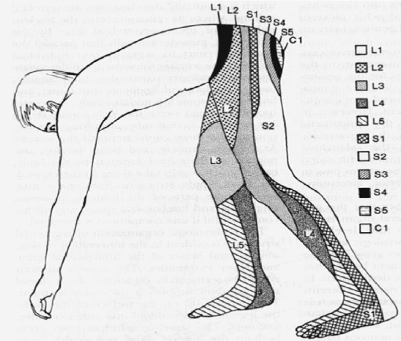
Anatomical diagram depicting a human dermatome map of the lumbosacral and coccygeal regions, shown from a lateral, bent-over perspective to highlight the sensory distribution along the lower extremities and posterior trunk. The illustration uses distinct shading patterns and alphanumeric labels to delineate specific cutaneous areas innervated by individual spinal nerve roots. Key segments include the lumbar dermatomes L1 through L5, sacral dermatomes S1 through S5, and the coccygeal dermatome C1. Visually, L1 covers the inguinal and hip region, L2-L3 transition down the anterior and lateral thigh, and L4-L5 extend down the lower leg to the dorsal foot. Sacral segments S1 and S2 cover the posterior leg and heel, while S3-S5 and C1 are concentrated in the perineal and gluteal regions. This medical illustration is a standard educational tool for neurological examinations, aiding in the localization of spinal nerve root compression or radiculopathy based on sensory loss patterns.
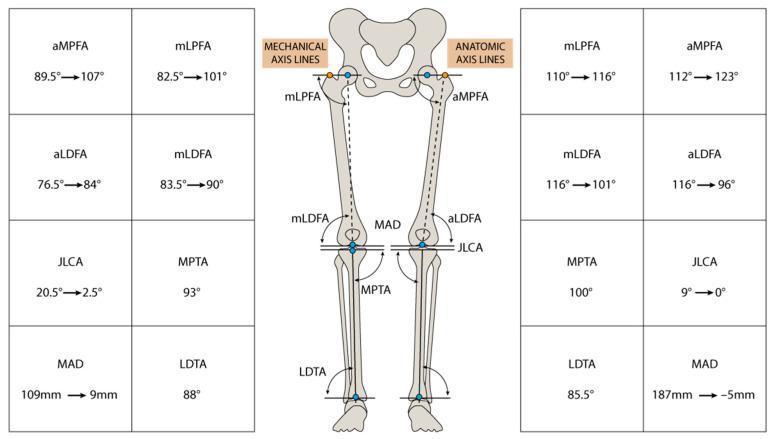
An educational musculoskeletal diagram and comparison chart illustrating lower limb alignment and surgical planning for deformity correction. The central figure displays an anteroposterior skeletal diagram of the pelvis and lower limbs with superimposed mechanical and anatomical axis lines. Key radiological parameters are labeled, including mechanical axis deviation (MAD), joint line convergence angle (JLCA), and various femoral and tibial angles (aMPFA, mLPFA, aLDFA, mLDFA, MPTA, and LDTA). Surrounding the diagram are tables documenting preoperative and postoperative values for each limb, represented by a transition from an initial value to a corrected value (e.g., MAD 109mm to 9mm on the left). The diagram visualizes the intersection of mechanical axes (hip center to ankle center) and anatomical axes (through the shafts) with articular surface tangents. This educational tool is used in orthopedic surgery and physical medicine to evaluate 'windswept' or varus/valgus deformities and to quantify the outcomes of corrective osteotomy.
This medical engineering diagram illustrates the biomechanical modeling and boundary conditions of a bionic lower limb prosthesis. The 3D geometric model is segmented into a dark gray upper knee mount, a contoured light blue calf component, and a tan-colored anatomical foot plate. The image depicts a finite element analysis (FEA) setup with specific load parameters. A blue marker 'A' indicates a 'Fixed Support' at the proximal interface. Red circular arrows 'B' and 'C' designate the mechanical rotational joints at the knee and ankle, respectively, both set with a 20-degree rotation limit to simulate physiological range of motion. A yellow downward arrow 'D' represents standard earth gravity (9806.6 mm/s²) acting upon the calf segment. This visualization is intended for orthopedic and rehabilitative engineering education, focusing on the dynamic characteristics and neural control development for advanced limb replacement systems using biomimetic principles and additive manufacturing materials.
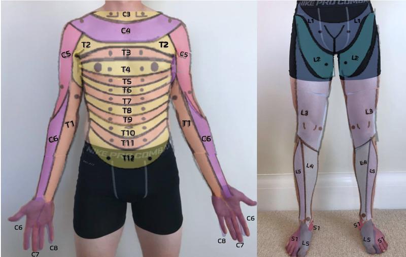
This clinical dermatome map, presented as an anatomical diagram overlaid on a human subject, illustrates the cutaneous sensory distribution of spinal nerves. The anterior trunk displays a sequential horizontal banding pattern representing thoracic dermatomes T2 through T12, with T4 roughly at the nipple line and T10 at the umbilicus. The upper extremities show cervical and upper thoracic distribution: C5 (lateral upper arm), C6 (lateral forearm and thumb), C7 (middle finger), C8 (medial hand and little finger), and T1 (medial forearm and arm). The lower extremity map details the lumbar and sacral levels: L1 (inguinal region), L2 (anterior mid-thigh), L3 (distal thigh and knee), L4 (medial lower leg), L5 (lateral lower leg and dorsal foot), and S1 (lateral foot and small toe). This resource is designed for primary care musculoskeletal medicine to assist in the neurological examination of sensory deficits, radiculopathy, or spinal cord injury level localization.
lower limb cutaneous nerve distribution peripheral innervation
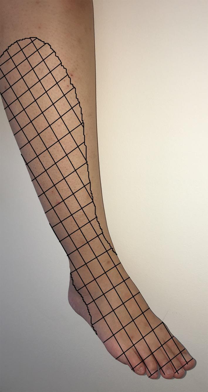
This clinical photograph with a graphical overlay illustrates the sensory distribution of the common peroneal nerve (common fibular nerve) on the right lower limb. The image displays a lateral view of a human leg and foot, with a black grid pattern demarcating the specific cutaneous regions affected by sensory loss or paresthesia. The grid covers the lateral aspect of the shin, extending distally across the anterior ankle and over the entire dorsal surface of the foot, including the phalanges. This visualization represents the typical dermatomal and peripheral nerve distribution associated with common peroneal nerve injury, often seen in clinical scenarios such as compression neuropathies, postoperative complications (e.g., following tourniquet use or arthroscopy), or direct trauma to the fibular neck. The educational focus is on identifying the anatomical boundaries of superficial and deep peroneal nerve sensory innervation, which is critical for differentiating peripheral nerve lesions from radiculopathy in neurology and orthopedics.
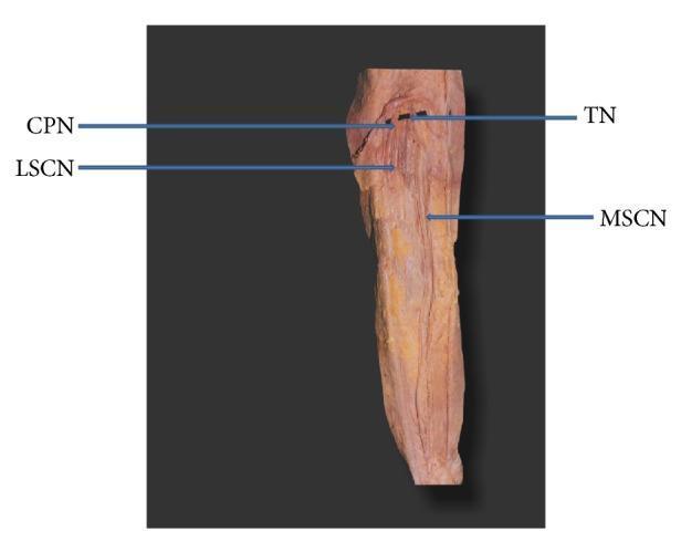
This clinical photograph displays a gross anatomical dissection of the posterior lower limb, specifically focusing on the nerves of the popliteal fossa and the proximal leg. The image illustrates a specific anatomical variation where the medial sural cutaneous nerve (MSCN) continues distally as the sural nerve without joining a peroneal communicating branch. Key labeled structures include the tibial nerve (TN) and common peroneal nerve (CPN) branching proximally. Arising from the CPN is the lateral sural cutaneous nerve (LSCN), which descends toward the lateral aspect of the gastrocnemius. The MSCN is shown originating from the tibial nerve and coursing vertically down the midline of the posterior leg superficial to the gastrocnemius muscle. This specimen serves as an educational example of peripheral nerve anatomy and sural nerve formation patterns, which is clinically significant for nerve conduction studies, sural nerve biopsies, and regional anesthesia blocks.
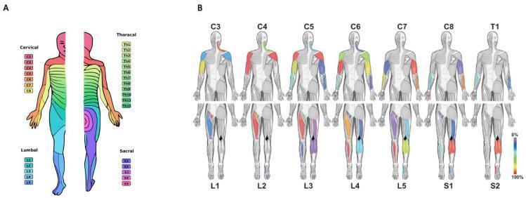
This medical illustration displays the sensory and motor innervation patterns of the human spinal nerves. Panel A is a dermatome map showing the horizontal and longitudinal sensory distribution of 31 pairs of spinal nerves (Cervical C2-C8, Thoracic Th1-Th12, Lumbar L1-L5, and Sacral S1-S5) across both anterior and posterior views of the human body. Panel B illustrates myotomal distributions through 14 distinct anatomical figures. The top row tracks cervical and first thoracic (C3-T1) root contributions to upper limb musculature, while the bottom row tracks lumbosacral (L1-S2) root contributions to the lower limbs. Specific muscle groups are highlighted with a color-coded heat map (0-100%) to represent response rates during intraoperative nerve root stimulation. This educational graphic serves as a clinical reference for mapping peripheral neurological deficits to their corresponding spinal levels, useful in neurology, physiatry, and neurosurgery for diagnosing nerve root entrapment or spinal cord injuries.
Dermatomes of the Lower Limb
What is a Dermatome?
Key principle: Dermatome overlap is extensive - sensory loss from a single root lesion (radiculopathy) is always smaller than the anatomical dermatome, because adjacent roots cover most of the same territory. The "autonomous zone" (area with no overlap) is small.
Dermatomal Map - Lower Limb
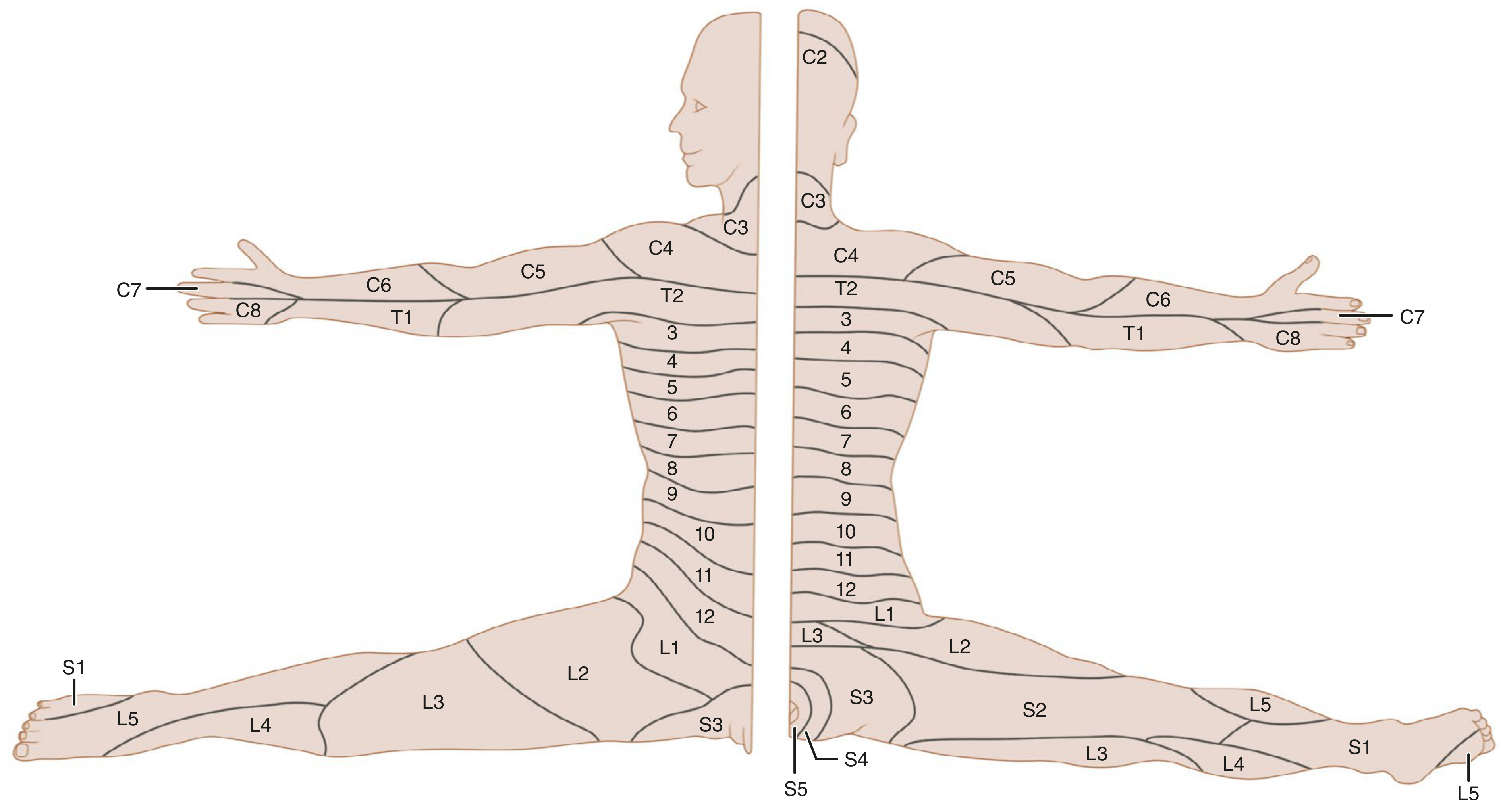
Segmental Levels and Their Territories
| Root | Cutaneous Territory | Key Landmark |
|---|---|---|
| L1 | Inguinal region, upper medial thigh | Femoral pulse / inguinal ligament |
| L2 | Anterior and medial thigh (upper) | Medial mid-thigh |
| L3 | Anterior and medial thigh (lower), medial knee | Medial aspect of thigh, knee |
| L4 | Medial lower leg, medial malleolus, medial foot | Knee; medial leg |
| L5 | Lateral lower leg, dorsum of foot, 1st webspace (big toe) | Lateral calf; dorsal foot |
| S1 | Lateral foot, small toe, heel, posterior calf | Lateral foot; Achilles; posterior calf |
| S2 | Posterior thigh, posterior calf (narrow strip) | Posterior thigh |
| S3 | Inner thigh / proximal posterior thigh | "Saddle" medial zone |
| S4-S5 | Perineum, perianal area | Perineum |
Root-by-Root Detail
L1
- Inguinal region and hip crease
- Supplied via the iliohypogastric and ilioinguinal nerves (terminal branches of L1)
- Lateral genitalia also receives L1 via the genitofemoral nerve (L1-L2)
L2
- Anterior and medial thigh (upper half)
- Main carrier: lateral femoral cutaneous nerve (L2-L3) for the lateral thigh, femoral nerve (L2-L4) for anterior/medial thigh
- Clinical test: sensation at medial mid-thigh
L3
- Lower anterior and medial thigh; crosses to medial knee
- Main carrier: femoral nerve (medial cutaneous nerve of thigh); obturator nerve (L2-L4) for medial thigh
- Hip adduction (obturator) lost at L3
L4
- Medial lower leg, medial malleolus, medial foot (great toe side)
- Main carrier: saphenous nerve (terminal sensory branch of femoral nerve, L3-L4)
- Patellar reflex (knee jerk) = L4
- Clinical test: sensation at medial malleolus; foot dorsiflexion (tibialis anterior, L4-L5)
L5
- Lateral lower leg (peroneal region), dorsum of foot, 1st-2nd webspace
- Main carrier: superficial peroneal (fibular) nerve (dorsal foot), deep peroneal nerve (1st webspace)
- Foot drop (weak dorsiflexion + eversion) is the hallmark of L5 lesion
- No standard reflex for L5 (hamstring jerk is unreliable)
- Clinical test: big toe extension (extensor hallucis longus)
S1
- Lateral foot and small toe; posterior calf; heel
- Main carrier: sural nerve (lateral foot/leg), lateral plantar nerve (sole)
- Achilles reflex (ankle jerk) = S1
- Clinical test: foot eversion (peroneus longus/brevis); plantar flexion
S2
- Posterior thigh; narrow strip of posterior calf
- Main carrier: posterior femoral cutaneous nerve (S1-S3)
S3-S5
- Perineum, perianal and perivulvar/scrotal skin ("saddle area")
- Anal wink reflex = S3-S4-S5
- S2-S4 control rectal sphincter tone
Reflexes as Dermatome Anchors (High-Yield)
| Reflex | Root Level |
|---|---|
| Patellar (knee jerk) | L4 |
| Hamstring | L5-S1 (unreliable) |
| Achilles (ankle jerk) | S1 |
| Anal wink | S3-S4 |
| Bulbocavernosus | S3-S4 |
| Cremasteric | L1-L2 |
Motor Levels (for correlation)
| Root | Movement Lost |
|---|---|
| L1-L2 | Hip flexion (iliopsoas) |
| L3 | Hip adduction |
| L4 | Hip abduction; knee extension (quadriceps) |
| L4-L5 | Foot dorsiflexion (tibialis anterior) |
| L5-S1 | Foot eversion; big toe extension |
| S1-S2 | Plantar flexion (gastrocnemius/soleus) |
| S2-S4 | Rectal/urethral sphincter tone |
- Rosen's Emergency Medicine, Table 35.4-35.6
Axial Line Concept
- Campbell's Operative Orthopaedics 15th Ed, eFig. 67.2
Radicular vs. Peripheral Nerve Lesions
| Feature | Radiculopathy (root lesion) | Peripheral nerve lesion |
|---|---|---|
| Pattern | Dermatome (root territory) | Peripheral nerve territory |
| Example cause | Disc herniation | Peroneal nerve compression at fibular head |
| Overlap | Extensive - small autonomous zone | Less overlap |
| Distribution | Follows spinal segment (spiral in limb) | Follows anatomical nerve course |
When a nerve root is damaged (e.g., L4-L5 disc herniation), sensory loss is in the radicular pattern. When a peripheral nerve is cut (e.g., common peroneal nerve at fibular neck), loss follows the peripheral nerve pattern, which differs from the dermatome.
- General Anatomy and Musculoskeletal System, THIEME Atlas
Clinical Pearls
- L4-L5 disc is the most common level of lumbar herniation; L5 and S1 are the most commonly affected roots.
- L5 radiculopathy = foot drop + numbness dorsal foot + no standard reflex loss (ankle jerk is preserved).
- S1 radiculopathy = weak plantar flexion + lateral foot numbness + absent ankle jerk.
- Cauda equina syndrome (massive L3-L4 or L4-L5 disc, or tumor): bilateral leg weakness + saddle anaesthesia (S3-S5) + bladder/bowel dysfunction - a surgical emergency.
- Meralgia paraesthetica = lateral thigh burning/numbness from compression of the lateral femoral cutaneous nerve (L2-L3) at the inguinal ligament - pure sensory, no motor loss.
- A dermatome and a peripheral nerve territory are NOT the same: the saphenous nerve covers the medial leg (L3-L4 roots), while the sural nerve covers the lateral leg and foot (S1-S2 roots).