
Bronchial blockers 5 mark answer
bronchial blocker types Arndt Cohen Univent placement
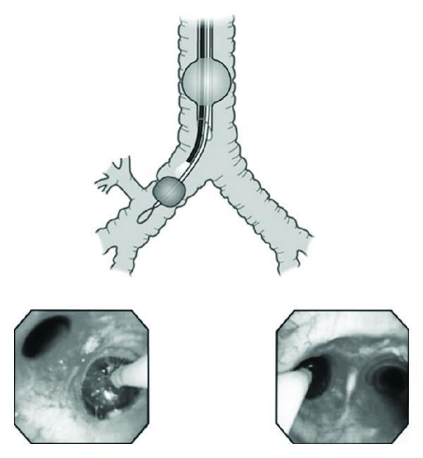
Educational medical graphic demonstrating the placement of a bronchial blocker for one-lung ventilation (OLV). The image consists of a central schematic illustration and two lower endoscopic views. The schematic depicts a single-lumen endotracheal tube (ETT) with its cuff inflated in the trachea above the carina. Through this ETT, a bronchial blocker is advanced into the right main bronchus; its distal balloon is inflated to occlude the airway, approximately 10mm below the carina. The left endoscopic image provides a distal view within the bronchus, showing the blocker's tip and guidewire loop. The right endoscopic image provides a proximal view from the trachea, showing the carina and the blocker entering the left main bronchus. This visual resource illustrates the proper depth and orientation required to achieve lung isolation while maintaining airway access via a single-lumen tube, a critical technique in thoracic anesthesia and surgery.
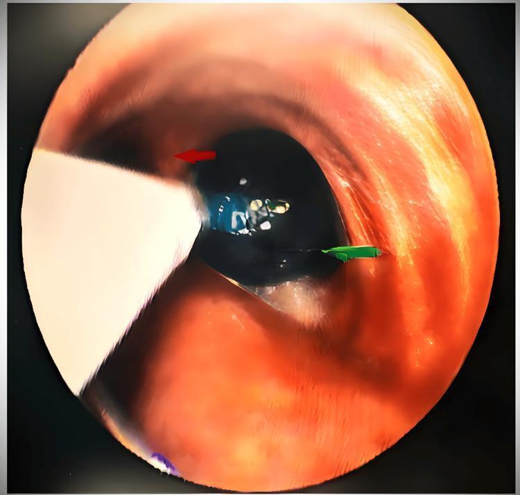
This clinical diagnostic image shows a bronchoscopic view of the tracheobronchial tree during the placement of an EZ bronchial blocker. The visual focuses on the carina (indicated by a red arrow), which serves as the primary anatomical landmark for positioning. A Y-shaped bronchial blocker is seen 'stranded' or seated directly on the carina, with its distal bifurcations extending into the left and right mainstem bronchi. Within the right bronchus, a green arrow points to a distended, inflated blue cuff that occupies the lumen. This configuration is used in thoracic anesthesia to achieve one-lung ventilation (OLV) by mechanically occluding a specific bronchus. The image demonstrates the correct endoscopic verification of device placement, ensuring the blocker is securely anchored at the carina to prevent intraoperative dislodgement and to facilitate isolated lung collapse for surgical procedures such as bronchogenic cyst excision.
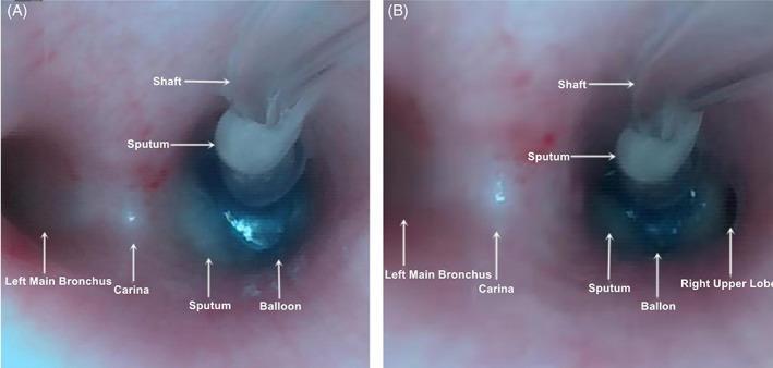
Fiberoptic bronchoscopy images demonstrating the placement of a bronchial blocker (BB) at the carina for one-lung ventilation. Both images (A) and (B) show the bronchial tree landmarks including the carina, the left main bronchus, and the blocker's shaft and balloon cuff. In image (A), the inflated blue balloon demonstrates optimal position, achieving complete occlusion of the right main bronchus. In image (B), the balloon is malpositioned or under-inflated, resulting in incomplete occlusion and visible patency of the right upper lobe bronchus. White, viscous sputum is present near the blocker shaft and within the bronchial lumen in both views. This comparison serves as an educational tool for verifying bronchial blocker placement and identifying common causes of failed lung isolation, such as cuff displacement or insufficient inflation, which can be correlated with bedside lung ultrasound findings.
Bronchial Blockers
Definition
- Miller's Anesthesia, 10e | Morgan and Mikhail's Clinical Anesthesiology, 7e | Barash Clinical Anesthesia, 9e
Types of Bronchial Blockers
| Device | Key Feature |
|---|---|
| Univent (Torque Control Blocker) | Blocker housed within a modified single-lumen tube (SLT); silicon tube with high friction coefficient for better torque control |
| Arndt Wire-Guided Blocker (Cook Critical Care) | Wire loop snare; fiberscope passed through loop to guide placement; 7 Fr and 5 Fr (pediatric); spherical or elliptical cuff |
| Cohen Flexitip Blocker (Cook Critical Care) | Rotating wheel deflects soft tip >90°; pear-shaped HVLP cuff; distinctive blue cuff visible on FOB |
| Fuji Uniblocker | Can be placed extra-luminally (exterior to SLT); useful in pediatrics and tracheostomies |
| EZ-Blocker (Teleflex) | 7-Fr, 4-lumen, Y-shaped catheter; distal limbs straddle carina; color-coded balloons (blue/yellow) for each bronchus |
- Miller's Anesthesia, 10e, p. 7161-7165
Placement Technique
- A single-lumen tracheal tube (ETT ≥ 7.0 mm ID for 9-Fr blockers) is placed first.
- The blocker is passed coaxially (through the ETT) or, for Cohen/Fuji, separately extraluminally through the glottis.
- A flexible fiberoptic bronchoscope (FOB) guides the blocker into the target bronchus.
- Cuff is inflated (4-8 mL air) under direct FOB visualization - HVLP (high-volume low-pressure) cuff.
- Position must be reconfirmed after turning the patient to lateral decubitus position.
- The SLT lumen (1.4-1.6 mm) allows limited suctioning or O₂ insufflation to the collapsed lung.
- Morgan & Mikhail, 7e, p. 1034
Indications / Preferred Over DLT
-
Difficult airway - awake nasotracheal or orotracheal intubation with SLT, BB added afterward
-
Existing tracheostomy - BB placed via existing stoma
-
Need for postoperative mechanical ventilation - no tube exchange required (avoids airway compromise)
-
Critically ill patients already on a ventilator requiring OLV
-
Previous contralateral lung resection - selective lobar blockade improves oxygenation
-
Pediatric patients - 5 Fr Arndt or Fogarty embolectomy catheter used
-
Selective lobar collapse - more precise than DLT
-
Miller's Anesthesia, 10e, p. 7161 | Barash, 9e, p. 3200
Advantages vs. Disadvantages
| Advantages | Disadvantages |
|---|---|
| No tube exchange needed if postop ventilation required | Slower, often incomplete lung collapse (small central lumen) |
| Can achieve lobar (not just whole-lung) isolation | More prone to dislodgement than DLT |
| Suitable for difficult/tracheostomy airways | Cannot independently ventilate collapsed lung |
| Smaller SLT used; less airway trauma | Larger ETT needed (≥7 mm ID) for coaxial placement |
| Useful when airway anatomy is distorted | Suctioning through 1.4 mm lumen is very limited |
- Morgan & Mikhail, 7e, p. 1034 | Barash, 9e, p. 3200
DLT vs. Bronchial Blocker - Key Comparisons
-
DLT preferred when: both lungs must be ventilated independently; protection of dependent lung from life-threatening contamination; tumour occupying main bronchus; sleeve resection; bilateral procedures (double lung transplant, bilateral sympathectomy).
-
BB preferred when: difficult airway, tracheostomy, anticipated postop ventilation, pediatric patients, selective lobar blockade required.
-
Peak airway pressure should be kept < 30 cmH₂O when using BB to prevent gas leak past the HVLP cuff.
-
Barash, 9e, p. 3200
Complications
- Dislodgement - most common; more frequent than with DLT; can lead to loss of lung isolation
- Tracheal obstruction - if inflated balloon migrates above carina → inability to ventilate → hypoxia, cardiorespiratory arrest; requires immediate deflation
- Failure to achieve lung separation - due to abnormal anatomy or inadequate bronchial seal
- Inclusion into stapling line - BB or Arndt wire loop stapled during lobectomy → requires surgical re-exploration; prevent by withdrawing the blocker a few centimeters before stapling
- Balloon failure to deflate - reported with Fuji blocker; may require removal of BB together with ETT during extubation
- Miller's Anesthesia, 10e, p. 7167
- P - Placement through SLT under FOB guidance
- L - Lung collapse distal to inflated cuff
- A - Arndt/Cohen/Univent/EZ-Blocker are types
- C - Confirm position after lateral positioning
- E - Exchange of tube not needed postoperatively (key advantage)