
Can you explain about pharyngeal apparatus
Reading File
The pharyngeal apparatus (also called the branchial apparatus) is a series of structures that develop in the embryonic head and neck region, giving rise to many important adult structures of the face, neck, and oral cavity.
Searching sources...
Finding Sources
Finding Sources
Reading File
Reading File
Reading File
Loading Image
Reading File
Loading Image
Reading File
Now I have comprehensive material. Let me compile the full, well-organized explanation.
Pharyngeal Apparatus
The pharyngeal apparatus consists of pharyngeal arches, pouches, grooves (clefts), and membranes - a series of embryonic structures that develop in the head and neck region and give rise to many adult structures of the face, neck, and associated glands.
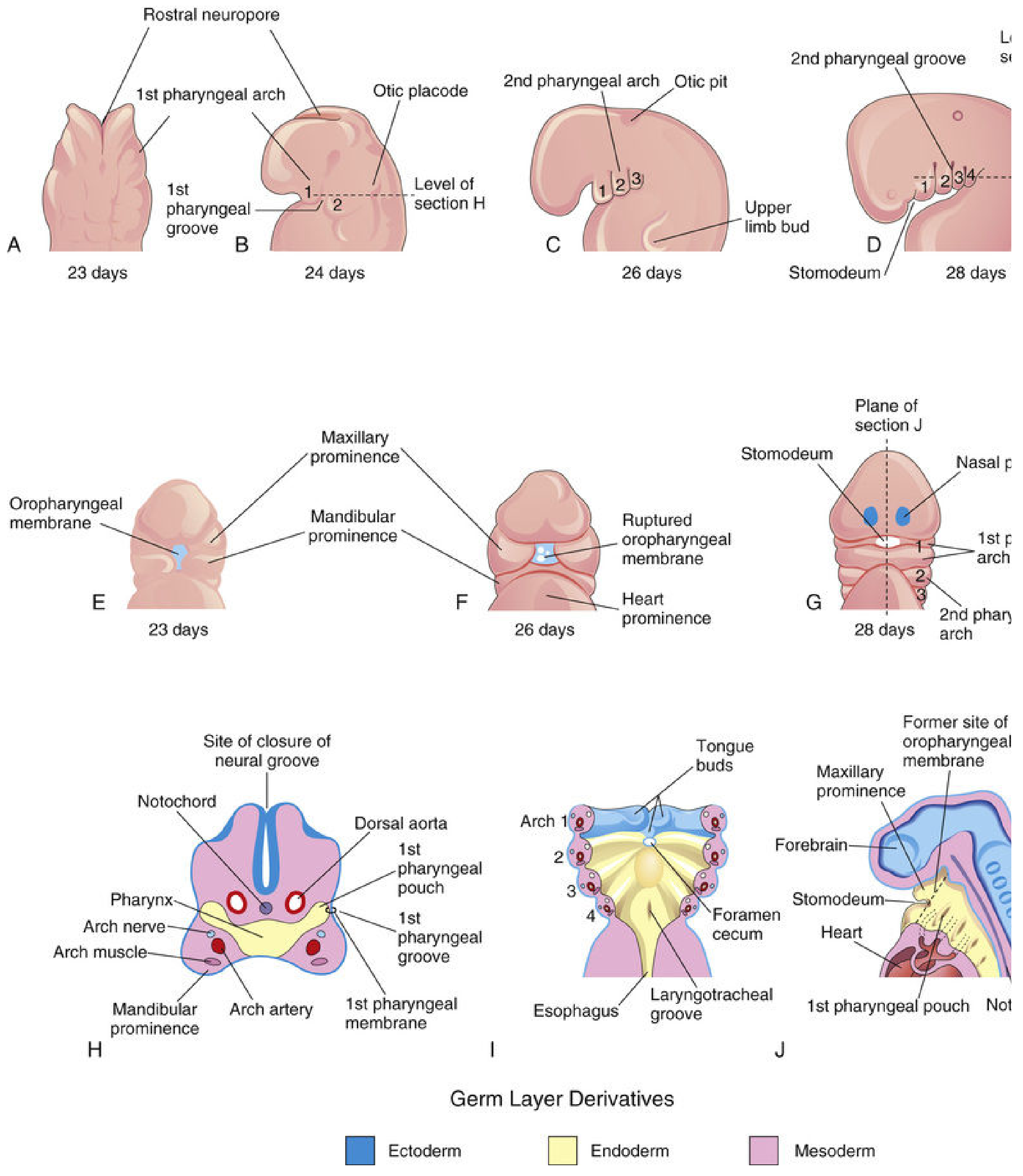
Fig. 9.1 - Development of the pharyngeal apparatus from 23 to 28 days (The Developing Human, Moore & Persaud)
1. Overview
The pharyngeal apparatus begins forming in the 4th week of embryonic development. Neural crest cells migrate into the future head and neck regions, and the structures that form are analogous to the gill apparatus (branchial apparatus) of fish - hence the older term "branchial apparatus."
The four main components are arranged around the primordial pharynx (derived from the cranial part of the foregut):
| Component | Location |
|---|---|
| Pharyngeal arches | External ridges, craniocaudal |
| Pharyngeal pouches | Internal, endodermal outpocketings |
| Pharyngeal grooves (clefts) | External, ectodermal inpocketings between arches |
| Pharyngeal membranes | Thin plates between each groove and pouch |
2. Pharyngeal Arches
By the end of the 4th week, 4 pairs of arches are visible externally. The 5th and 6th arches are rudimentary and not visible on the surface. Sonic hedgehog (Shh) and Dlx2 signaling govern their formation and patterning.
The first arch separates into two prominences:
- Maxillary prominence - forms the maxilla, zygomatic bone, part of the vomer
- Mandibular prominence - forms the mandible and squamous temporal bone
Composition of Each Arch
Each arch has a core of mesenchyme covered externally by ectoderm and internally by endoderm. The mesenchyme is mostly derived from neural crest cells (migrating in during week 4), with the original mesodermal core contributing muscle primordia.
Each typical arch contains:
- An artery - arising from the truncus arteriosus, passing around the primordial pharynx to the dorsal aorta
- A cartilaginous rod - forming the skeletal framework
- A muscular component - differentiating into head and neck muscles
- A nerve - sensory and motor, derived from neuroectoderm of the primordial brain
Derivatives of Each Arch
| Arch | Cartilage / Bone | Muscles | Nerve |
|---|---|---|---|
| 1st (Mandibular) | Meckel's cartilage → mandible, malleus, incus, sphenomandibular ligament | Muscles of mastication, mylohyoid, ant. digastric, tensor tympani, tensor veli palatini | CN V (Trigeminal) |
| 2nd (Hyoid) | Reichert's cartilage → stapes, styloid process, stylohyoid ligament, lesser horn & upper body of hyoid | Muscles of facial expression, stapedius, stylohyoid, post. digastric, platysma | CN VII (Facial) |
| 3rd | Greater horn & lower body of hyoid | Stylopharyngeus | CN IX (Glossopharyngeal) |
| 4th | Laryngeal cartilages (thyroid, etc.) | Pharyngeal constrictors, cricothyroid, levator veli palatini | CN X (Superior laryngeal br.) |
| 6th | Laryngeal cartilages (cricoid, arytenoids) | Intrinsic laryngeal muscles (except cricothyroid) | CN X (Recurrent laryngeal br.) |
3. Pharyngeal Pouches
There are 4 pairs of pharyngeal pouches - outpocketings of endoderm on the lateral walls of the primordial pharynx. Their derivatives are important glands and structures of the neck and ear.
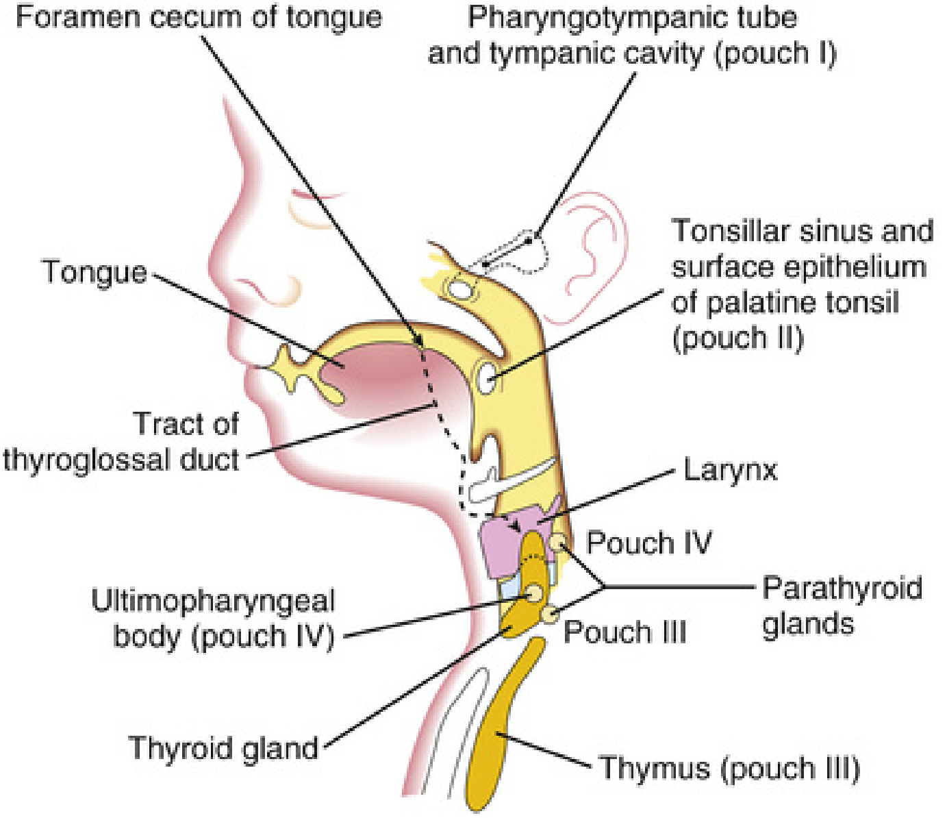
Fig. 9.8 - Adult derivatives of the pharyngeal pouches (The Developing Human)
Derivatives of Each Pouch
Pouch 1:
- Elongates into the tubotympanic recess, which gives rise to the tympanic cavity (middle ear) and the pharyngotympanic (Eustachian) tube
- The endoderm at its tip contacts the first pharyngeal groove ectoderm to form the tympanic membrane
Pouch 2:
- Largely obliterated as the palatine tonsil develops
- A remnant persists as the tonsillar sinus (fossa)
- At ~20 weeks, mesenchyme differentiates into lymphoid tissue forming the lymphatic nodules of the palatine tonsil
- Germinal centers form in the neonatal period
Pouch 3:
- Dorsal part → inferior parathyroid glands
- Ventral part → thymus (a primary lymphoid organ)
- The thymus is bilobed, with each lobe having its own blood supply and nerve supply
Pouch 4:
- Dorsal part → superior parathyroid glands
- Ventral part / ultimopharyngeal body → parafollicular (C) cells of the thyroid gland (calcitonin-secreting)
Key memory point: The inferior parathyroid glands originate from pouch 3, and the superior ones from pouch 4. Because pouch 3 derivatives travel further caudally with the thymus, the inferior glands end up lower in the neck than the superior glands.
4. Pharyngeal Grooves (Clefts)
These are ectodermal inpocketings between the arches on the outer surface of the embryo. There are 4 pairs.
- Only the 1st pharyngeal groove contributes to an adult structure - it gives rise to the external acoustic meatus (external ear canal)
- Grooves 2, 3, and 4 are normally obliterated when the 2nd arch (hyoid arch) overgrows them, fusing with the cardiac eminence and forming a smooth neck contour
Clinical Note - Branchial Cyst / Sinus
If the ectodermal remnants of grooves 2-4 are not completely obliterated, a branchial cyst or sinus can persist, typically appearing as a cyst or opening along the anterior border of the sternocleidomastoid muscle in the neck.
5. Pharyngeal Membranes
These are thin bilaminar plates where the ectoderm of a groove meets the endoderm of a pouch, with minimal mesenchyme in between.
- The 1st pharyngeal membrane contributes to the tympanic membrane (eardrum) - this is the only one to persist as an adult structure
- The others normally degenerate
6. The Stomodeum and Oropharyngeal Membrane
The stomodeum (primordial mouth) is a slight depression of the surface ectoderm. It is separated from the primordial pharynx by the oropharyngeal membrane (ectoderm externally + endoderm internally). This membrane ruptures at approximately 26 days, opening communication between the pharynx and the amniotic cavity. Failure to rupture leads to orofacial defects.
7. Thyroid Gland Development (Related)
The thyroid does not arise from a pouch but from a midline endodermal thickening at the foramen cecum of the tongue (between the tuberculum impar and the copula). It descends in the neck via the thyroglossal duct, passing ventral to the hyoid bone and laryngeal cartilages, and reaches its final position by 7 weeks. The thyroglossal duct normally degenerates; its proximal remnant persists as the foramen cecum on the dorsum of the tongue.
Summary Table
| Structure | Origin | Adult Derivative |
|---|---|---|
| Arch 1 | Neural crest mesenchyme | Jaw bones, malleus, incus, muscles of mastication |
| Arch 2 | Neural crest mesenchyme | Stapes, styloid, hyoid (lesser horn), facial muscles |
| Arch 3 | Neural crest mesenchyme | Hyoid (greater horn), stylopharyngeus |
| Arch 4 & 6 | Neural crest mesenchyme | Laryngeal cartilages, laryngeal muscles |
| Pouch 1 | Endoderm | Middle ear, Eustachian tube |
| Pouch 2 | Endoderm | Palatine tonsil, tonsillar sinus |
| Pouch 3 | Endoderm | Inferior parathyroid, thymus |
| Pouch 4 | Endoderm | Superior parathyroid, C cells (via ultimopharyngeal body) |
| Groove 1 | Ectoderm | External acoustic meatus |
| Membrane 1 | Ectoderm + Endoderm | Tympanic membrane |
- The Developing Human: Clinically Oriented Embryology (Moore, Persaud & Torchia), Chapter 9
This is a shared conversation. Sign in to Orris to start your own chat.