
Great saphenous vein histology
Reading File
This is a visual/histology question - image retrieval will be critical. Let me search the library now.
Finding Sources
Finding Sources
Finding Sources
Finding Sources
Finding Sources
Reading File
Excellent! Found the key histology source. Let me read the relevant sections from "Histology A Text and Atlas" which specifically mentions the great saphenous vein.
Reading File
I have the key text. Now let me read the specific great saphenous vein section and fetch the relevant images.
Reading File
Loading Image
Now let me fetch the specific great saphenous vein photomicrograph and the large vein image.
Loading Image
Reading File
I now have everything needed for a comprehensive answer - the specific GSV photomicrograph, the medium-sized vein diagram, and the detailed textbook descriptions. Let me compile the full answer.
Great Saphenous Vein - Histology
The great saphenous vein (GSV) is the longest vein in the body. It is classified as a muscular vein - a special subcategory distinct from typical medium or large veins - because it contains an unusually abundant amount of smooth muscle in all three tunics. This distinguishes it from most other veins of comparable caliber.
Photomicrograph of the Great Saphenous Vein (H&E, x380)
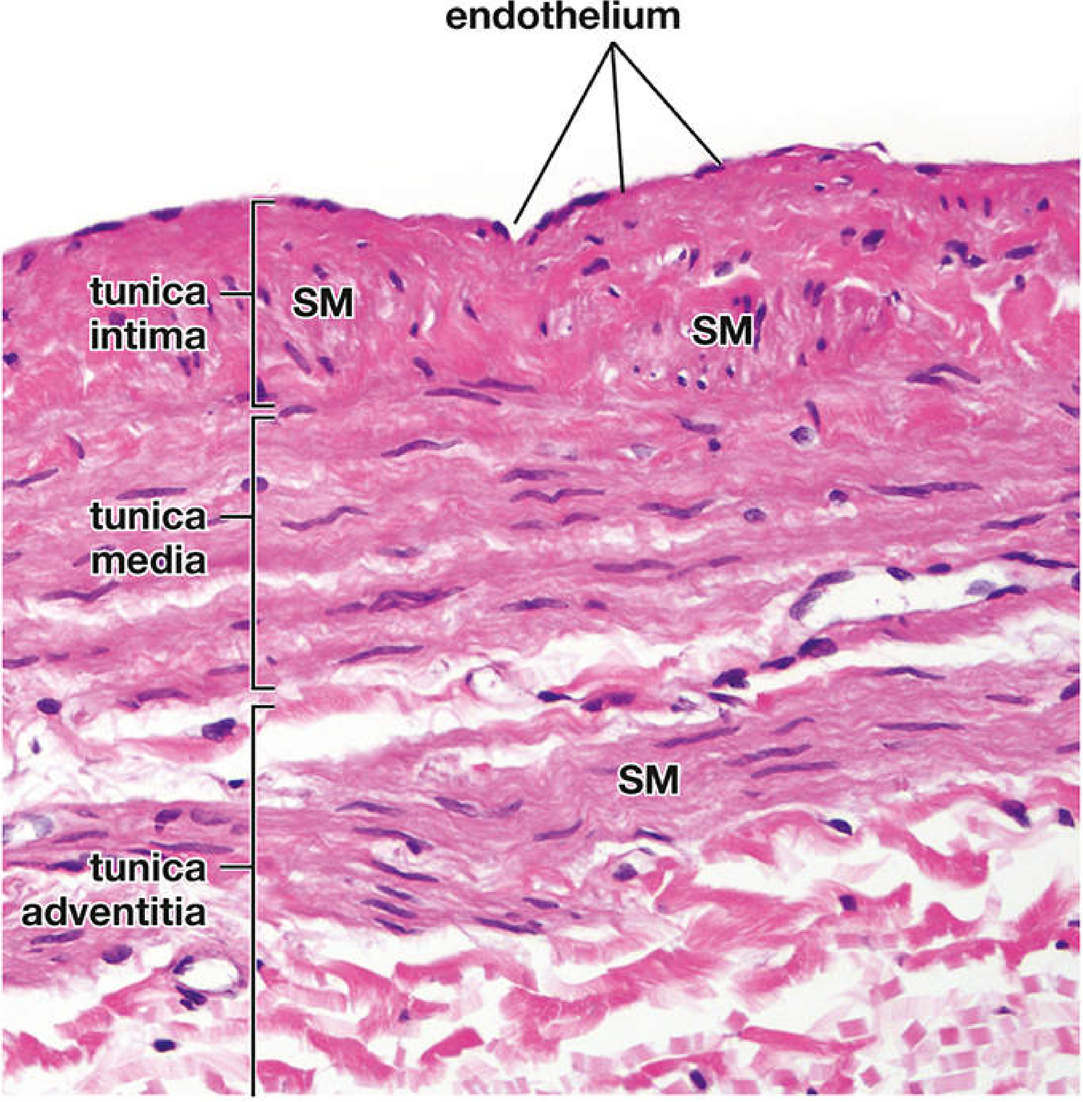
Figure 13.28 from Histology: A Text and Atlas (Ross & Pawlina). SM = longitudinal smooth muscle bundles. Courtesy of Dr. Joseph J. Maleszewski. x380.
Wall Structure - Layer by Layer
1. Tunica Intima
- Lined by endothelium with its basal lamina (innermost surface)
- Thicker than in typical medium-sized veins - the defining histological feature
- Contains numerous longitudinal smooth muscle bundles (SM) separated by connective tissue fibers - this is unique to the GSV
- A thin, poorly developed internal elastic membrane separates the intima from the media (often discontinuous or indistinct)
- Subendothelial connective tissue is present
2. Tunica Media
- Contains a relatively thick layer of circularly arranged smooth muscle cells - thicker than in most comparably-sized veins
- This circular smooth muscle layer is responsible for venomotor tone
- Interspersed collagen and elastic fibers are present between smooth muscle cells
3. Tunica Adventitia
- Well-developed and prominent
- Contains additional smooth muscle fibers arranged in spiral, oblique, and longitudinal bundles
- Collagen and elastic fiber networks are abundant
- Fibroblasts and vasa vasorum (small vessels supplying the wall) are present
Comparison: Typical Medium-Sized Vein vs. Great Saphenous Vein
| Feature | Typical Medium Vein | Great Saphenous Vein |
|---|---|---|
| Tunica intima | Thin, minimal SM | Thick; prominent longitudinal SM bundles |
| Internal elastic membrane | May be present (thin) | Thin, poorly developed, often absent |
| Tunica media | Few circular SM layers | Relatively thick circular SM |
| Tunica adventitia | Thickest layer; mostly collagen/elastic | Well-developed; additional longitudinal/spiral SM bundles |
| Valves | Present | Present (bicuspid valves along course) |
| Classification | Medium vein | Muscular vein |
Schematic: Medium-Sized Vein Wall (for reference)
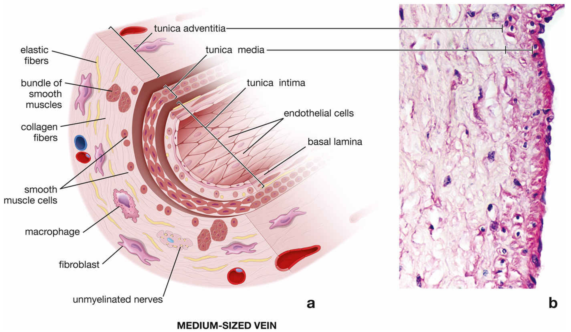
Figure 13.24 from Histology: A Text and Atlas. The GSV differs from this typical pattern by having abundant SM in the intima and adventitia.
Valves
The GSV contains bicuspid (two-leaflet) venous valves at intervals along its course. Each valve consists of:
- Two semilunar leaflets (cusps) formed by folds of the tunica intima
- A core of connective tissue covered on both surfaces by endothelium
- Most numerous in the lower limb, where they prevent retrograde blood flow against gravity
- The valve cusps face the lumen; backpressure forces them closed
Clinical Relevance
- CABG (Coronary Artery Bypass Graft) Surgery: The GSV is harvested as a conduit for bypass grafting. Its abundant smooth muscle makes it relatively durable. When arterial grafts (e.g., internal thoracic artery) are insufficient or multiple grafts are needed, the GSV is used for autotransplantation.
- Varicose veins: Incompetent valves combined with venous wall weakness lead to vein dilation, tortuosity, and valve failure - a common disorder of the GSV.
- DVT and superficial thrombophlebitis: The subcutaneous position and muscular wall characteristics influence the GSV's vulnerability to inflammation and thrombosis.
Source: Histology: A Text and Atlas with Correlated Cell and Molecular Biology (Ross & Pawlina), 9th ed., pp. 1122-1123 - Great Saphenous Vein section (Fig. 13.28).
This is a shared conversation. Sign in to Orris to start your own chat.