
Diagnostic findings for thyroid as well as step wise treatment approach
Reading File
The query is broad — "thyroid" covers hypothyroidism, hyperthyroidism, thyroid nodules, thyroid cancer, thyroiditis, etc. Let me clarify before diving in.
Asking for Preferences
Which thyroid condition are you asking about?
Finding Sources
Finding Sources
Finding Sources
Finding Sources
Good. Now let me read the key sections in parallel — Family Medicine textbook for hypothyroidism/hyperthyroidism/thyroiditis, Robbins for thyroiditis pathology, Tietz for thyroid cancer labs, and Sabiston for nodules/cancer surgery.
Reading File
Reading File
Reading File
Reading File
Good data coming in. Let me now read the detailed treatment sections from the Family Medicine textbook (hypothyroidism + thyroiditis sections), plus the Tietz lab section on thyroid cancer, and the Sabiston nodule/cancer section for diagnostic workup.
Reading File
Reading File
Reading File
I have a flowchart image URL. Let me fetch it along with getting the cancer treatment section and recent pubmed evidence — all in parallel.
Loading Image
Reading File
Searching PubMed
thyroid disorders diagnosis management guidelines
I have rich data from the textbooks. Let me now read the thyroid cancer treatment section from Sabiston plus get more detail on hypothyroidism treatment.I have sufficient data to deliver a comprehensive, well-structured answer. Let me now synthesize.
Thyroid Disorders: Diagnostic Findings & Stepwise Treatment
1. HYPOTHYROIDISM
Diagnostic Findings
| Test | Finding |
|---|---|
| Serum TSH | Elevated (primary hypothyroidism) or low/normal (central hypothyroidism) |
| Free T4 (FT4) | Low (overt); normal (subclinical) |
| Free T3 (FT3) | Low (late disease, preferential T3 expression) |
| Anti-TPO antibodies | Elevated in Hashimoto's thyroiditis |
| Anti-thyroglobulin antibodies | Elevated in autoimmune thyroiditis |
| Lipid panel | Hyperlipidemia (secondary dyslipidemia) |
| CBC | Normocytic or macrocytic anaemia |
Clinical features: Fatigue, cold intolerance, weight gain, constipation, dry skin/hair, bradycardia, delayed deep tendon reflexes, myxoedema in severe cases.
Subclinical hypothyroidism: TSH elevated, FT4 normal — treat if TSH >10 mIU/L, or if symptomatic/pregnant.
Stepwise Treatment of Hypothyroidism
Step 1 — Confirm diagnosis
- Serum TSH + FT4. If TSH elevated + FT4 low → overt hypothyroidism.
- Check anti-TPO to identify autoimmune (Hashimoto) etiology.
Step 2 — Initiate levothyroxine (LT4)
- Starting dose: 1.6 mcg/kg/day in healthy adults; lower (25–50 mcg/day) in elderly or cardiac patients.
- Administer on an empty stomach, 30–60 min before food.
- Pregnancy: start promptly and target TSH 0.1–2.5 mIU/L.
Step 3 — Monitor and titrate
- Recheck TSH at 6–8 weeks after initiation or dose change.
- Target TSH: 0.5–2.5 mIU/L (standard); 0.1–1.5 mIU/L in pregnancy.
- Dose adjustments of 12.5–25 mcg increments.
Step 4 — Long-term management
- Annual TSH monitoring once stable.
- Dose often needs adjustment with pregnancy, weight change, or interacting drugs (calcium, iron, PPI — all reduce LT4 absorption).
Myxoedema coma (emergency):
- IV LT4 200–400 mcg loading dose, then 50–100 mcg/day IV.
- IV hydrocortisone (rule out adrenal insufficiency before giving T4).
- Supportive care (ventilation, warming, fluids).
Source: Textbook of Family Medicine 9e, Tietz Textbook of Laboratory Medicine 7th Ed
2. HYPERTHYROIDISM / GRAVES DISEASE
Diagnostic Findings
| Test | Finding |
|---|---|
| Serum TSH | Suppressed (<0.1 mIU/L; in Graves' often <0.01 mIU/L, may be unmeasurable) |
| Free T4 (FT4) | Elevated |
| Free T3 (FT3) | Elevated (T3 toxicosis: FT3 high with normal/low FT4) |
| TSH-receptor stimulating antibodies (TRAb/TSH-RS Abs) | Positive in Graves disease (highly specific) |
| Radioiodine uptake (RAIU) | High uptake = Graves/toxic nodule; Low uptake = thyroiditis/factitious |
| Thyroid scintigraphy (¹²³I) | Diffuse uptake in Graves; focal "hot" nodule in toxic adenoma |
| Thyroid ultrasound | Diffuse enlargement + hypervascularity in Graves; nodule in toxic adenoma |
| Anti-TPO antibodies | Often elevated in Graves |
Clinical features (hypermetabolic state): Tachycardia, wide pulse pressure, systolic hypertension, fever, tremor, warm moist skin, anxiety, diarrhoea, weight loss. In Graves: goiter, exophthalmos, pretibial myxoedema (triad).
Stepwise Treatment of Hyperthyroidism
Step 1 — Symptomatic control
- Beta-blocker (propranolol 20–40 mg TID or atenolol 50–100 mg/day) — immediate control of palpitations, tremor, anxiety, tachycardia.
- This is a temporizing agent while etiology is confirmed.
Step 2 — Establish etiology
- TSH + FT4/FT3 + TRAb + RAIU scan.
- Distinguish Graves, toxic nodule, thyroiditis, factitious.
Step 3 — Antithyroid drugs (ATD)
- Methimazole (MMI) — preferred in most patients; 10–40 mg/day initially.
- Propylthiouracil (PTU) — preferred in 1st trimester pregnancy and thyroid storm; 100–200 mg TID. (Risk of hepatotoxicity limits long-term use.)
- Monitor: CBC + LFTs (agranulocytosis risk — educate patient to report sore throat/fever).
- Continue 12–18 months; check remission with TRAb level.
Step 4 — Definitive therapy (choose one):
| Option | Indication | Notes |
|---|---|---|
| Radioiodine (¹³¹I) | Graves, toxic nodule/MNG in non-pregnant adults | Most common choice; may worsen Graves ophthalmopathy; leads to permanent hypothyroidism |
| Total thyroidectomy | Large goiter, compressive symptoms, active ophthalmopathy, pregnancy, malignancy concern | Requires pre-op euthyroid state with ATDs; risk of hypoparathyroidism & RLN injury |
| Long-term ATD | Patient preference, mild disease, elderly/high surgical risk | Indefinite use acceptable; remission occurs in ~30–40% |
Step 5 — Post-definitive management
- After ¹³¹I or thyroidectomy: permanent LT4 replacement.
- Monitor TSH every 6–12 weeks until stable, then annually.
Thyroid storm (emergency):
- PTU 500–1000 mg loading, then 250 mg q4h
- Lugol's iodine (1 hour after ATD)
- Hydrocortisone IV + beta-blockers
- Antipyretics (avoid aspirin — displaces T4 from binding proteins)
Source: Textbook of Family Medicine 9e, Robbins & Kumar Basic Pathology
3. THYROIDITIS
Types & Distinguishing Features
| Type | Cause | Pain | TSH | FT4 | Antibodies | RAIU |
|---|---|---|---|---|---|---|
| Hashimoto's (chronic lymphocytic) | Autoimmune (CD8+ T cells, anti-TPO, anti-TG) | No | Elevated (hypothyroid) | Low | Anti-TPO, anti-TG strongly + | Normal/low |
| Subacute granulomatous (de Quervain) | Viral (post-URI) | Yes | Low initially | High | Negative | Very low (pathognomonic) |
| Painless/postpartum | Autoimmune | No | Low then high | High then low | Anti-TPO positive | Low |
| Acute suppurative | Bacterial infection | Yes | Usually normal | Normal | Negative | Variable |
Key diagnostic pearl: In thyroiditis-related thyrotoxicosis, the T4:T3 ratio is elevated (high T4, relatively low T3), unlike Graves where both are high — reflecting stored hormone release rather than new synthesis.
Stepwise Treatment of Thyroiditis
Hashimoto's Thyroiditis:
- If euthyroid — no treatment; annual TSH monitoring.
- If hypothyroid — LT4 replacement (as above).
- If subclinical hypothyroidism (TSH 4.5–10) — treat if symptomatic, pregnant, or planning pregnancy.
Subacute (de Quervain) Thyroiditis:
- Thyrotoxic phase — NSAIDs (ibuprofen 400–600 mg TID) for pain; beta-blockers for symptoms. No ATD (not de novo synthesis).
- Moderate-severe pain → prednisone 40 mg/day, taper over 6–8 weeks.
- Hypothyroid phase — temporary LT4 if symptomatic; usually self-limiting.
- Recovery phase — stop LT4 gradually; confirm euthyroidism.
Postpartum Thyroiditis:
- Thyrotoxic phase — beta-blocker if symptomatic; resolves spontaneously.
- Hypothyroid phase — LT4 if symptomatic or planning conception.
- Recheck TSH at 12 months postpartum; 20–30% develop permanent hypothyroidism.
Source: Robbins, Cotran & Kumar Pathologic Basis of Disease; Tietz Textbook of Laboratory Medicine 7th Ed; Textbook of Family Medicine 9e
4. THYROID NODULES
Diagnostic Algorithm
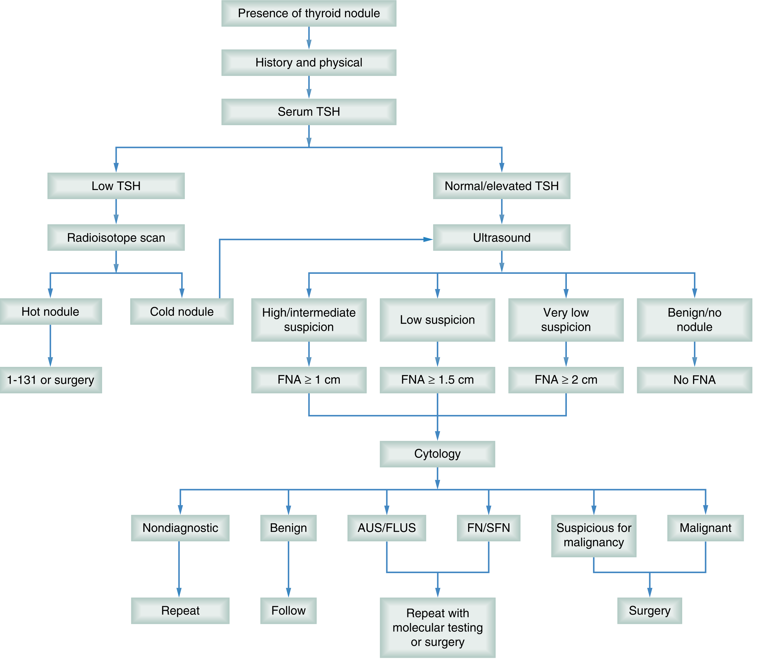
High-risk clinical features suggesting malignancy:
- Age <20 or >70 years, male sex
- Rapid growth, hoarseness, dysphagia
- Firm, fixed, immobile nodule >3–4 cm
- Cervical lymphadenopathy
- History of head/neck irradiation
- Family history of thyroid cancer or MEN2
Sonographic Risk Stratification (ATA 2015)
| Ultrasound Pattern | Malignancy Risk | FNA Threshold |
|---|---|---|
| High suspicion (solid hypoechoic + microcalcifications + irregular margins + taller-than-wide) | >70–90% | ≥1 cm |
| Intermediate suspicion (hypoechoic, solid) | 10–20% | ≥1 cm |
| Low suspicion (isoechoic/hyperechoic solid) | 5–10% | ≥1.5 cm |
| Very low suspicion (spongiform, partially cystic) | <3% | ≥2 cm |
| Benign pattern (purely cystic) | <1% | No FNA |
Bethesda Classification (FNA Cytology)
| Category | Malignancy Risk | Management |
|---|---|---|
| I — Nondiagnostic | N/A | Repeat FNA |
| II — Benign | 0–3% | Follow-up ultrasound |
| III — AUS/FLUS | 6–18% | Repeat FNA or molecular testing |
| IV — Follicular neoplasm | 10–40% | Molecular testing or surgery (hemithyroidectomy) |
| V — Suspicious for malignancy | 45–75% | Surgery |
| VI — Malignant | 97–99% | Surgery |
Source: Sabiston Textbook of Surgery
5. THYROID CANCER
Types & Key Diagnostic Markers
| Cancer Type | Frequency | Key Features | Markers |
|---|---|---|---|
| Papillary thyroid carcinoma (PTC) | ~85–90% | Nuclear features: ground-glass (Orphan Annie) nuclei, pseudoinclusions; psammoma bodies; lymphatic spread | Thyroglobulin (Tg); BRAF/RET mutations |
| Follicular thyroid carcinoma (FTC) | ~5–10% | Capsular ± vascular invasion required for diagnosis; hematogenous spread | Thyroglobulin (Tg); PAX8/PPARG fusion |
| Medullary thyroid carcinoma (MTC) | ~3–5% | Arises from parafollicular C cells; amyloid deposits; sporadic (70%) or familial/MEN2 (30%) | Serum calcitonin (diagnostic + surveillance); CEA; RET mutation |
| Anaplastic thyroid carcinoma (ATC) | <2% | Highly aggressive, uniformly lethal; arises from de-differentiation of PTC/FTC via TP53 loss | Rapid growing mass; FNA shows pleomorphic giant cells |
Stepwise Treatment of Differentiated Thyroid Cancer (PTC/FTC)
Step 1 — Surgery
- Hemithyroidectomy (lobectomy): Low-risk, unifocal PTC <4 cm, no extrathyroidal extension.
- Total thyroidectomy: Tumors >4 cm, bilateral disease, extrathyroidal extension, nodal/distant metastases, or high-risk features.
- Lymph node dissection: Central neck (Level VI) for PTC; lateral neck dissection if lateral nodes involved.
Step 2 — Radioactive iodine (RAI / ¹³¹I) ablation
- For intermediate/high-risk patients after total thyroidectomy.
- Purpose: Ablate remnant thyroid tissue, facilitate Tg surveillance, treat occult metastases.
- Preparation: Hypothyroid withdrawal or recombinant TSH stimulation (rhTSH/Thyrogen).
Step 3 — TSH suppression therapy
- LT4 at doses to suppress TSH:
- High-risk: TSH <0.1 mIU/L
- Low-risk: TSH 0.5–2 mIU/L (avoid over-suppression)
Step 4 — Surveillance
- Serum thyroglobulin (Tg) ± anti-Tg antibodies every 6–12 months.
- Neck ultrasound at 6–12 months post-surgery, then annually.
- Whole-body scan if Tg rising or structural recurrence suspected.
Step 5 — Recurrent/metastatic disease
- Repeated RAI if still iodine-avid.
- Kinase inhibitors (sorafenib, lenvatinib) for RAI-refractory differentiated thyroid cancer.
Medullary Thyroid Cancer:
- Total thyroidectomy + central neck dissection.
- Screen for RET mutation (germline) and MEN2-associated tumors (pheochromocytoma, hyperparathyroidism).
- Surveillance: serum calcitonin + CEA.
- Targeted therapy: Vandetanib or cabozantinib for advanced/metastatic MTC.
Anaplastic Thyroid Cancer:
- Multidisciplinary urgent approach.
- Surgery (if resectable) + external beam radiation + chemotherapy.
- BRAF V600E mutation present in ~40%: dabrafenib + trametinib combination approved.
- Prognosis: median survival <6 months.
Source: Sabiston Textbook of Surgery; Robbins, Cotran & Kumar Pathologic Basis of Disease; Tietz Textbook of Laboratory Medicine 7th Ed
Recent Guidelines (2023–2025)
| Guideline | PMID |
|---|---|
| 2023 European Thyroid Association Clinical Practice Guidelines for thyroid nodule management | PMID 37358008 |
| 2025 American Thyroid Association Management Guidelines for Adult Patients with Differentiated Thyroid Cancer | PMID 40844370 |
| NCCN Guidelines: Thyroid Carcinoma, Version 1.2025 | PMID 40639400 |
| Diagnostic tests for medullary thyroid carcinoma: umbrella review | PMID 36877452 |
Summary: Core Lab Panel by Condition
| Condition | TSH | FT4 | FT3 | Anti-TPO | TRAb | Tg | Calcitonin |
|---|---|---|---|---|---|---|---|
| Primary hypothyroidism | ↑ | ↓ | ↓ (late) | ± | — | — | — |
| Hashimoto's | ↑ | ↓ | ↓ | ↑↑ | — | — | — |
| Graves disease | ↓↓ | ↑ | ↑ | + | ↑↑ | — | — |
| Subacute thyroiditis | ↓ | ↑ | Normal | — | — | ↑ (early) | — |
| PTC/FTC (post-op) | Suppressed | — | — | — | — | Surveillance | — |
| MTC | Normal | Normal | Normal | — | — | — | ↑↑ |
- All thyroid evaluation starts with serum TSH — it is the single most sensitive screening test.
- Hypothyroidism is treated with LT4 titrated to TSH; Hashimoto's is the most common cause.
- Hyperthyroidism requires identifying the etiology (Graves vs. toxic nodule vs. thyroiditis) before choosing definitive therapy (RAI, surgery, or long-term ATD).
- Thyroiditis treatment depends on the phase — thyrotoxic phases are managed symptomatically (no ATDs), hypothyroid phases with LT4.
- Thyroid nodules follow a TSH → ultrasound → FNA → Bethesda classification pathway; molecular testing is increasingly used for indeterminate cytology.
- Thyroid cancer treatment is risk-stratified: surgery is the cornerstone; RAI, TSH suppression, and kinase inhibitors follow based on histology and risk.
This is a shared conversation. Sign in to Orris to start your own chat.