A patient is in ICU and his chest is full of rhonchi and the mucus is overloaded in the alveoli. The patient's saturation is about 74. We just gave a patient mucomelt tablet with nebulization and hydrocort. The patient having history of hypertension and the patient oxygen saturation is near about 80%. What was the best treatment to clear the alveoli mucus secretion in ICU management with the suitable drugs? Explain very precisely with diagrams and the work of mechanism that they are working on ICU management. And the other drugs that are used for a COPD patient in ICU management, also suggest me that.

Reading File
Finding Sources
Finding Sources
Finding Sources
Finding Sources
Finding Sources
Searching Images

mucolytic mechanism of action airway secretion COPD bronchospasm

Searching Images

N-acetylcysteine NAC mechanism mucolytic disulfide bond mucus

Searching Images

bronchodilator salbutamol ipratropium mechanism of action airway smooth muscle

Searching Images

corticosteroid hydrocortisone anti-inflammatory mechanism COPD lungs

Loading Image
Loading Image
Searching Images

COPD acute exacerbation ICU ventilation oxygen therapy management algorithm

Loading Image
Loading Image
Now I have comprehensive information to compose a detailed, well-illustrated answer. Let me build the complete response.

ICU Management of Mucus Overload with Respiratory Failure in a COPD Patient

Clinical Snapshot

  • SpO₂: 74–80% → Severe hypoxemia (normal ≥ 95%)
  • Findings: Bilateral rhonchi, alveolar mucus impaction
  • Comorbidity: Hypertension
  • Already given: Mucomelt (acetylcysteine) nebulisation + IV hydrocortisone
This is a severe acute exacerbation of COPD (AECOPD) with hypoxemic respiratory failure. The priority is: secure oxygenation → clear airways → relieve bronchospasm → treat underlying trigger.

STEP 1 — Airway Clearance: The Immediate Priority

🔬 Mechanism of Mucus Impaction in COPD

In COPD, excess mucus accumulates because:
  1. Goblet cell hyperplasia → overproduction of viscid mucus
  2. Disrupted mucociliary escalator → impaired clearance
  3. Inflammatory exudate floods alveoli → consolidation + rhonchi
  4. Disulfide bonds crosslink mucin glycoproteins → gel-like consistency

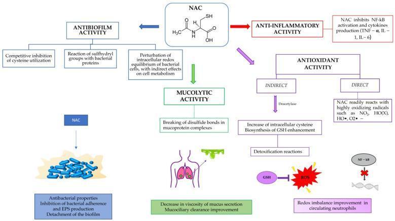
DRUG 1: N-Acetylcysteine (NAC / Mucomelt) — ✅ Already Given

NAC Mechanism of Action
Mechanism:
ActionHow it works
MucolyticFree sulfhydryl (–SH) groups break disulfide bonds in mucin glycoprotein complexes → reduces viscosity → improves mucociliary clearance
Antioxidant (Direct)Directly scavenges free radicals: NO₂, HO•, O₂•⁻
Antioxidant (Indirect)Deacetylase → cysteine → glutathione (GSH) synthesis → protects alveolar epithelium
Anti-inflammatoryInhibits NF-κB → suppresses TNF-α, IL-1, IL-6
AntibiofilmDisrupts bacterial biofilms; inhibits EPS production
ICU Dosing:
  • Nebulisation: 300 mg in 3 mL NS via jet nebuliser every 6–8 hours
  • IV form (for paracetamol toxicity): 150 mg/kg loading dose
  • Oral: 600 mg twice daily (maintenance)
Outcome: Decreases mucus viscosity, restores mucociliary escalator function, and clears alveolar impaction.

DRUG 2: Hydrocortisone (IV Corticosteroid) — ✅ Already Given

Mechanism:
Hydrocortisone binds to intracellular glucocorticoid receptors (GR-α) → GR-α/ligand complex translocates to nucleus → binds glucocorticoid response elements (GRE) on DNA:
Hydrocortisone → GR-α complex → Nucleus
       ↓
  Trans-activation:               Trans-repression:
  Annexin-1 (anti-inflammatory)    NF-κB ↓ → IL-1, IL-6, IL-8, TNF-α ↓
  MAPK phosphatase-1               AP-1 ↓ → MMP-9 ↓ (less tissue damage)
  β₂-adrenoceptor upregulation     Mucin gene MUC5AC expression ↓
Clinical effects in AECOPD:
  • Reduces airway oedema → less obstruction
  • Decreases eosinophilic inflammation → faster FEV₁ recovery
  • Reduces treatment failure rate and length of hospital stay
  • Upregulates β₂-receptor sensitivity (synergy with bronchodilators)
ICU Dosing:
  • IV Hydrocortisone: 100–200 mg every 6–8 hours (or methylprednisolone 1–2 mg/kg/day for 5 days)
  • Do NOT use >7–14 days → myopathy, hyperglycaemia, hypertension risk
Important for your patient: Since he has hypertension, monitor BP carefully — mineralocorticoid effects of hydrocortisone can cause sodium retention and BP rise. Consider switching to methylprednisolone (no mineralocorticoid effect) after stabilisation.

STEP 2 — Oxygen Therapy (SpO₂ 74–80% → Life-threatening)

Target: SpO₂ 88–92% in COPD (NOT 100% — hypercapnic drive risk)
DeviceFiO₂Use
Venturi mask (24–28%)0.24–0.28First-line controlled O₂ in COPD
Non-rebreather maskUp to 0.85If SpO₂ < 80% — temporary
High-Flow Nasal Cannula (HFNC)Up to 1.0Preferred in hypoxemic respiratory failure
NIV (BiPAP)TitratedBest choice in AECOPD with hypercapnia
In this patient with SpO₂ 74–80%: Start with NIV (BiPAP) — IPAP 12–16 cmH₂O, EPAP 4–6 cmH₂O. NIV reduces work of breathing, recruits atelectatic alveoli, and improves mucus clearance through positive pressure.

STEP 3 — Bronchodilators (Critical — Must Add)

DRUG 3: Salbutamol (Albuterol) — Short-Acting β₂ Agonist (SABA)

Mechanism:
Salbutamol → β₂ receptor (Gs protein)
         ↓
    Adenylyl cyclase activated
         ↓
    cAMP ↑ → Protein Kinase A (PKA) activated
         ↓
    Myosin Light Chain Kinase (MLCK) phosphorylated → INACTIVE
         ↓
    Airway smooth muscle RELAXATION → Bronchodilation
    (also: ↑ mucociliary beat frequency → mucus clearance)
Additional benefit: β₂ stimulation activates chloride channels → fluid secretion into airway lumen → lubricates mucus plug → eases expectoration.
ICU Dosing: 2.5 mg via nebuliser every 20 minutes × 3 doses, then every 1–4 hours

DRUG 4: Ipratropium Bromide — Short-Acting Muscarinic Antagonist (SAMA)

Mechanism:
Parasympathetic overactivity in COPD:
ACh → M₃ receptor on smooth muscle → bronchospasm + mucus secretion ↑

Ipratropium blocks M₃ receptors →
  • Bronchodilation (smooth muscle relaxation)
  • Reduced mucus secretion from submucosal glands
  • Reduced cholinergic bronchospasm
ICU Dosing: 0.5 mg nebulised every 4–6 hours (combine with salbutamol in same nebuliser = Combivent/Duolin)
Salbutamol + Ipratropium combination is superior to either agent alone in acute COPD exacerbation and is the standard of care in ICU.

STEP 4 — Antibiotics (Treat Infectious Trigger)

Most AECOPD requiring ICU admission are triggered by bacterial infection (Haemophilus influenzae, Streptococcus pneumoniae, Moraxella catarrhalis, Gram-negatives in severe cases).

DRUG 5: Antibiotics

SeverityDrugDose
Moderate AECOPDAmoxicillin-clavulanate625 mg PO/IV TDS
Severe/ICUCo-amoxiclav + Azithromycin ORIV
Pseudomonas riskPiperacillin-tazobactam + CiprofloxacinIV
Penicillin allergyLevofloxacin 500 mg OD IV5–7 days

STEP 5 — Additional ICU Drugs for COPD

COPD Exacerbation Management Framework

DRUG 6: Aminophylline (IV Methylxanthine) — Second-line

Mechanism:
  • Inhibits phosphodiesterase → cAMP ↑ → bronchodilation
  • Adenosine receptor antagonism → reduced bronchoconstriction
  • Improves diaphragmatic contractility (important in fatigued respiratory muscles)
  • CNS respiratory centre stimulation → helps in hypercapnia
ICU Dosing: Loading dose 5 mg/kg IV over 30 min → Maintenance 0.5 mg/kg/hr infusion Caution: Narrow therapeutic window (10–20 µg/mL). Monitor levels. Arrhythmia risk.

DRUG 7: Magnesium Sulfate (IV)

Mechanism:
  • Natural calcium antagonist → smooth muscle relaxation → bronchodilation
  • Inhibits mast cell degranulation → reduces histamine release
  • Inhibits ACh release at neuromuscular junction → relaxes bronchial smooth muscle
ICU Dosing: 2 g IV over 20 minutes (single dose) Use in: Severe bronchospasm not responding to β₂ agonists

DRUG 8: Carbocysteine / Erdosteine (Mucoregulators)

Mechanism:
  • Carbocysteine: Stimulates serous cell secretion, thins mucus, restores the IgA content of bronchial secretions
  • Erdosteine: Pro-drug → active metabolites with free thiol groups → breaks disulfide bonds (similar to NAC) + antioxidant + inhibits bacterial adhesion
Dosing: Carbocysteine 750 mg TDS orally; Erdosteine 300 mg BD

DRUG 9: Furosemide (If Fluid Overload Component)

In your patient with hypertension, there may be a component of pulmonary oedema compounding the mucus overload.
Mechanism: Loop diuretic → inhibits Na⁺/K⁺/2Cl⁻ co-transporter in Loop of Henle → diuresis → reduces pulmonary venous congestion → clears alveolar fluid
ICU Dosing: 40–80 mg IV stat, then reassess

DRUG 10: Doxapram (Respiratory Stimulant) — If NIV Fails

Mechanism: Stimulates peripheral chemoreceptors (carotid body) → increases respiratory drive → increases tidal volume and respiratory rate
Use: When NIV is unavailable or not tolerated, as bridge to prevent intubation Dose: 1.5–4 mg/kg/hr IV infusion

INTERVENTIONAL / NON-PHARMACOLOGICAL ICU MANAGEMENT

InterventionMechanism/Purpose
Chest physiotherapy (percussion + postural drainage)Mechanically mobilises mucus from peripheral airways to central airways for suction/expectoration
Endotracheal suctioningDirect removal of mucus via ETT if intubated
Bronchoscopy with BALIn refractory mucus plugging — direct washout of alveolar secretions
Mucolytic nebulisationHypertonic saline (3–7%) increases osmotic gradient → draws fluid into mucus → reduces viscosity
HydrationIV fluids maintain mucociliary clearance; dehydration thickens mucus
PositioningSemi-recumbent 30–45° → reduces aspiration, improves diaphragmatic excursion

Complete ICU Drug Regimen Summary

PriorityDrugRouteMechanism
🔴 ImmediateControlled O₂ + NIV (BiPAP)MaskOxygenation + alveolar recruitment
🔴 ImmediateSalbutamol 2.5 mg + Ipratropium 0.5 mgNebuliserBronchodilation (β₂ + M₃ block)
🔴 ImmediateNAC (Mucomelt) 300 mgNebuliserMucolytic — breaks disulfide bonds
🟠 UrgentHydrocortisone/Methylprednisolone IVIVAnti-inflammatory, oedema reduction
🟠 UrgentAntibiotics (co-amoxiclav ± azithromycin)IVTreat bacterial trigger
🟡 AdjunctAminophyllineIV infusionBronchodilation + diaphragm support
🟡 AdjunctMagnesium sulfate 2 gIVBronchodilation (refractory spasm)
🟡 AdjunctCarbocysteine/ErdosteineOralMucoregulatory, maintenance
🟡 AdjunctFurosemide (if fluid overload)IVDecongest pulmonary circulation
⚪ If neededDoxapramIV infusionRespiratory stimulant (NIV failure)

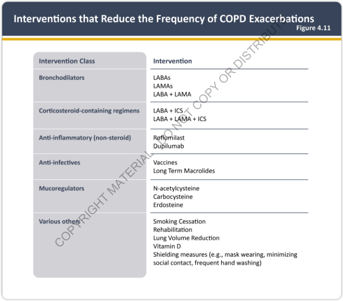
COPD Exacerbation-Reducing Interventions (Evidence-Based)

Interventions that Reduce COPD Exacerbation Frequency — GOLD 2025
As shown above from the GOLD 2025 Report, long-term prevention of exacerbations includes:
  • LABAs (salmeterol, formoterol) + LAMAs (tiotropium, glycopyrronium)
  • ICS + LABA combinations (budesonide/formoterol)
  • Triple therapy (LABA + LAMA + ICS) for high-risk patients
  • Roflumilast (PDE4 inhibitor) — for chronic bronchitis phenotype
  • Mucoregulators: NAC, carbocysteine, erdosteine (reduce exacerbation frequency)
  • Macrolides (azithromycin 250 mg OD) — long-term anti-inflammatory

Hypertension Consideration

Since the patient has hypertension:
  • Avoid high-dose salbutamol without monitoring → tachycardia, BP fluctuation
  • Prefer methylprednisolone over hydrocortisone (no mineralocorticoid effect → less sodium retention)
  • Magnesium sulfate is actually beneficial — mild antihypertensive effect
  • Avoid aminophylline toxicity → arrhythmias worsen hypertensive cardiac risk

Summary: The cornerstone of ICU management in this patient is the combination of NIV for oxygenation + salbutamol/ipratropium for bronchospasm + NAC nebulisation for mucolysis + systemic corticosteroids for inflammation + antibiotics for infection. Chest physiotherapy and bronchial hygiene should run in parallel. Mucomelt (NAC) was correctly initiated and works by cleaving the disulfide crosslinks in mucin — this reduces gel viscosity and restores mucociliary clearance, directly targeting the alveolar mucus overload described.

A 69-year-old patient come in OPD with WBC 9.6 and RBC was 3.10 and hemoglobin was 8.1 gram per deciliter and the platelets was 1.5 lakhs. And the patient come in OPD with complaint of severe hypogastric pain with epigastric region per abdomen pain. When the RBS was done, it was 138 mg per deciliter. When the liver profile was done, serum bilirubin and the total serum bilirubin was 8.86, direct was 7.02 and the indirect was 1.84. Serum protein of albumin, globulin or albumin globulin ratio are normal, but the SGOT, SGPT was 397 SGOT and 286 SGPT. And the serum alkaline phosphate was 205. The kidney shows hyponatremia and hypokalemia and the signs of serum sodium was 134 and the serum potassium was 3.25. And the patient having fever and the patient's eyes was yellow. What was the best treatment and the best test to perform for the patient to deep down into the disease and what was the best treatment for the patient to have a treatment with diagrams and the reason for the treatment and suggest with the best treatment for the patient with diagrams and the treatment.

Reading File
Finding Sources
Finding Sources
Searching Images

obstructive jaundice differential diagnosis liver bile duct cancer pancreas

Finding Sources
Finding Sources
Searching Images

ERCP bile duct stone choledocholithiasis endoscopy treatment

Reading File
Searching Images

jaundice classification types hepatocellular obstructive pre-hepatic bilirubin pathway diagram

Searching Images

cholangitis ascending treatment antibiotics ERCP biliary drainage

Finding Sources
Loading Image
Loading Image
Loading Image
Loading Image
Searching Images

bilirubin metabolism liver bile pathway conjugation unconjugated direct indirect

Loading Image
Now I have everything needed. Let me construct the complete, comprehensive answer.

Clinical Analysis: 69-Year-Old with Jaundice, Abdominal Pain & Fever

Step 1 — Interpreting the Lab Data (Pattern Recognition)

ParameterPatient ValueNormalInterpretation
WBC9.6 × 10³/µL4–11High-normal → infection/inflammation
RBC3.10 × 10⁶/µL4.5–5.5 (M)Low → Anaemia
Haemoglobin8.1 g/dL13.5–17.5Moderate anaemia
Platelets1.5 lakhs (150,000)1.5–4 lakhsLow-normal → chronic disease
RBS138 mg/dL<140Borderline — monitor
Total Bilirubin8.86 mg/dL< 1.2Severely elevated
Direct Bilirubin7.02 mg/dL< 0.3Markedly elevated → conjugated
Indirect Bilirubin1.84 mg/dL< 1.0Mildly elevated
SGOT (AST)397 U/L< 40Very high → hepatocellular damage
SGPT (ALT)286 U/L< 40Very high → hepatocellular damage
Alkaline Phosphatase205 U/L44–147Elevated → biliary/cholestatic
Serum Na⁺134 mEq/L135–145Hyponatraemia
Serum K⁺3.25 mEq/L3.5–5.0Hypokalaemia
Albumin/Globulin ratioNormalNormalChronic liver disease not yet causing synthetic failure

Step 2 — Diagnosis: What is This Pattern?

🔑 The Key Triad Present:

Fever + Jaundice + Abdominal Pain (Epigastric + Hypogastric) = Charcot's Triad → Ascending Cholangitis until proven otherwise
This is a mixed hepatocellular + cholestatic (obstructive) jaundice pattern:
Direct Bili >>> Indirect Bili   →  Obstructive / Post-hepatic component
SGOT & SGPT very high           →  Hepatocellular damage (secondary to back-pressure)
ALP elevated                    →  Biliary obstruction / cholestasis
Fever + Pain                    →  Biliary sepsis / ascending cholangitis
Anaemia (Hb 8.1)                →  Chronic illness OR haemolysis from biliary disease
Hyponatraemia + Hypokalaemia    →  Systemic sepsis, poor oral intake, vomiting

Most Probable Diagnoses (in order):

RankDiagnosisWhy
1stCholedocholithiasis with Ascending CholangitisCharcot's triad + predominantly direct hyperbilirubinaemia + elevated ALP + age 69
2ndCarcinoma of the Head of PancreasAge, obstructive pattern, ALP ↑, direct bili ↑, weight loss (inferred)
3rdCholangiocarcinoma (bile duct cancer)Obstructive jaundice in elderly
4thAcute Cholecystitis with CBD involvementAbdominal pain + fever
5thAcute Viral Hepatitis (HAV, HEV, HBV)High SGOT/SGPT, but less explains direct bili dominance

Bilirubin Metabolism — Why Direct Bili is High

Bilirubin Hepatic Conjugation Pathway — Harrison's Internal Medicine
Normal pathway:
  1. Unconjugated bilirubin (UCB) bound to albumin → transported to liver sinusoids
  2. Taken up by hepatocytes via OATP1B1/OATP1B3 transporters
  3. Bound to GST (glutathione S-transferase) intracellularly
  4. UGT1A1 enzyme conjugates UCB → BMG (monoglucuronide) → BDG (diglucuronide)
  5. MRP2 excretes conjugated bilirubin into bile canaliculus → bile duct → duodenum
In this patient: A blockage anywhere in the bile duct system (stone, tumour, stricture) → conjugated (direct) bilirubin cannot exit → backs up into blood → direct hyperbilirubinaemia + jaundice + dark urine + pale stools.

Step 3 — Best Investigations to Perform

Tier 1: Immediate (Emergency)

InvestigationPurposeWhat to Expect
Abdominal Ultrasound (USG)First-line: CBD dilation, gallstones, liver echogenicityDilated CBD > 7mm, stones, ductal dilation
Blood cultures × 2Sepsis/ascending cholangitis workupGram-negative bacteraemia (E. coli, Klebsiella)
Urine R/E + C/SRule out UTI as pain source; urobilinogenElevated urobilinogen if hepatic
Coagulation profile (PT/INR)Obstructive jaundice impairs Vit K absorption (fat-soluble)PT prolonged if obstruction prolonged
Amylase / LipaseRule out gallstone pancreatitisElevated if pancreatitis present
Serum LDH + Reticulocyte countAssess haemolytic contribution to anaemiaElevated in haemolysis
Peripheral blood smearType of anaemiaNormocytic in chronic disease

Tier 2: Diagnostic Imaging

TestAccuracyIndication
MRCP (Magnetic Resonance Cholangiopancreatography)>95%Best non-invasive test — shows CBD, IHBD, pancreatic duct, stones, strictures
EUS (Endoscopic Ultrasound)>90%CBD stones missed on USG; periampullary tumours
CT Abdomen with contrast (triphasic)HighMass lesions — pancreatic cancer, cholangiocarcinoma, liver mets
ERCP (Endoscopic Retrograde Cholangiopancreatography)Gold standardDiagnostic AND therapeutic — used when obstruction confirmed

ERCP Fluoroscopy showing Bile Duct Stones with Filling Defects:

ERCP showing CBD stones (red arrows) with upstream biliary dilation
ERCP fluoroscopy: Radiolucent filling defects (red arrows) in the dilated CBD = choledocholithiasis with upstream biliary obstruction.

CT scan showing double duct sign (pancreatic cancer):

CT double duct sign — simultaneous dilation of CBD and pancreatic duct
CT abdomen: Double duct sign — simultaneous CBD and pancreatic duct dilation suggests carcinoma of the head of pancreas.

Tier 3: Tumour Markers & Serology

TestPurpose
CA 19-9Pancreatic/biliary cancer marker
CEAColorectal/GI malignancy
AFP (Alpha-fetoprotein)Hepatocellular carcinoma
Hepatitis B surface antigen (HBsAg)Viral hepatitis cause
Anti-HCVHepatitis C
Anti-HAV IgM / Anti-HEV IgMAcute viral hepatitis
ANA / ANCA / AMAAutoimmune hepatitis / PSC / PBC

Step 4 — Treatment Plan

🚨 Immediate Resuscitation

1. IV access × 2 (large bore)
2. IV fluid resuscitation — Normal saline 0.9% to correct hyponatraemia
3. IV potassium supplementation — correct hypokalaemia (KCl 20 mEq in 500 mL NS)
4. Nil by mouth (NPO)
5. Monitor vitals every 15–30 minutes
6. Check for Reynolds' Pentad: Charcot's triad + Shock + Confusion → ICU immediately

DRUG 1: IV Antibiotics — Treat Ascending Cholangitis / Biliary Sepsis

Mechanism of Biliary Infection: Most ascending cholangitis is caused by Gram-negative bacilli (E. coli, Klebsiella, Pseudomonas) and anaerobes (Bacteroides fragilis), which ascend from the duodenum through the Sphincter of Oddi into the bile duct when obstruction occurs.
Bile duct obstruction
      ↓
Bile stasis → bacterial overgrowth
      ↓
Gram-negatives multiply → endotoxin (LPS) release
      ↓
Systemic bacteraemia → SIRS → Sepsis → Fever + rigors
      ↓
If untreated: Reynolds' Pentad → Multi-organ failure → Death
Antibiotic Regimens:
SeverityFirst ChoiceAlternative
Mild–Moderate CholangitisPiperacillin-Tazobactam 4.5 g IV 8-hourlyCo-amoxiclav 1.2 g IV 8-hourly
Severe / SepsisMeropenem 1 g IV 8-hourly + Metronidazole 500 mg IV 8-hourlyImipenem or Cefoperazone-sulbactam
Penicillin allergyCiprofloxacin 400 mg IV 12-hourly + Metronidazole
Duration: Until biliary drainage achieved + 5–7 days post-procedure

DRUG 2: Ursodeoxycholic Acid (UDCA) — Choleretic / Hepatoprotective

Mechanism:
  • Replaces toxic hydrophobic bile acids with hydrophilic UDCA → less hepatocyte membrane damage
  • Stimulates bile secretion (choleresis) → flushes bile ducts
  • Has anti-apoptotic effect on hepatocytes (inhibits cytochrome C release)
  • Immunomodulatory — reduces aberrant MHC class I expression on bile duct epithelium
Dose: 10–15 mg/kg/day in 2–3 divided doses orally Use: Bridge therapy during workup; long-term in primary biliary cholangitis (PBC)

DRUG 3: Vitamin K (IV/IM) — Correct Coagulopathy

Mechanism: Obstructive jaundice → bile cannot reach intestine → fat malabsorption → Vitamin K deficiency → factors II, VII, IX, X not carboxylated → prolonged PT/INR → bleeding risk
  • Phytomenadione (Vitamin K1): 10 mg IV/IM once daily × 3 days
  • Monitor INR; if not correcting → fresh frozen plasma (FFP) before any procedure

DRUG 4: Analgesics — Manage Biliary Colic

DrugMechanismDose
Diclofenac 75 mg IMCOX inhibitor → prostaglandin inhibition → pain relief; also reduces Sphincter of Oddi spasmFirst-line for biliary colic
Hyoscine butylbromide 20 mg IVAnticholinergic → smooth muscle relaxation → relieves biliary spasmAdd for spasm
Tramadol 50–100 mg IV slowµ-opioid agonist + SNRI → pain reliefAvoid morphine (increases Sphincter of Oddi tone)
⚠️ Do NOT use morphine in biliary colic — it contracts the Sphincter of Oddi and worsens pain/obstruction.

DRUG 5: Proton Pump Inhibitor (PPI) — Epigastric Protection

With elevated liver enzymes, fever, and possible biliary/pancreatic involvement, PPI is protective.
  • Pantoprazole 40 mg IV once daily or
  • Omeprazole 20–40 mg IV once daily

DRUG 6: Antiemetics + Electrolyte Correction

  • Ondansetron 4–8 mg IV PRN for nausea/vomiting
  • IV Potassium — KCl in NS drip (never IV push → arrhythmia)
  • IV Sodium correction — target rise < 10–12 mEq/day to avoid osmotic demyelination syndrome

Step 5 — The Definitive Treatment: ERCP with Biliary Drainage

Per Harrison's (22nd ed): "If the suspicion for a bile duct stone is high and urgent treatment is required (as in a patient with obstructive jaundice and biliary sepsis), ERCP is the procedure of choice since it remains the gold standard for diagnosis and allows for immediate treatment."

ERCP Procedure Steps:

Ascending Cholangitis — ERCP showing pus draining from papilla (A) and biliary stent placed (B)
Left (A): Major duodenal papilla discharging purulent bile = ascending cholangitis. Right (B): Post-ERCP with biliary stent in situ providing drainage.

What ERCP Does:

Duodenoscope → passed through mouth → stomach → duodenum
      ↓
Cannulation of Ampulla of Vater (sphincterotome)
      ↓
Endoscopic Sphincterotomy (cut the sphincter to widen opening)
      ↓
Balloon sweep / Dormia basket extraction of CBD stones
      ↓
Biliary stent placement if stone cannot be removed (temporary drainage)
      ↓
Bile flows freely → bilirubin falls → fever resolves → liver enzymes normalise

ERCP Image with Stone Extraction:

ERCP endoscopic view: large pigmented CBD stones being removed with balloon catheter
Endoscopic view during ERCP: Dark greenish-brown bile duct stones being extracted using a balloon catheter after sphincterotomy.

Step 6 — After Stabilisation: Elective Cholecystectomy

Once infection is controlled and bilirubin normalises (typically 4–6 weeks after ERCP):
  • Laparoscopic cholecystectomy to remove the gallbladder (source of stones)
  • Without cholecystectomy, stones recur in 30–50% within 3 years

Step 7 — If Malignancy Found (Pancreatic/Cholangiocarcinoma)

If CT/MRCP/EUS reveals a mass:
StageTreatment
ResectableWhipple's procedure (pancreaticoduodenectomy) for pancreatic head cancer
UnresectablePalliative ERCP + metal stent for biliary drainage + Chemotherapy (Gemcitabine + Nab-paclitaxel)
CholangiocarcinomaSurgery if resectable; photodynamic therapy + stenting if not

CT showing pancreatic head mass with obstructed CBD:

CT abdomen: Mass at pancreatic head (arrow) blocking CBD — classic obstructive jaundice pattern

Complete Management Summary

PATIENT PRESENTS: Jaundice + Fever + Epigastric/Hypogastric Pain
                         ↓
              ┌─────── CHARCOT'S TRIAD ───────┐
              Fever    Jaundice    Abdominal Pain
                         ↓
         IMMEDIATE: Resuscitation + Antibiotics
         IV fluids | IV Pip-Taz | Analgesics | K+ replacement
                         ↓
              USG ABDOMEN (CBD dilation? Stones?)
                         ↓
         MRCP / CT ABDOMEN (confirm anatomy + rule out mass)
                         ↓
              ┌──────────────────────────────┐
              │                              │
         CHOLEDOCHOLITHIASIS          MALIGNANCY SUSPECTED
              │                              │
          ERCP + Sphincterotomy         ERCP + Stent
          + Stone Extraction            + Biopsy/EUS-FNA
          + CBD Clearance               + CT Staging
              │                              │
         Elective Laparoscopic        Whipple / Chemo
         Cholecystectomy              / Palliative Care

Electrolyte Management

ProblemCauseTreatment
Hyponatraemia (Na 134)Vomiting, NPO, systemic sepsisNS 0.9% IV; raise Na max 10 mEq/day
Hypokalaemia (K 3.25)Vomiting, poor intake, diuresisKCl 40–60 mEq/day IV in divided doses
Anaemia (Hb 8.1)Chronic disease ± haemolysisIf symptomatic: packed RBC transfusion; target Hb > 8

Why SGOT/SGPT Are So High (397/286)

These elevated transaminases reflect secondary hepatocellular damage from:
  1. Back-pressure of bile into hepatocytes → hepatocyte necrosis
  2. Cholestatic hepatotoxicity from retained bile acids
  3. Ischaemic hepatitis if sepsis reduces liver perfusion
The ALP elevation (205 U/L) reflects activation of biliary ductal epithelial cells releasing alkaline phosphatase under pressure — this is the cholestatic signature. ALP is typically more elevated in purely obstructive causes, while SGOT/SGPT dominate in hepatocellular disease. This patient has both, confirming a mixed picture where obstruction → secondary hepatocellular damage.
Harrison's Principles of Internal Medicine 22E, Biliary Tract Disease section (p. 2533)Clinical Gastrointestinal Endoscopy, Choledocholithiasis section
This is a shared conversation. Sign in to Orris to start your own chat.