
Chemosis
Reading File
Chemosis is edema (swelling) of the conjunctiva, causing a jelly-like protrusion of the ocular surface. Let me search the textbooks for detailed coverage.
Searching sources...
Finding Sources
Finding Sources
Good results. Now let me read the dedicated chemosis section from Wills Eye Manual and the Kanski section on conjunctival reaction/chemosis causes.
Reading File
Reading File
Reading File
Excellent content. Let me fetch the relevant chemosis image and also get the CCF/orbital content.
Loading Image
Loading Image
Reading File
I now have comprehensive material. Here is the full answer:
Chemosis
Chemosis is edema (fluid accumulation) of the bulbar conjunctiva, producing a translucent, gelatinous swelling that can protrude through the eyelids. The word comes from Greek cheme (gaping) + osis (condition).
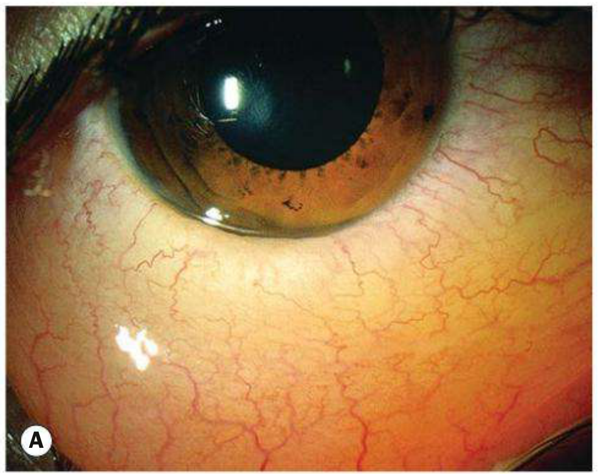
Moderate chemosis in acute allergic conjunctivitis (Kanski's):
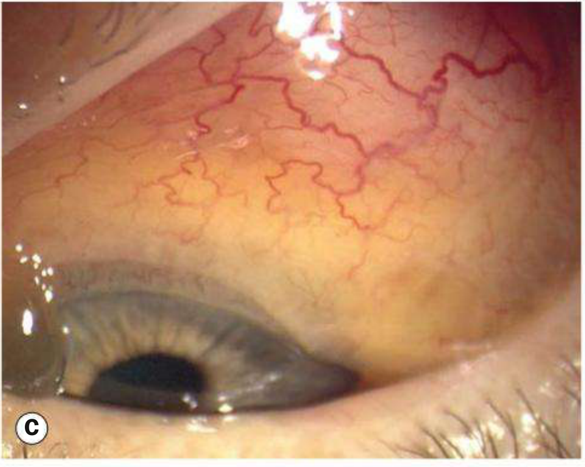
Chemosis visible from below (Kanski's, Fig. 6.2C):
Pathophysiology
Chemosis results from increased fluid in the subconjunctival space due to one or more of the following mechanisms:
| Mechanism | Example |
|---|---|
| Increased vascular permeability | Hypersensitivity (IgE-mediated), infection |
| Increased venous pressure | Carotid-cavernous fistula (CCF), SVC syndrome, right heart failure |
| Decreased plasma oncotic pressure | Nephrotic syndrome, hypoalbuminemia |
| Local inflammation/venous obstruction | Orbital disease, post-surgical |
Causes
Acute Chemosis
- Allergic (most common) - acute pollen exposure (type I hypersensitivity); hallmark of acute allergic conjunctivitis in children, frequently dramatic; resolves within hours as vascular permeability normalizes
- Severe infective conjunctivitis - bacterial or viral
- Angioneurotic edema (hereditary or acquired C1-esterase inhibitor deficiency)
Subacute / Chronic Chemosis
Local causes:
- Thyroid eye disease (Graves' orbitopathy)
- Chronic allergic conjunctivitis
- Ocular or eyelid surgery (post-operative)
- Trauma
- Carotid-cavernous fistula (CCF) - chemosis + proptosis + red eye + pulsating exophthalmos + bruit; arterialized conjunctival vessels are the hallmark
Systemic causes:
- Increased systemic vascular permeability: allergic conditions, meningitis, vasculitis
- Increased venous pressure: SVC syndrome, right-sided heart failure
- Decreased plasma oncotic pressure: nephrotic syndrome, hypoalbuminemia
- Drugs (e.g., topical eye drops)
- Venous congestion (e.g., carotid-cavernous fistula)
- Myxedema
Clinical Features
- Appearance: Translucent, boggy, jelly-like swelling of the bulbar conjunctiva; may herniate between the eyelids
- Symptoms: Usually asymptomatic unless severe; foreign body sensation, red eye; itching and watering in allergic cases
- Associated signs (depending on cause): proptosis, lid edema, conjunctival injection, restricted eye movements, bruit (CCF)
Key Associations to Remember
| Cause | Key Distinguishing Feature |
|---|---|
| Acute allergy (pollen) | Children, spring/summer, dramatic chemosis, resolves in hours |
| Carotid-cavernous fistula | Proptosis + chemosis + arterialized (corkscrew) vessels + pulsatile exophthalmos |
| Thyroid eye disease | Lid lag, lid retraction, restrictive myopathy, bilateral |
| Nephrotic syndrome | Bilateral periorbital edema + generalized edema, low albumin |
| SVC syndrome | Facial/neck edema, collateral veins on chest |
| Gonorrheal conjunctivitis | Hyper-acute purulent discharge, preauricular lymphadenopathy |
Management
Treatment is directed at the underlying cause:
- Acute allergic: Cool compresses; resolves spontaneously; topical adrenaline 0.1% (a single drop) can reduce extreme chemosis; antihistamine drops for seasonal/perennial forms
- Infective: Appropriate antimicrobial therapy
- Post-surgical: Lubricant eye drops, time; severe cases may need temporary tarsorrhaphy
- CCF: Endovascular embolization
- Systemic disease: Treat the underlying condition (e.g., diuretics for heart failure, immunosuppression for nephrotic syndrome)
Sources: Kanski's Clinical Ophthalmology, 10th ed.; The Wills Eye Manual; Localization in Clinical Neurology, 8th ed.
Blepharitis
Searching sources...
Finding Sources
Finding Sources
Reading File
Reading File
Good content. Now let me get the Kanski table of blepharitis characteristics, the angular blepharitis section, and the signs sections.
Reading File
Reading File
Excellent. Now let me get the rest of Kanski's content - signs, complications, treatment - and the angular blepharitis section.
Reading File
Let me get the Kanski treatment and complications section which was in the truncated portion.
Reading File
Loading Image
Loading Image
I now have comprehensive material from both Kanski's and Wills Eye Manual. Here is the full structured answer:
Blepharitis
Blepharitis is chronic inflammation of the eyelid margins. It is one of the most common ocular conditions, causing persistent discomfort and irritation with a characteristic pattern of remissions and exacerbations. There is a well-known poor correlation between symptom severity and objective signs.
Classification
Blepharitis is divided into anterior and posterior forms, with considerable overlap ("mixed blepharitis"):
| Feature | Anterior Blepharitis | Posterior Blepharitis |
|---|---|---|
| Location | Base of eyelashes, anterior lid margin | Meibomian glands, posterior lid margin |
| Subtypes | Staphylococcal, Seborrhoeic | Meibomian gland dysfunction (MGD) |
| Key deposit | Hard scales / collarettes (Staph); soft greasy scales (Seb) | Oily/turbid/toothpaste-like meibomian secretion |
| Lash changes | Madarosis, trichiasis, poliosis (more marked) | Mild lash changes |
| Cysts | Hordeolum (stye) | Chalazion (meibomian cyst) |
| Corneal signs | Punctate erosions, infiltrates, vascularization | More pronounced - punctate erosions, infiltrates |
| Tear film | Mild instability | Foamy, unstable - dry eye more marked |
| Associated skin disease | Staphylococcal: atopic dermatitis; Seborrhoeic: seborrhoeic dermatitis | Acne rosacea |
Pathophysiology
Anterior - Staphylococcal
An abnormal cell-mediated immune response to S. aureus cell wall components produces lid inflammation. This is more common and severe in atopic dermatitis patients. Bacterial toxins directly damage the ocular surface.
Anterior - Seborrhoeic
Strongly associated with generalized seborrhoeic dermatitis (scalp, nasolabial folds, retroauricular skin, sternum).
Posterior - Meibomian Gland Dysfunction (MGD)
Bacterial lipases (from S. aureus and others) break down meibum triglycerides into free fatty acids (FFAs). FFAs:
- Raise the melting point of meibum → inspissation, gland plugging
- Promote S. aureus growth
- Disrupt the lipid layer of the tear film → increased tear evaporation and osmolarity → unstable tear film and dry eye
Demodex
- Demodex folliculorum longus - anterior blepharitis
- Demodex folliculorum brevis - posterior blepharitis
Collarettes (cylindrical collections of keratin, waste, mite eggs around lash bases) are considered pathognomonic of Demodex infestation.
Clinical Features
Symptoms
- Burning, grittiness, mild photophobia, crusting of lid margins
- Symptoms worse in the morning (vs. dry eye alone which worsens through the day)
- Stinging may be more prominent in posterior disease
- Contact lens intolerance
- Usually bilateral and symmetrical, no visual disturbance
Signs - Anterior (Staphylococcal)
Blepharitis with lash collarettes (Wills Eye Fig. 5.8.1):
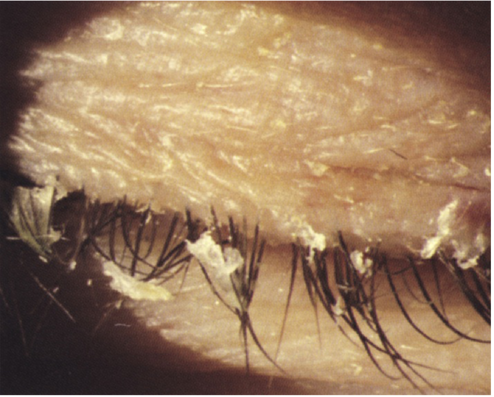
- Hard scales and crusting at the base of lashes
- Collarettes (cylindrical collections around lash bases) - pathognomonic
- Mild papillary conjunctivitis, chronic conjunctival hyperaemia
- Long-standing: scarring/notching of lid margin, madarosis (lash loss), trichiasis, poliosis (white lashes)
Signs - Anterior (Seborrhoeic)
- Hyperaemic, greasy anterior lid margins
- Soft scales; lashes stuck together
Signs - Posterior (MGD)
Meibomitis with inspissated meibomian glands (Wills Eye Fig. 5.8.2):
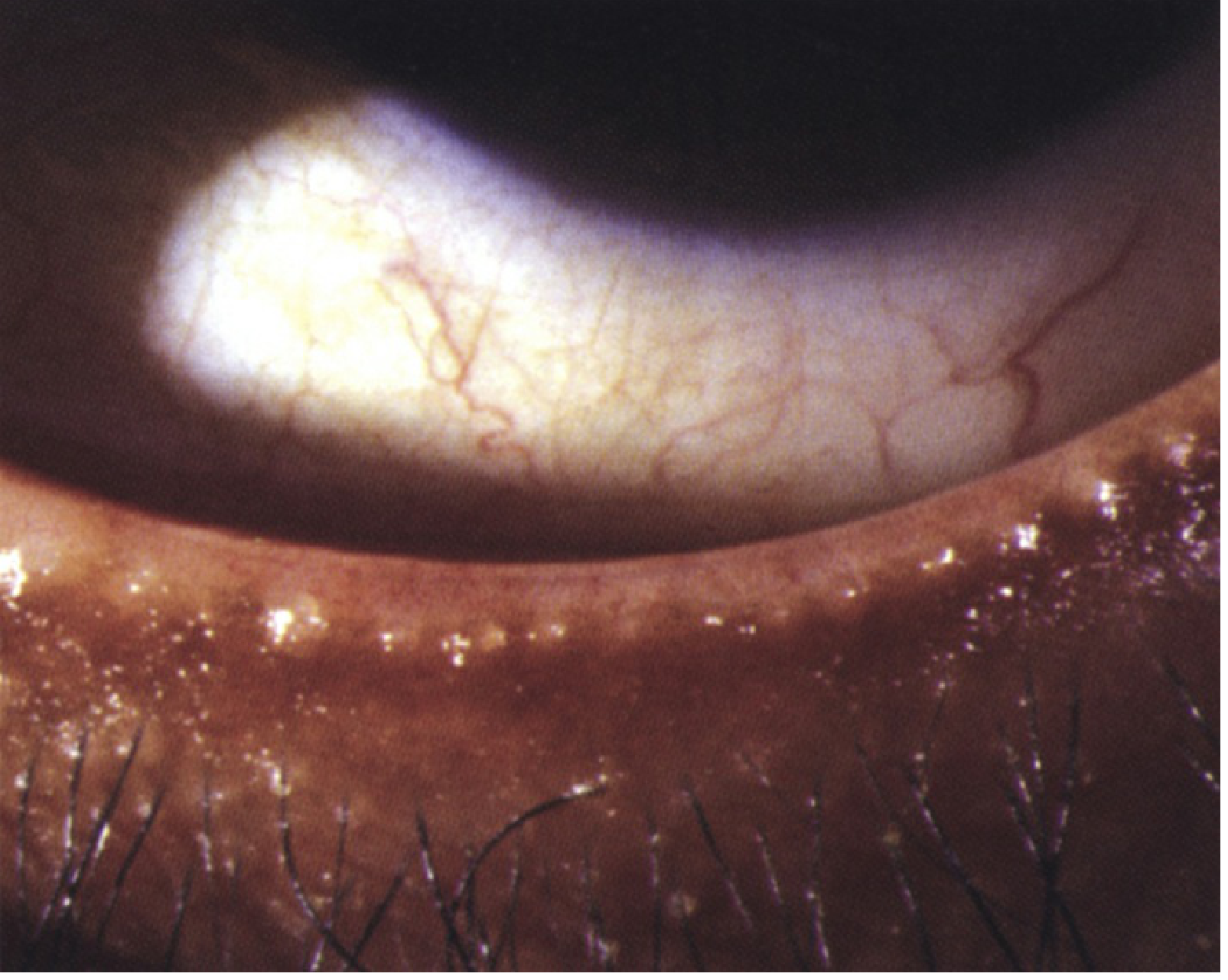
- Capping of meibomian gland orifices with oil globules
- Pouting, recession, or plugging of gland orifices
- Hyperaemia and telangiectasis of the posterior lid margin
- Expression of turbid or toothpaste-like meibomian secretion on pressure
- Lid transillumination: gland loss and cystic ductal dilatation
- Oily, foamy, unstable tear film; froth at lid margins and inner canthi
Complications / Associated Conditions
- Dry eye syndrome (all types, most severe in posterior)
- Corneal changes: superficial punctate keratopathy (SPK), peripheral infiltrates, vascularization (pannus), phlyctenules
- Chalazion / hordeolum
- Atopic keratoconjunctivitis (in patients with atopic dermatitis)
- Acne rosacea (posterior)
- Blepharokeratoconjunctivitis (chronic progression)
Red flag: Intractable, unilateral, or asymmetric blepharitis (especially upper vs. lower eyelid asymmetry) is a rare but important presentation of sebaceous cell carcinoma of the eyelid - requires appropriate clinical workup. - Wills Eye Manual
Angular Blepharitis
- Involves the canthal area (medial or lateral angle)
- Caused by Moraxella lacunata (most common) or S. aureus
- Associated with angular conjunctivitis
- Treatment: topical tetracycline or chloramphenicol
Treatment
Step-by-step (Wills Eye Manual)
- Eyelid hygiene (cornerstone) - lid margin scrubs twice daily with commercial eyelid scrub or dilute baby shampoo on a washcloth
- Warm compresses - 5-10 minutes, 2-4 times daily (softens inspissated meibum)
- Artificial tears - preservative-free, 4-8 times/day if dry eye component
- Topical antibiotics - erythromycin ointment or azithromycin gel-drop to lid margins at bedtime (moderately severe disease)
- Oral tetracyclines - doxycycline 100 mg twice daily (unresponsive cases, rosacea); azithromycin 500 mg/day x 3 days for 3 cycles with 7-day intervals is an alternative
- Omega-3 supplementation - improves meibomian gland function
- Cyclosporine 0.05%/0.09% or lifitegrast 5% drops twice daily for the inflammatory/dry eye component
- Demodex-directed therapy - tea-tree oil lid scrubs or hypochlorous acid lid cleansing agent for minimum 6 weeks if collarettes present and prior treatment failed
- Advanced meibomian gland treatments - thermal pulsation + expression, intense pulsed light (IPL), microblepharoexfoliation, meibomian gland probing
Note: Tetracycline derivatives (doxycycline) are contraindicated in pregnancy, breastfeeding, and children ≤8 years. Use erythromycin 200 mg twice daily as an alternative. - Wills Eye Manual
Follow-up
Review at 2-4 weeks. Lid hygiene and warm compresses may be reduced to once daily as the condition improves but are typically needed indefinitely - blepharitis is rarely cured, only controlled.
Sources: Kanski's Clinical Ophthalmology, 10th ed.; The Wills Eye Manual, 8th ed.
This is a shared conversation. Sign in to Orris to start your own chat.