
Heart failure approach
Reading File
Searching sources...
Finding Sources
Searching PubMed
"heart failure" management guidelines
Now let me read the main textbook content from Braunwald's (the definitive cardiology reference) and Family Medicine simultaneously:
Reading File
Reading File
Good content from Family Medicine. Let me now get the full heart failure section including diagnosis, workup, and management:
Reading File
Finding Sources
The offset I read for Family Medicine was the reference section. Let me read the actual clinical content that was in the para_line_ranges:
Reading File
Finding Sources
Excellent - that gave me very rich content. I now have comprehensive material to provide a full heart failure approach. Let me compile the complete answer:
Loading Image
I now have all the material needed for a comprehensive answer. Here is the full Heart Failure approach:
Heart Failure - Clinical Approach
Definition
Heart failure (HF) is a clinical syndrome in which the heart cannot meet the metabolic requirements of the body at normal filling pressures. It is not synonymous with congestive HF - pulmonary congestion may be absent; patients may present with dyspnea and reduced exercise capacity without overt fluid overload. The preferred term is simply "heart failure."
Classification
By Ejection Fraction
| Type | EF | Mechanism |
|---|---|---|
| HFrEF (HF with reduced EF) | < 45-50% | Impaired LV contraction (systolic dysfunction) |
| HFpEF (HF with preserved EF) | > 45-50% | Impaired LV filling/relaxation (diastolic dysfunction) |
HFpEF is nearly as common as HFrEF and carries similar morbidity.
NYHA Functional Classification
| Class | Symptoms |
|---|---|
| I | No symptoms with ordinary activity |
| II | Slight limitation - comfortable at rest, symptomatic with moderate exertion |
| III | Marked limitation - comfortable at rest, symptomatic with minimal exertion |
| IV | Symptoms at rest |
ACC/AHA Staging (A-D)
- Stage A: High risk, no structural disease, no symptoms
- Stage B: Structural disease, no symptoms (e.g., asymptomatic LV dysfunction post-MI)
- Stage C: Structural disease + prior or current symptoms
- Stage D: Refractory HF requiring advanced/specialized interventions
Etiology & Precipitants
Common causes of LV remodeling:
- Coronary artery disease / myocardial infarction (most common in developed countries)
- Hypertension
- Valvular heart disease
- Diabetes mellitus
- Cardiomyopathy (dilated, hypertrophic, restrictive)
- Congenital heart disease
- Alcohol, anemia, thyroid disease
High-output precipitants (CO > 4 L/min/m²): hyperthyroidism, Paget disease, AV fistula, severe anemia
Common decompensation triggers: dietary sodium/fluid non-compliance, medication non-adherence, infection, arrhythmia (especially new AF), ACS, uncontrolled hypertension, NSAID use.
Pathophysiology
The modern model centers on LV remodeling (stretching and dilation → reduced function), driven by neurohormonal activation regardless of the initial insult:
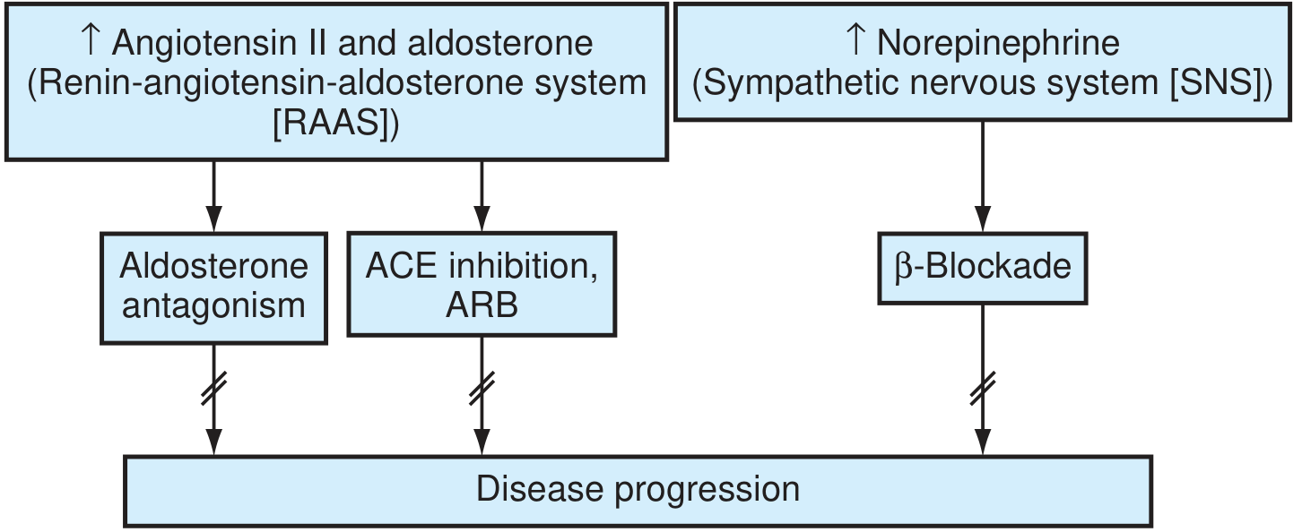
The two key axes: RAAS (Angiotensin II + Aldosterone) and SNS (Norepinephrine) - pharmacotherapy targets both to halt disease progression.
Key mechanisms:
- Angiotensin II: promotes myocyte apoptosis, hypertrophy, ventricular fibrosis, and aldosterone release
- Aldosterone: augments RAAS harmful effects, promotes adverse remodeling; "escapes" ACE inhibition over time - hence MRA needed in addition to ACEi/ARB
- Catecholamines (SNS): downregulate β-adrenoreceptors, directly toxic via cAMP-dependent calcium overload, increase MVO2, precipitate arrhythmias, induce LVH
- Cellular: increased MMPs → fibrosis and collagen deposition; altered calcium flux; metabolic shift to glycolysis
Clinical Features
Symptoms
- Left-sided: dyspnea on exertion, orthopnea, paroxysmal nocturnal dyspnea (PND), fatigue, reduced exercise tolerance
- Right-sided: peripheral edema, ascites, RUQ discomfort (hepatic congestion), early satiety, weight gain
Signs
- Elevated JVP, hepatojugular reflux
- S3 gallop (ventricular filling sound - classic for systolic HF)
- S4 gallop (atrial kick into stiff ventricle - suggests diastolic dysfunction)
- Displaced point of maximal impulse (PMI) - cardiomegaly
- Bibasilar crackles, pleural effusion (typically right > left)
- Pitting pedal/ankle edema, ascites
- Cool extremities, narrow pulse pressure (low output states)
- Cheyne-Stokes respiration (in severe HF)
Workup
Initial Investigations
| Test | Finding/Purpose |
|---|---|
| ECG | LVH, prior MI (Q waves), arrhythmia (AF), bundle branch block, ischemia |
| Chest X-ray | Cardiomegaly (CTR > 0.5), pulmonary venous congestion, Kerley B lines, bat-wing edema, pleural effusion |
| BNP / NT-proBNP | Key biomarker - elevated in HF; helps distinguish cardiac vs. non-cardiac dyspnea; guides therapy titration |
| Echocardiogram | Defines EF (HFrEF vs HFpEF), regional wall motion abnormalities, valvular disease, pericardial effusion - most important diagnostic test |
| CBC | Anemia (high-output HF, worsening HF) |
| BMP/CMP | Renal function (baseline before ACEi/diuretics), electrolytes, liver function |
| Thyroid function | Exclude hypo/hyperthyroidism |
| Lipids, fasting glucose | Cardiovascular risk |
| Urinalysis | Exclude renal disease |
Additional Studies (selected patients)
- Cardiac MRI: tissue characterization, infiltrative disease, myocarditis, sarcoidosis
- Cardiac catheterization: CAD assessment when revascularization is a consideration; hemodynamic evaluation
- Nuclear stress test/stress echo: myocardial viability, ischemia
- Holter/ambulatory monitor: arrhythmia screening
- Endomyocardial biopsy: suspected myocarditis, specific cardiomyopathies (per AHA/ACC/ESC guidance)
Management
General Principles
- Treat underlying cause
- Address precipitants
- Reduce symptoms
- Slow disease progression
- Reduce hospitalizations and mortality
Non-Pharmacologic
- Sodium restriction: < 2 g/day (evidence mixed for strict restriction, but reasonable in symptomatic HF)
- Fluid restriction: ~1.5-2 L/day in moderate-severe HF
- Daily weight monitoring: patient weighs every morning; seek care if weight gain > 2 lbs/day or 5 lbs/week
- Exercise: cardiac rehabilitation in stable HF - improves functional capacity and quality of life
- Alcohol abstinence (especially alcoholic cardiomyopathy)
- Smoking cessation
- Vaccination: influenza annually, pneumococcal
Pharmacotherapy - HFrEF (Evidence-Based)
The "four pillars" of disease-modifying therapy in HFrEF:
1. ACE Inhibitor (or ARB if intolerant)
- Mechanism: blocks RAAS, reduces Ang II-mediated apoptosis and fibrosis, lowers preload and afterload
- Evidence: Reduces mortality and hospitalization (CONSENSUS, SOLVD, TRACE trials)
- Agents: enalapril, lisinopril, ramipril, captopril
- Dose: titrate to target doses used in trials
- ARBs (e.g., valsartan, losartan): use when ACEi not tolerated due to cough; do NOT combine ACEi + ARB (increased adverse effects without benefit)
- ARNI (sacubitril/valsartan, Entresto): superior to enalapril in reducing CV death and HF hospitalization (PARADIGM-HF trial) - preferred over ACEi in patients who can tolerate and afford it (Class I recommendation)
2. Beta-Blocker
- Mechanism: blocks SNS toxicity, prevents catecholamine-induced myocyte damage, reduces arrhythmia risk, improves LV remodeling
- Evidence: Three agents proven to reduce mortality: carvedilol (COPERNICUS, US Carvedilol), metoprolol succinate (MERIT-HF), bisoprolol (CIBIS-II)
- Important: Start at low dose in stable (not acutely decompensated) HF and titrate up slowly
- Note: Carvedilol preferred in HF with diabetes (neutral metabolic effects vs. metoprolol per GEMINI trial)
3. Mineralocorticoid Receptor Antagonist (MRA)
- Mechanism: aldosterone "escapes" ACEi suppression - selective blockade is needed; reduces fibrosis and adverse remodeling
- Evidence: Spironolactone (RALES trial) - 30% mortality reduction in NYHA III-IV HFrEF; Eplerenone (EMPHASIS-HF) - reduced mortality and hospitalization in NYHA II HFrEF
- Monitoring: watch for hyperkalemia and renal impairment; avoid if K+ > 5.0 or GFR < 30
- Agents: spironolactone 25-50 mg/day; eplerenone 25-50 mg/day
4. SGLT2 Inhibitor
- Mechanism: reduces HF hospitalizations and CV death through cardiorenal mechanisms independent of glycemic effect
- Evidence: dapagliflozin (DAPA-HF) and empagliflozin (EMPEROR-Reduced) - significant reduction in HF hospitalizations and CV death in HFrEF, regardless of diabetes status
- Agents: dapagliflozin 10 mg/day, empagliflozin 10 mg/day
- Now Class I recommendation in HFrEF
Diuretics (Symptom Control)
- Loop diuretics (furosemide, torsemide, bumetanide): first-line for fluid overload/congestion; no proven mortality benefit but essential for symptom relief
- Torsemide has more predictable bioavailability than furosemide
- Titrate to achieve euvolemia (target: JVP normal, no peripheral edema, no orthopnea)
- Monitor electrolytes and renal function
Other Agents
| Drug | Indication |
|---|---|
| Hydralazine + Isosorbide dinitrate | Class I in Black patients with HFrEF on ACEi/ARB + beta-blocker; also use when ACEi/ARB/ARNI not tolerated (e.g., renal failure) |
| Ivabradine | HR > 70 bpm in sinus rhythm despite max beta-blocker; reduces HF hospitalization |
| Digoxin | Rate control in HF + AF; reduces HF hospitalization but no mortality benefit; narrow therapeutic window |
| Vericiguat | NYHA II-IV HFrEF who had recent HF event; reduces CV death/HF hospitalization |
HFpEF Management
- No proven mortality-reducing pharmacotherapy (contrast with HFrEF)
- Control heart rate and blood pressure aggressively
- Diuretics for congestion/fluid overload
- Treat underlying causes: hypertension, coronary disease, AF, obesity, sleep apnea
- SGLT2 inhibitors (empagliflozin - EMPEROR-Preserved trial): reduced HF hospitalizations in HFpEF - Class IIa
Device Therapy
| Device | Indication |
|---|---|
| ICD (Implantable Cardioverter-Defibrillator) | EF ≤ 35% despite ≥ 3 months GDMT, NYHA Class II-III; expected survival > 1 year |
| CRT (Cardiac Resynchronization Therapy) | EF ≤ 35% + LBBB with QRS ≥ 150 ms + NYHA II-IV; improves EF, symptoms, survival |
| CRT-D | Combination CRT + defibrillator |
| LVAD (Left Ventricular Assist Device) | NYHA IV refractory HF - bridge to transplant or destination therapy |
| Heart transplantation | End-stage HF refractory to maximal therapy |
Acute Decompensated HF (ADHF)
"LMNOP" mnemonic:
- Lasix (furosemide IV) - aggressive diuresis
- Morphine (use cautiously; reduces preload, anxiolytic - not routinely recommended)
- Nitrates (IV nitroglycerin or nitroprusside for hypertensive HF; powerful preload/afterload reduction)
- Oxygen / NIV (supplemental O2 to maintain SaO2 > 94%; CPAP/BiPAP reduces intubation need)
- Positioning (sit upright)
Additional ADHF management:
- Identify and treat precipitant
- Vasopressors/inotropes (dobutamine, dopamine, milrinone) in cardiogenic shock or low output state
- Avoid negative inotropes, NSAIDs
- Ultrafiltration for diuretic-refractory congestion
- Consider hemodynamic monitoring (Swan-Ganz) in refractory cases
Monitoring
- BNP/NT-proBNP: trending down indicates effective therapy; rising signals decompensation
- Renal function and electrolytes: monitor 1-2 weeks after ACEi/ARB/MRA initiation or dose change
- Echocardiogram: reassess EF after 3-6 months of optimal medical therapy (EF may recover)
- Weight monitoring: patient self-monitoring daily
Prognosis
- Mortality remains ~50% at 5 years from diagnosis
- 1-month rehospitalization rate ~25%
- Poor prognostic markers: low EF, elevated BNP, low sodium, worsening renal function, NYHA class IV, cardiac cachexia, inability to tolerate GDMT
Sources: Textbook of Family Medicine 9e, pp. 689-738; Braunwald's Heart Disease, 2-Volume Set (Cardiovascular Medicine)
This is a shared conversation. Sign in to Orris to start your own chat.