
Tympanosclerosis
tympanosclerosis tympanic membrane
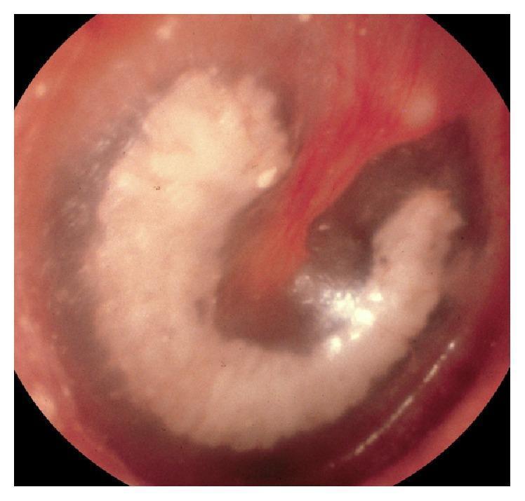
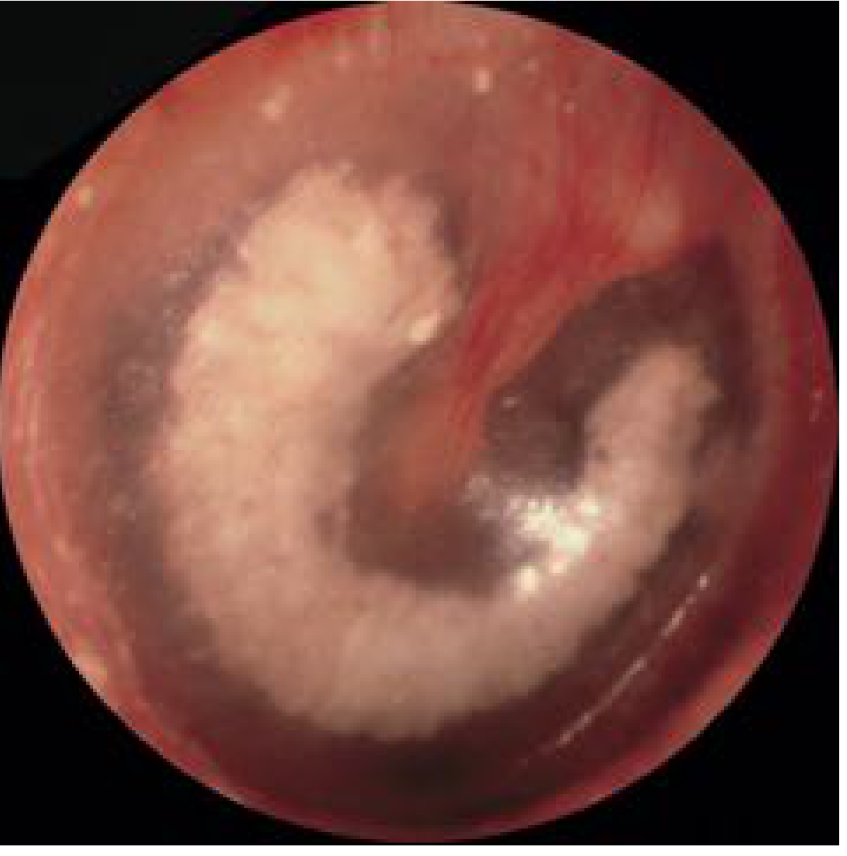
This clinical photograph shows an endoscopic view of a right tympanic membrane demonstrating significant tympanosclerosis. The primary finding is a dense, chalky white, semi-circular plaque within the substance of the eardrum. This sclerotic lesion is prominent in the pars tensa and extends superiorly to involve the pars flaccida. The plaque partially surrounds and obscures the proximal portion of the malleus manubrium, which appears hyperemic (reddened). The remaining portions of the tympanic membrane appear diffusely thickened and opaque, though the middle ear space remains aerated without signs of active effusion or infection. This visual represents the hyalinization and calcification of the fibrous layer of the tympanic membrane, a common sequela of chronic otitis media or previous ventilation tube insertion, which can lead to conductive hearing loss by reducing membrane compliance.
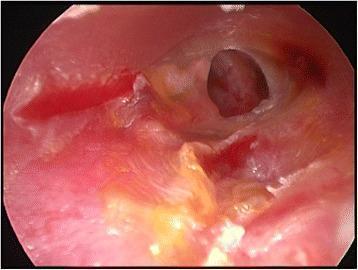
An endoscopic clinical photograph of a left tympanic membrane exhibiting a medium-sized subtotal perforation. The tympanic membrane itself shows signs of significant pathological changes, including diffuse erythema (inflammation) and yellowish discoloration, likely representing tympanosclerosis or chronic inflammatory exudate. The perforation is centrally located with irregular margins, through which the dark middle ear cavity and portions of the promontory are visible. The surrounding external auditory canal tissue is also notably erythematous and hyperemic. There is a small amount of white squamous debris or purulent discharge visible at the inferior portion of the canal. This image serves as a diagnostic visual for chronic otitis media and provides the preoperative anatomical context for surgical interventions such as myringoplasty or tympanoplasty.
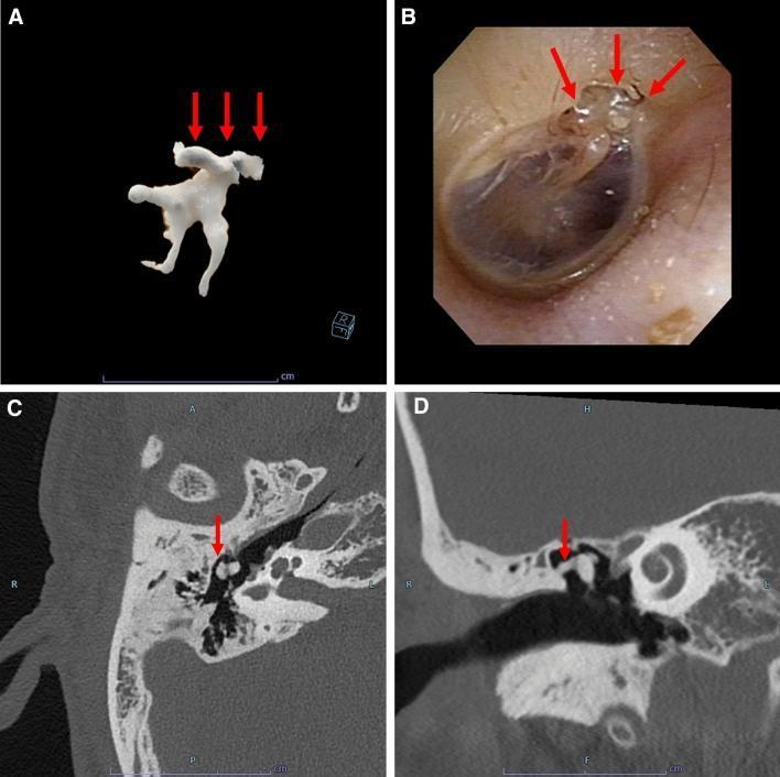
A multi-panel clinical and diagnostic figure demonstrating tympanosclerosis in the middle ear of an adult patient. (A) 3D volumetric reconstruction of the ossicular chain, showing irregular, calcified deposits (red arrows) integrated into the superior aspect of the ossicles. (B) Otoscopic clinical photograph of the tympanic membrane, revealing characteristic white, chalky plaques (red arrows) indicative of myringosclerosis/tympanosclerosis in the superior portion of the drum. (C) Axial high-resolution computed tomography (CT) scan and (D) coronal CT scan of the temporal bone. Both radiological views demonstrate discrete, irregular, high-density (hyperattenuating) calcifications within the tympanic cavity, specifically localized around the malleus and incus (red arrows). These findings represent the hyalinization and subsequent calcification of connective tissue within the middle ear and tympanic membrane, typically a sequela of chronic otitis media. The density of the lesions on CT is comparable to that of cortical bone.
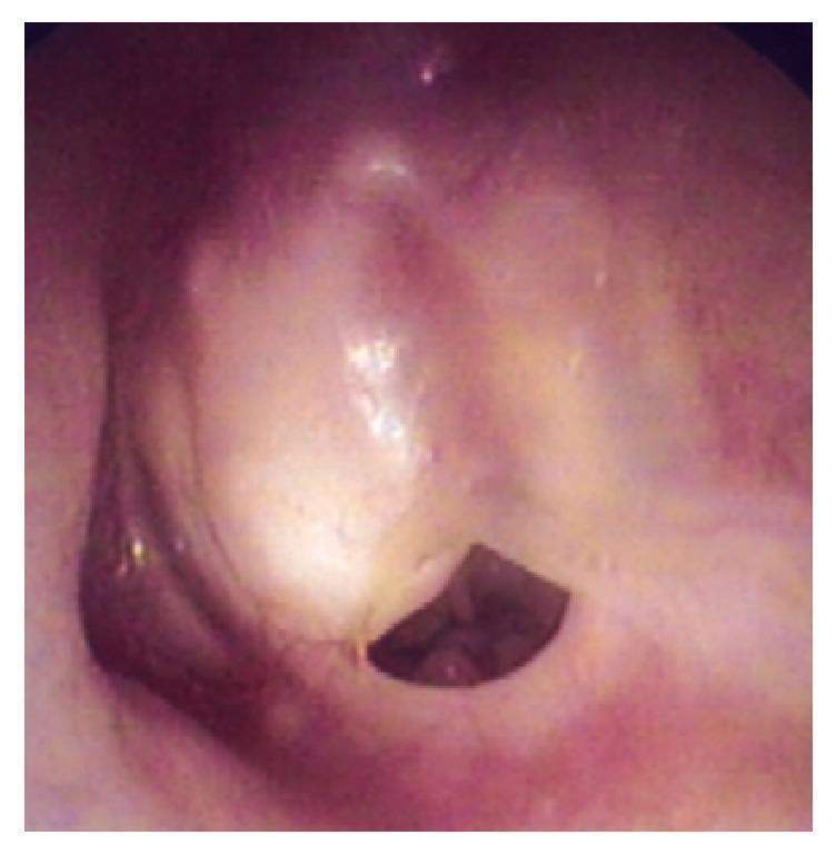
This clinical photograph, obtained via otomicroscopy, shows the left tympanic membrane. The membrane exhibits significant pathological changes, characterized by a thickened, opaque, and whitish appearance consistent with tympanosclerosis or chronic inflammatory changes. A well-defined, subtotal perforation is visible in the inferior portion of the membrane. The edges of the perforation appear dry and stable, with no evidence of acute infection, active otorrhea, or visible discharge. The surrounding external auditory canal tissue appears healthy and non-inflamed. Clinically, this visual finding is associated with a history of chronic otitis media and may be an otoscopic manifestation of underlying middle ear pathology, such as a cholesterol granuloma as indicated by the patient's clinical history of hearing loss and otalgia.
tympanosclerosis
Tympanosclerosis
Definition
- Cummings Otolaryngology, p. 2693
- Shambaugh Surgery of the Ear, p. 419
Clinical Appearance
Incidence / Epidemiology
| Context | Incidence |
|---|---|
| Children (age 4-15 yrs), average 4-yr follow-up | ~10% (Daly) |
| Chronic otitis media overall | 9-38% |
| After ventilation tube (grommet) insertion | ~59% |
| After myringotomy alone | ~13% |
| After tympanostomy tubes (meta-analysis of 134 studies) | 32% vs 10% controls |
| At surgery for chronic otitis media | ~20% (Kinney, n=1495) |
| Middle ear found at surgery | ~2.5% |
- Cummings Otolaryngology, p. 2694; Shambaugh Surgery of the Ear, p. 419
Pathogenesis
-
Inflammatory/infectious pathway: Recurrent acute otitis media (AOM) or otitis media with effusion (OME) causes a destructive process within middle ear connective tissue. Bacterial enzymes (proteinases, collagenases) directly degrade collagen, leading to collagen degeneration and subsequent dystrophic calcification.
-
Mechanical pathway: Eustachian tube obstruction alone (without infection) can produce tympanosclerosis in animal models - physical deformation of the membrane may be sufficient to trigger plaque formation.
- Specific bacteria: H. pylori and Chlamydia pneumoniae have been identified in tympanosclerotic ears
- Genetic basis: The LP/J mouse and Enpp1asj mutant mice develop spontaneous tympanosclerosis, suggesting a genetic predisposition
- Cummings Otolaryngology, p. 2694 (Fig. 140.19)
Histopathology
-
Acellular hyaline degeneration of the fibrous layer of the TM and middle ear mucosa
-
Calcification is present in most instances
-
Plaques are limited to the lamina propria when within the TM
-
Collagen and fibrous tissue with hyaline degeneration under microscopy
-
Deposition of hyaline occurs in concentric layers (similar to the rings of an onion), potentially reaching several millimeters in thickness
-
Osteoneogenesis can occur, causing ossicular fixation - most frequently at the heads of malleus and incus in the attic
-
Cummings Otolaryngology, pp. 2693-2694
Clinical Significance & Hearing Loss
Myringosclerosis (TM only)
- Usually clinically insignificant - causes little or no hearing impairment in most patients
- Smaller plaques can be left alone
- Extensive plaques involving a large area of TM remnant should be removed and replaced with a graft during tympanoplasty
Middle Ear Tympanosclerosis
-
Can cause ossicular fixation and significant conductive hearing loss
-
With roughly equal frequency, the disease fixes:
- The stapes in the oval window region
- The incus and malleus in the attic (epitympanum)
- Both simultaneously
-
Suspect ossicular fixation from tympanosclerosis when: conductive hearing loss + history of chronic ear infections + visible TM tympanosclerosis
-
Cummings Otolaryngology, p. 3145; Shambaugh, p. 419
Diagnosis
- Otoscopy/microscopy: Chalky white plaques on TM; reduced TM mobility on pneumatic otoscopy
- Audiometry: Conductive hearing loss (air-bone gap) when ossicles are involved
- CT temporal bone: Hyperattenuating (calcified) deposits within the tympanic cavity, around the ossicles - density comparable to cortical bone on imaging
- Malleus fixation may be suspected preoperatively by observing reduced or absent movement of the umbo and anterior process on pneumatic otoscopy
Management
Conservative
- Isolated TM plaques (myringosclerosis) without hearing loss: observation only
- TM tympanosclerosis does not typically progress to hearing impairment in the majority of cases
Surgical - Indications
Surgical Options
| Situation | Procedure |
|---|---|
| Incus + malleus head fixation in attic (intact ossicular chain) | Remove incus + head of malleus, reconstruct between malleus handle/TM and stapes |
| Attic wall approach | Remove outer attic wall, mobilize malleus and incus (risk of refixation) |
| Stapes involvement | Stapes mobilization or stapedectomy |
| TM plaque during tympanoplasty | Excise plaque if extensive; replace with graft |
Surgical Outcomes (Stapes Surgery)
-
Results are not as good as in otosclerosis:
- Post-op air-bone gap ≤20 dB achieved in ~70-72% of cases (vs. >90% in otosclerosis)
- Risk of sensorineural (dead ear) complication higher than in other middle ear disease: 4-5% reported
- Tendency for refixation of ossicles by fibrous tissue or new bone
-
Two-stage procedures (stapedectomy + total ossicular reconstruction) have reported excellent results in 79% (Smyth), but long-term outcomes vary considerably
-
Scott-Brown's ORL, Vol 2, p. 2082-2087; Cummings Otolaryngology, p. 2694
Key Points Summary
| Feature | Detail |
|---|---|
| Pathology | Acellular hyaline + calcified deposits in TM/middle ear submucosa |
| Cause | Resolved otitis media, trauma, grommet insertion |
| Most common site | TM (myringosclerosis) - usually benign |
| Dangerous site | Ossicles (attic/oval window) - causes conductive HL |
| Appearance | Chalky white semicircular/horseshoe plaque on TM |
| Reversibility | Irreversible |
| Treatment of choice | Surgery (ossiculoplasty/stapedectomy) if significant CHL |
| Surgery risk | Sensorineural loss 4-5%; refixation tendency |