
Diagnosis
vitiligo face neck depigmented patches poliosis
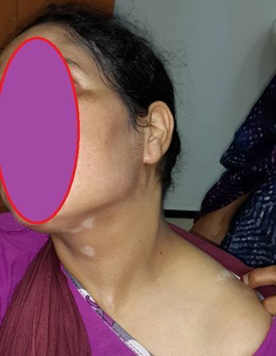
This clinical photograph shows the profile of a 31-year-old female patient with dermatological manifestations often associated with Vogt-Koyanagi-Harada (VKH) syndrome. Visible on the neck and upper clavicular region are several well-circumscribed, depigmented (achromic) patches characteristic of vitiligo. The lesions vary in size, with discrete macules on the neck and more confluent patches near the shoulder. Additionally, the hair at the scalp margin exhibits localized depigmentation, consistent with poliosis (white hair). The clinical significance of these visual findings, when combined with the patient's history of panuveitis and auditory symptoms, supports the diagnosis of an autoimmune multi-system disorder affecting melanocyte-containing tissues. This image serves as a clinical example of the integumentary signs in systemic autoimmune disease, targeted at medical students and dermatology residents specializing in pigmentary disorders and their extra-cutaneous associations.
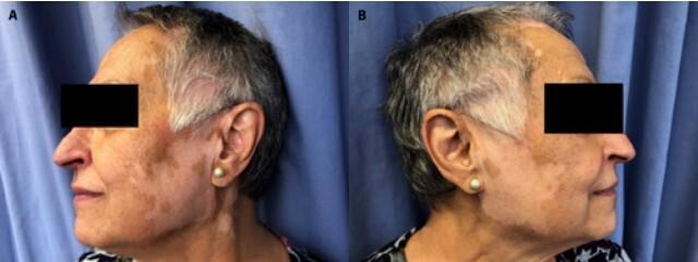
Clinical photograph of a patient's face and neck, shown in bilateral lateral profiles, illustrating features of drug-induced vitiligo. The image displays multiple sharply demarcated, achromic (depigmented) macules and patches. These lesions are distributed symmetrically across the cheeks, preauricular areas, ears, and lateral neck. A significant finding is leukotrichia, where the depigmentation extends to involve the hair follicles along the temporal hairline and sideburns, resulting in localized white hair. The surrounding skin appears normally pigmented, creating a high-contrast border characteristic of vitiligo. This visual demonstrates common cutaneous side effects associated with targeted cancer therapies like ribociclib and letrozole. The clinical presentation is essential for medical students and dermatologists to distinguish between primary vitiligo and drug-induced leukoderma in oncological patients.
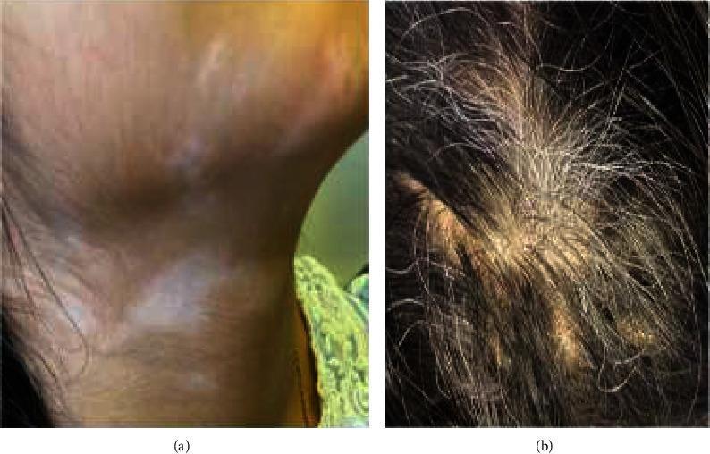
This composite clinical photograph displays dermatological and trichological manifestations of Vogt-Koyanagi-Harada (VKH) syndrome in a pediatric patient. Figure (a) illustrates cutaneous hypopigmentation, consistent with vitiligo, appearing as irregular, milky-white depigmented patches on the neck that contrast with the surrounding normally pigmented skin. Figure (b) shows poliosis (graying of the hair), characterized by a localized patch of depigmented, white/gray hair shafts amidst otherwise dark terminal scalp hair. These findings represent the integumentary phase of VKH syndrome, a multi-system autoimmune disorder targeting melanocytes. Educational focus is on identifying systemic clinical signs—such as vitiligo, poliosis, and alopecia—that occur alongside granulomatous panuveitis in the diagnosis of complete or incomplete VKH syndrome. These visible markers are essential for clinical classification and monitoring treatment response to immunosuppressive therapy.
Diagnosis: Vitiligo (with consideration of Vogt-Koyanagi-Harada Syndrome)
Clinical Findings in the Video
| Finding | Location |
|---|---|
| Well-demarcated, milky-white achromic macules and patches | Right alar base / nasolabial fold |
| Large confluent depigmented patch | Right lateral neck extending to jawline and post-auricular area |
| Multiple discrete depigmented macules | Left lateral neck and submandibular region |
| Poliosis (localized patch of white hair) | Anterior scalp/hairline |
Primary Diagnosis: Vitiligo
- Well-defined, milky-white, achromic macules and patches (not merely hypopigmented)
- Smooth, convex margins
- Distribution involving the face, neck, and scalp - among the most commonly affected sites
- Poliosis (white hair within lesional areas) - a recognized clinical sign of vitiligo
Important Red Flag: Consider Vogt-Koyanagi-Harada (VKH) Syndrome
- Ocular symptoms: Blurred vision, eye pain, photophobia, or redness (panuveitis / bilateral granulomatous uveitis is the hallmark)
- Auditory symptoms: Tinnitus, hearing loss (sensorineural), or dysacousia
- Neurological symptoms: Headache, meningismus, CSF pleocytosis (meningoencephalitis phase)
- Other skin findings: Alopecia, in addition to vitiligo/poliosis
Differential Diagnosis
| Condition | Features in common | Distinguishing features |
|---|---|---|
| Vitiligo (primary) | Achromic patches, poliosis | No systemic involvement |
| VKH syndrome | Vitiligo + poliosis | + uveitis, hearing loss, meningitis |
| Alezzandrini syndrome | Unilateral facial vitiligo + poliosis | Ipsilateral retinal degeneration + ipsilateral deafness; extremely rare (<10 cases) |
| Waardenburg syndrome | Poliosis, depigmentation | Congenital, heterochromia, congenital deafness |
| Pityriasis alba | Facial hypopigmentation | Ill-defined, not completely achromic; seen in children with atopy |
| Tinea versicolor | Neck/trunk hypopigmentation | KOH positive; not truly achromic |
| Post-inflammatory leukoderma | Depigmented patches | History of prior skin inflammation |
Recommended Workup
- Ophthalmology referral - slit lamp exam to rule out uveitis (VKH)
- Audiometry - screen for sensorineural hearing loss
- Thyroid function (TSH, anti-TPO) - 13-19% of vitiligo patients have autoimmune thyroid disease
- ANA, anti-dsDNA - to exclude lupus-associated depigmentation
- Blood glucose - type 1 diabetes association
- Wood's lamp examination - confirms true achromia (brilliant white fluorescence vs. dull in hypopigmentation)
- Skin biopsy (if uncertain) - absent melanocytes on DOPA stain