
Tract of goll and burdach
I'll look this up in the medical library right away.
Searching sources...
Reading File
Finding Sources
Finding Sources
Good - found the key sections. Now let me read them in parallel.
Reading File
Reading File
Excellent detail. Let me now read the continuation of the Neuroanatomy textbook section and also fetch the diagram image.
Reading File
Loading Image
Now let me fetch the spinal cord cross-section somatotopic diagram as well.
Loading Image
I now have comprehensive information. Here is the full answer:
Tracts of Goll and Burdach (Posterior Columns / Dorsal Columns)
The Tract of Goll (fasciculus gracilis) and the Tract of Burdach (fasciculus cuneatus) are the two subdivisions of the posterior (dorsal) columns of the spinal cord. Together they form the first leg of the Posterior Column-Medial Lemniscal (PCML) pathway, the main route for conscious proprioception, vibration sense, discriminative touch, and 2-point discrimination.
Names and Eponyms
| Eponym | Anatomical Name | Meaning |
|---|---|---|
| Tract of Goll | Fasciculus gracilis | "gracilis" = thin/slender |
| Tract of Burdach | Fasciculus cuneatus | "cuneatus" = wedge-shaped |
Location in the Spinal Cord
Both tracts sit in the dorsal funiculus (posterior white matter) of the spinal cord:
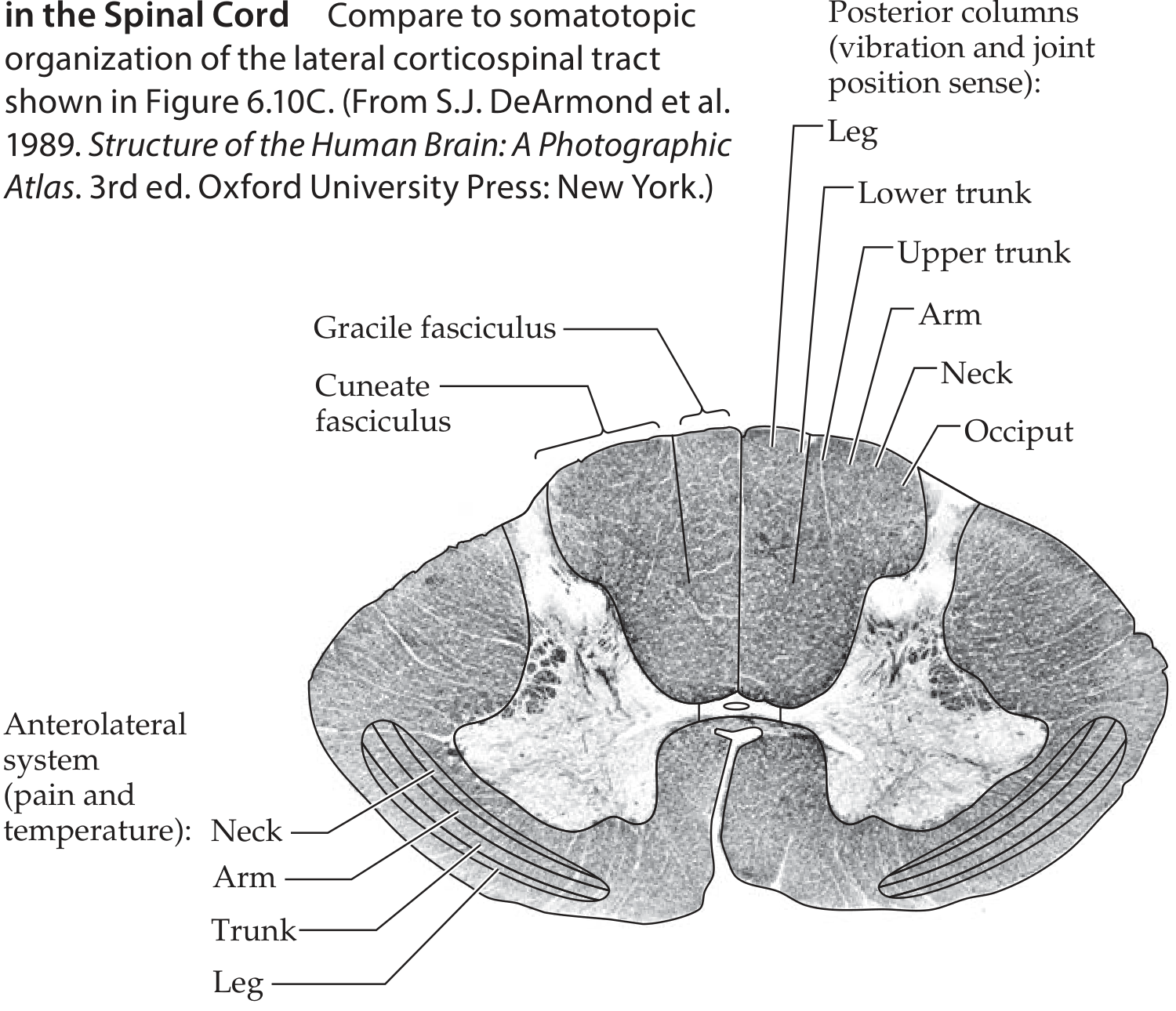
- Fasciculus gracilis (Goll) - medial position
- Fasciculus cuneatus (Burdach) - lateral position (lateral to gracilis)
Somatotopic Organization
This is a high-yield point: fibers are arranged medial to lateral corresponding to sacral to cervical levels. As fibers ascend, incoming fibers from higher levels push existing fibers medially.
| Fasciculus | Spinal levels | Body region |
|---|---|---|
| Gracilis (Goll) - medial | Sacral, lumbar, lower 6 thoracic (below T6) | Legs and lower trunk |
| Cuneatus (Burdach) - lateral | Upper 6 thoracic and all cervical (above T6) | Upper trunk, arms, neck |
Key point: The fasciculus cuneatus is only present above T6 because below that level, there is no upper limb input. So at lumbar and sacral levels, only the gracilis exists; the cuneate fasciculus appears from mid-thoracic levels upward.
Neurons and Course (3-Neuron Pathway)
1st Order Neuron (Peripheral to Spinal Cord)
- Cell body in the dorsal root ganglion (DRG)
- Large-diameter, heavily myelinated A-alpha and A-beta fibers
- Enter the spinal cord via the medial dorsal root entry zone
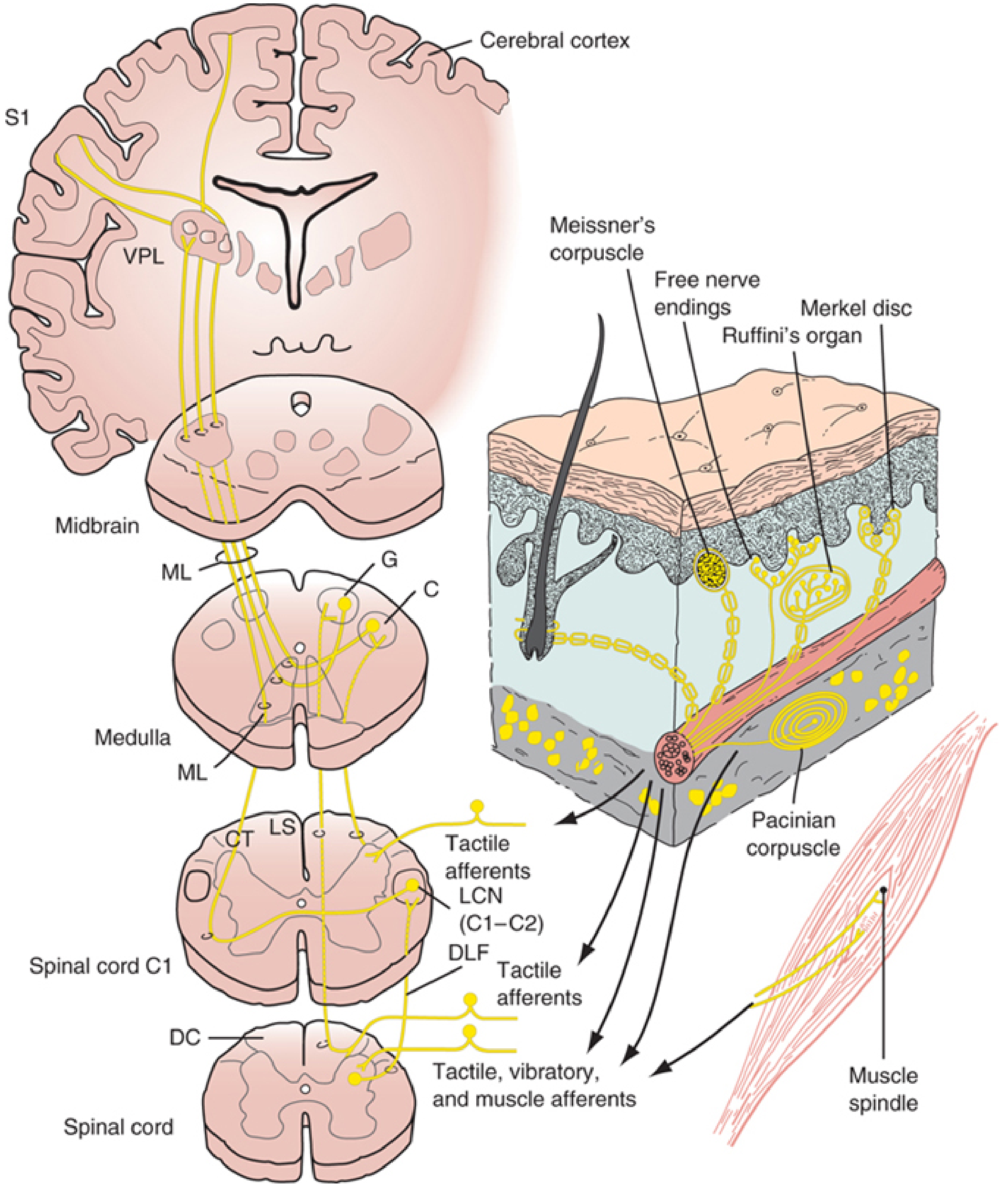
- Axons ascend ipsilaterally in the posterior column (do NOT synapse in the cord)
- Some collaterals enter the dorsal horn gray matter to synapse on interneurons/motor neurons (contributes to spinal reflexes)
2nd Order Neuron (Spinal Cord to Thalamus)
- 1st order fibers synapse at the posterior column nuclei in the medulla:
- Gracilis → Nucleus gracilis
- Cuneatus → Nucleus cuneatus
- 2nd order axons then cross as internal arcuate fibers (decussation in the lower medulla)
- Form the medial lemniscus on the contralateral side
- The medial lemniscus initially has a vertical orientation in the medulla (feet ventral, "little person stands up"), then becomes horizontal in the pons and midbrain (legs lateral, arms medial - "little person lies down")
- Terminate in the ventral posterior lateral (VPL) nucleus of the thalamus
3rd Order Neuron (Thalamus to Cortex)
- VPL neurons project via the posterior limb of the internal capsule
- Reach the primary somatosensory cortex (S1) in the postcentral gyrus (Brodmann areas 3, 1, and 2)
Sensory Modalities Carried
| Modality | Receptor |
|---|---|
| Proprioception (conscious) | Muscle spindle primary afferents (Ia), Golgi tendon organ |
| Vibration sense | Pacinian corpuscles, Meissner's corpuscles, Merkel discs |
| Discriminative / fine touch | Meissner's corpuscles, Merkel discs, Ruffini endings |
| 2-point discrimination | Multiple mechanoreceptors |
| Stereognosis, graphesthesia | (cortical integration of above) |
Key Features to Remember
| Feature | Detail |
|---|---|
| Decussation level | Lower medulla (as internal arcuate fibers) |
| Ipsilateral ascent | Yes - fibers stay ipsilateral until medulla |
| Fiber type | Large myelinated (A-alpha, A-beta) |
| First synapse | Nucleus gracilis / nucleus cuneatus in medulla |
| Second synapse | VPL nucleus of thalamus |
| Final destination | Postcentral gyrus (S1), areas 3, 1, 2 |
| Cuneate fasciculus present from | T6 and above only |
Clinical Relevance - Posterior Column Syndrome
Damage to the posterior columns causes ipsilateral loss of:
- Proprioception (positive Romberg's sign)
- Vibration sense
- Fine touch and 2-point discrimination
- Sensory ataxia
Pain and temperature are SPARED (those travel in the anterolateral spinothalamic tract).
Classic causes of posterior column damage:
- Tabes dorsalis (neurosyphilis)
- Subacute combined degeneration (Vitamin B12 deficiency)
- Friedreich's ataxia
- Multiple sclerosis
- Cervical spondylosis
- Posterior spinal artery infarct
Sources: Neuroanatomy through Clinical Cases, 3rd Ed. | Localization in Clinical Neurology, 8th Ed.
Artificial kidney ist year MBBS level
Reading File
The artificial kidney (dialysis/haemodialysis) is a 1st year MBBS physiology topic. Let me search the library now.
Searching sources...
Finding Sources
Finding Sources
Found the Guyton and Hall section - perfect for 1st year MBBS. Let me read it in full.
Reading File
Reading File
Reading File
Finding Sources
Now let me get the diagram image from this section.
Reading File
Loading Image
I have everything needed for a thorough 1st year MBBS level answer. Here it is:
Artificial Kidney (Haemodialysis)
1st Year MBBS | Physiology - Renal System
(Guyton & Hall, Textbook of Medical Physiology)
Definition
An artificial kidney (dialyzer/haemodialyser) is a device used to remove metabolic waste products, excess electrolytes, and water from the blood when the kidneys are unable to do so - mimicking the excretory function of the natural kidneys.
Indications for Use
- Acute Kidney Injury (AKI) - to support the patient until kidneys recover
- End-Stage Renal Disease (ESRD) / Chronic Kidney Disease (CKD) - used chronically to maintain life when loss of kidney function is irreversible
- Certain poisonings (barbiturates, salicylates, methanol, etc.)
Over 800,000 patients in the United States currently receive some form of ESRD therapy. The vast majority are treated with dialysis due to the shortage of donor kidneys.
Basic Principle
The core principle is diffusion across a semipermeable membrane:
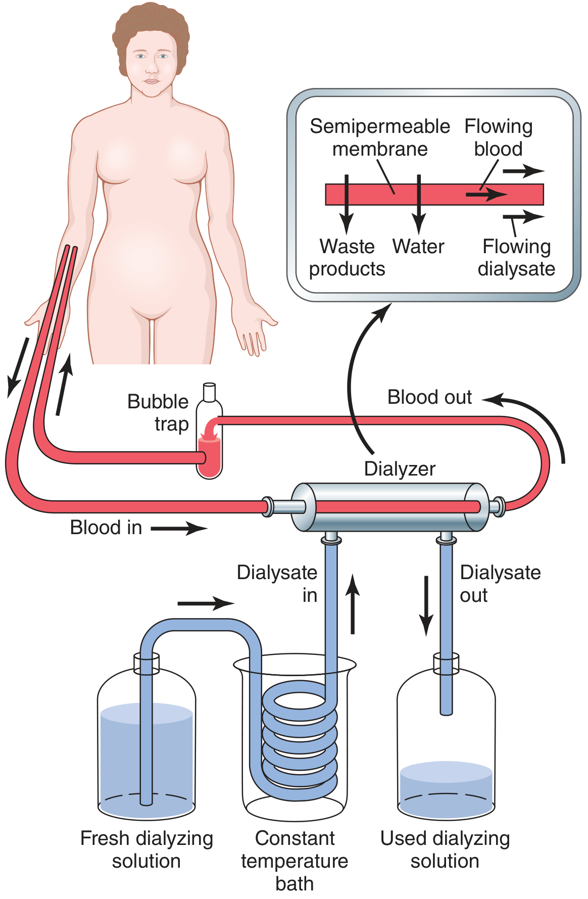
- Blood flows through minute channels bounded by a thin semipermeable membrane (e.g., cellophane/cuprophane)
- On the other side of the membrane flows the dialysate (dialyzing fluid)
- Waste substances in the blood (urea, creatinine, urate, phosphate, sulfate, excess K+) diffuse down their concentration gradient from blood into the dialysate
- Useful substances (glucose, bicarbonate) can be added to the dialysate to diffuse back into the blood if needed
- The membrane is porous enough to allow passage of all plasma constituents EXCEPT plasma proteins (too large to cross)
Components of the Artificial Kidney
| Component | Function |
|---|---|
| Dialyzer | Main unit housing the semipermeable membrane where exchange occurs |
| Semipermeable membrane | Allows diffusion of small solutes; blocks proteins |
| Blood circuit | Carries blood from patient → dialyzer → back to patient |
| Bubble trap | Removes air bubbles before blood returns to the patient |
| Fresh dialyzing solution reservoir | Provides clean dialysate continuously |
| Constant temperature bath | Warms dialysate to body temperature (~37°C) |
| Used dialyzing solution reservoir | Collects waste-laden dialysate |
Factors Affecting Rate of Solute Transfer
The rate at which a substance is cleared depends on:
- Concentration gradient - the greater the gradient, the faster the diffusion (maximum at start of dialysis)
- Permeability of the membrane to that particular solute
- Surface area of the dialyzing membrane (0.6 to 2.5 m²)
- Duration of contact between blood and dialysate
- Flow rates - increasing blood flow rate OR dialysate flow rate maintains a high concentration gradient and optimizes diffusion (this is why a flowing/continuous system is used)
In a flowing (continuous) system, the concentration gradient is not easily dissipated, making hemodialysis far more efficient than a static system.
The Dialyzing Fluid (Dialysate)
The composition of the dialysate is carefully adjusted - not simply equal to normal plasma - to drive appropriate movements of solutes:
| Constituent | Normal Plasma | Dialyzing Fluid | Uremic Plasma |
|---|---|---|---|
| Na+ (mEq/L) | 142 | 133 | 142 |
| K+ (mEq/L) | 4.2 | 1.0 (↓ to pull K+ out) | 7 (↑↑) |
| Ca2+ (mEq/L) | 3 | 3 | 2 |
| HCO3- (mEq/L) | 24 | 35.7 (↑ to correct acidosis) | 14 (↓↓) |
| Phosphate (mEq/L) | 3 | 0 (to remove excess phosphate) | 9 (↑↑) |
| Urea (mg/dL) | 14 | 0 (to maximise removal) | 200 (↑↑) |
| Creatinine (mg/dL) | 1 | 0 (to maximise removal) | 6 (↑↑) |
| Glucose (mg/dL) | 100 | 125 (slightly high to prevent hypoglycaemia) | 100 |
Key principle: Urea, creatinine, urate, sulfate, and phosphate are absent from the dialysate so there is maximum gradient driving their removal from the blood.
Operational Details
| Parameter | Value |
|---|---|
| Blood volume in machine at any time | < 500 mL |
| Blood flow rate | Several hundred mL/min |
| Total membrane surface area | 0.6 - 2.5 m² |
| Anticoagulant used | Heparin (infused as blood enters to prevent clotting) |
| Urea clearance by artificial kidney | 100-225 mL/min |
| Urea clearance by two normal kidneys | ~70 mL/min |
| Session duration | 4-6 hours/day, 3 times per week |
Effectiveness - Clearance
- The artificial kidney clears urea at 100-225 mL/min - about twice as fast as two normal kidneys (70 mL/min)
- However, because it is used only 4-6 hours, 3 times/week, the overall cumulative clearance is still considerably less than normal kidneys functioning continuously
Limitations of the Artificial Kidney
The artificial kidney cannot replace ALL functions of the natural kidneys:
- Cannot secrete erythropoietin → anaemia persists (chronic dialysis patients need EPO injections)
- Cannot perform endocrine functions (activation of Vitamin D, renin secretion)
- Cannot fully maintain fluid-electrolyte balance around the clock
- Patients still have significantly impaired overall health
- Patients on dialysis have higher risks of infections, cardiovascular disease
Hemofiltration (Additional Mechanism)
In addition to diffusion, bulk flow / hemofiltration can also occur:
- A hydrostatic pressure is applied to force fluid and solutes across the membrane
- Used when ultrafiltration (removal of excess water) is needed
Peritoneal Dialysis vs Haemodialysis
In peritoneal dialysis, the body's own peritoneal membrane acts as the semipermeable membrane, and dialysate is introduced into the peritoneal cavity instead of using an external machine.
Summary Box (Exam Points)
- Principle: Diffusion across semipermeable membrane down concentration gradient
- Membrane: Cellophane - allows small solutes, blocks plasma proteins
- What is removed: Urea, creatinine, K+, phosphate, sulfate, urate, excess water
- Anticoagulant used: Heparin
- Urea clearance: 100-225 mL/min (> normal kidneys' 70 mL/min)
- Schedule: 4-6 hrs, 3×/week
- Cannot replace: Erythropoietin secretion, Vitamin D activation
Source: Guyton and Hall Textbook of Medical Physiology, Chapter 32 (Renal Disease, Diuretics, and Micturition)
patient with irritable bowel syndrome is prescribed a cellulose containing dietary supplementation. Discuss the importance of dietary fiber in providing health benefits beyond basic nutritio
Searching sources...
Reading File
Finding Sources
Finding Sources
Excellent - multiple relevant sources found. Let me read the key sections in parallel.
Reading File
Reading File
Reading File
Reading File
Reading File
Finding Sources
Reading File
Reading File
Loading Image
I now have comprehensive information from multiple authoritative sources. Here is the complete answer:
Dietary Fiber in IBS - Health Benefits Beyond Basic Nutrition
Clinical Context: A patient with Irritable Bowel Syndrome (IBS) is prescribed cellulose-containing dietary supplementation. Cellulose is the classic insoluble dietary fiber - a polysaccharide of glucose residues linked β-1,4 bonds that human enzymes cannot digest.
1. What is Dietary Fiber?
Dietary fiber is the portion of the diet resistant to digestion by human digestive enzymes. It consists principally of plant-derived polysaccharide derivatives and lignin.
Classification
| Type | Components | Sources |
|---|---|---|
| Insoluble fiber | Cellulose, Hemicellulose, Lignin | Whole wheat, bran, vegetables |
| Soluble fiber | Pectins, Gums, Mucilages | Oats, legumes, apples, psyllium |
Cellulose specifically: a polysaccharide of glucosyl residues linked β-1,4 (unlike starch which is α-1,4) - humans lack the enzyme (cellulase) to cleave these bonds, so it passes undigested to the colon.
Fermentation properties of fiber types:
| Fiber Type | Water Solubility | % Fermented by Bacteria |
|---|---|---|
| Lignin | Poor | 0% |
| Cellulose | Poor | ~15% |
| Hemicellulose | Good | 56-87% |
| Mucilages/Gums | Good | 85-95% |
| Pectins | Good | 90-95% |
2. Mechanisms of Action - How Fiber Provides Health Benefits
A. Stool Bulking and Regulation of Transit
- Insoluble fiber like cellulose attracts water and increases stool bulk directly (physical water-binding)
- Fiber that is not fermented retains water in the colon, softening stool
- This reduces intracolonic pressure, which is why fiber is beneficial in diverticular disease - it softens stool, reduces pressure on the colonic wall and enhances expulsion of feces
- Every gram of wheat fiber ingested yields approximately 2.7 g of stool expelled
- Increased stool bulk reduces colonic transit time, decreasing mucosal contact with irritants
In IBS: Soluble fiber (e.g., psyllium) has high water-holding capacity that pulls water into the colon and improves stool consistency. Insoluble fiber (cellulose/wheat bran) can sometimes mechanically irritate the colon and may aggravate symptoms in IBS with constipation - this is an important clinical caveat for the prescribed supplementation.
B. Fermentation to Short-Chain Fatty Acids (SCFAs) - Key Metabolic Benefit
Dietary fiber and undigested sugars that reach the colon are metabolized by colonic bacteria to produce:
- Acetate (2 carbons)
- Propionate (3 carbons)
- Butyrate (4 carbons) - the most biologically active
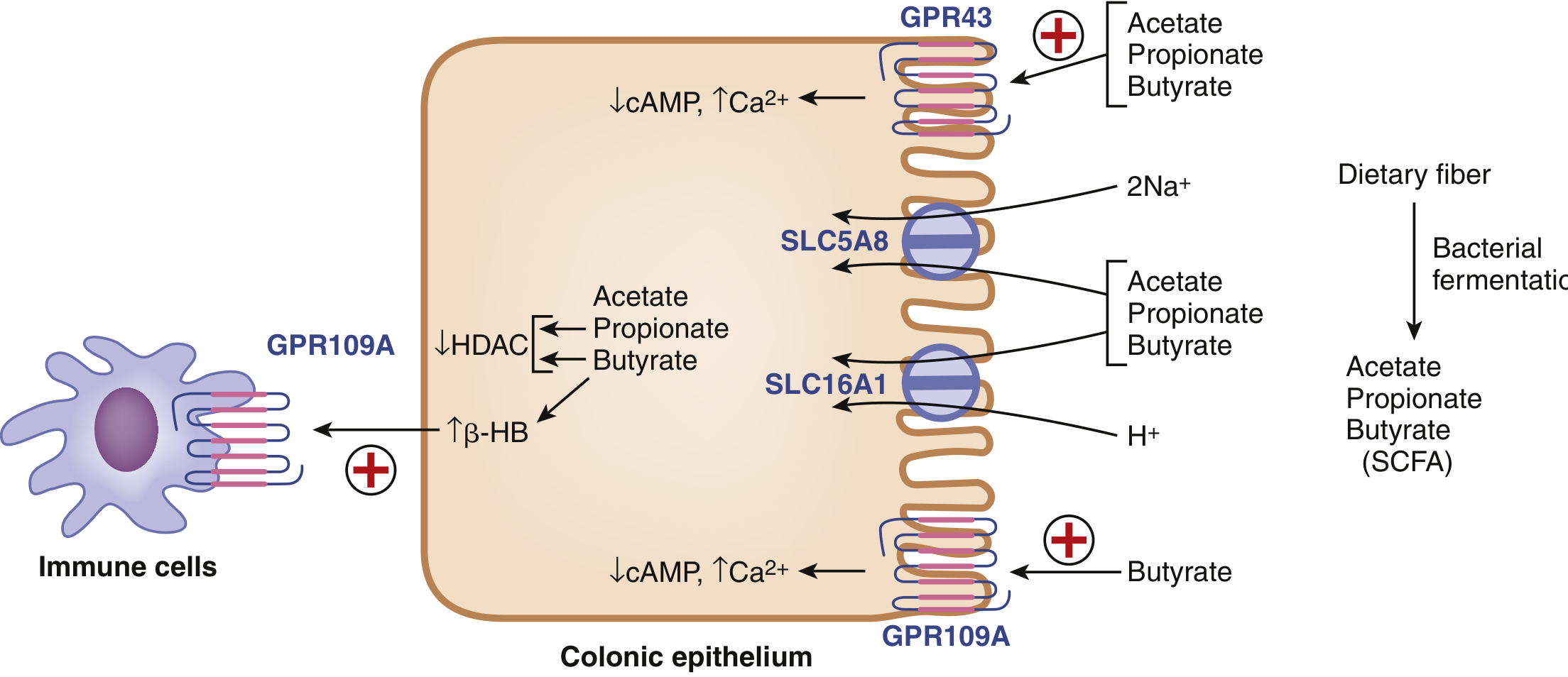
SCFAs are absorbed by colonocytes via:
- SLC16A1 (MCT1) - proton-coupled monocarboxylate transporter
- SLC5A8 (SMCT1) - sodium-coupled monocarboxylate transporter
Functions of SCFAs:
| SCFA | Key Action |
|---|---|
| Butyrate | Primary energy substrate for colonocytes; HDAC inhibitor (epigenetic regulation); generates β-hydroxybutyrate |
| Propionate | Energy substrate; inhibits HDAC |
| Acetate | Energy substrate; activates GPR43 |
| All 3 SCFAs | Activate GPR43 receptor (↓cAMP, ↑Ca²+) on colonocytes and immune cells |
| Butyrate specifically | Activates GPR109A receptor; modulates immune tolerance |
We may obtain as much as 10% of our total calories from compounds produced by bacterial digestion of substances in our digestive tract.
Fermentation of fiber has two net effects:
- Production of SCFAs that are trophic for colonic epithelium
- Increase in bacterial mass - contributing to increased stool volume
C. Gut Microbiome Modulation (Prebiotic Effect)
- Dietary fiber acts as a prebiotic - it selectively feeds beneficial colonic bacteria
- This promotes growth of health-promoting bacteria (Lactobacillus, Bifidobacteria)
- Butyrate from bacterial fermentation inhibits histone deacetylases (HDACs) - modulating the epigenetic profile and transcription of:
- Cell-cycle regulator p21
- G1-selective transcription factor CDX2
- Intracellular signaling kinase p38
- Butyrate also blocks development of dendritic cells, contributing to immune tolerance in the gut lamina propria - important for mucosal immunity in IBS
D. Cholesterol and Cardiovascular Benefit
- Soluble fiber (pectins, β-glucan from oats) lowers blood cholesterol by binding bile acids in the intestine
- Bound bile acids are excreted in feces instead of being reabsorbed via the enterohepatic circulation
- β-glucan from oats has been shown in studies to reduce cholesterol through reduction in bile acid reabsorption
- Dietary fiber is associated with a reduced incidence of coronary heart disease
E. Blood Glucose Regulation
- Fiber slows the absorption of glucose, reducing the glycemic index of meals
- Soluble fiber forms a viscous gel in the small intestine, physically slowing glucose absorption
- This blunts postprandial blood glucose spikes - beneficial in insulin resistance and type 2 diabetes prevention
F. Colorectal Cancer Protection
- Epidemiological, case-control, and animal studies suggest dietary fiber protects against colorectal cancer (CRC)
- Mechanisms include:
- Dilution of carcinogens by increased stool bulk
- Accelerated elimination by decreased transit time - reducing duration of mucosal contact
- Reduced concentration of secondary bile acids and fecal mutagens (wheat and rye fiber)
- Cellulose and hemicellulose decrease levels of bacterial metabolic enzymes (e.g., β-glucuronidase) - reducing activation of carcinogens
- Fiber components bind toxic and carcinogenic substances, reducing their contact with colonic mucosa
- SCFA production (especially butyrate) inhibits carcinogenesis by epigenetic mechanisms
G. Weight Management and Satiety
- Fiber increases satiety by slowing gastric emptying
- Reduces total caloric absorption
- Low-fiber, high-calorie "Western diets" are associated with obesity, colorectal disease, and metabolic syndrome
3. Relevance to IBS Specifically
| Fiber Type | Effect in IBS |
|---|---|
| Soluble fiber (psyllium) | Preferred in IBS - high water-holding capacity, improves stool consistency, well-tolerated |
| Insoluble fiber (cellulose/bran) | Can cause mechanical irritation of colon, aggravates bloating - use with caution in IBS-C |
- Soluble fiber consistently improves IBS symptoms in controlled trials; evidence for insoluble fiber is conflicting
- Fiber supplementation can cause abdominal distension, bloating, and flatulence especially in early weeks - gradual introduction over several weeks is advised
- Recommended daily intake: 25-30 g/day of soluble fiber for IBS/constipation patients
- Minimum effective dose to significantly alter bowel function: 12 g/day
Important clinical note: The prescription of cellulose (an insoluble fiber) in IBS should be monitored carefully. While it provides benefits via stool bulking, some IBS patients - particularly women with delayed colonic transit - may find insoluble fiber aggravates abdominal distension. In such cases, switching to soluble fiber supplementation (psyllium/ispaghula husk) may be more appropriate.
4. Recommended Daily Intake (2015 Dietary Guidelines)
| Group | Recommended Fiber/Day |
|---|---|
| Adult males 19-30 years | 34 g |
| Adult males 31-50 years | 30.8 g |
| Adult males >51 years | 28 g |
| Adult women 19-30 years | 28 g |
| Adult women 31-50 years | 25.2 g |
| Adult women >51 years | 22 g |
Summary
Dietary fiber, including cellulose, provides health benefits far beyond basic caloric nutrition:
- GI motility - bulks stool, reduces transit time, treats constipation
- SCFA production - nourishes colonocytes, modulates gene expression and immune function
- Prebiotic effect - supports healthy gut microbiome
- Cardiovascular - lowers LDL cholesterol via bile acid binding
- Metabolic - reduces glycemic index, aids weight control
- Oncologic - reduces colorectal cancer risk via multiple mechanisms
- Anti-inflammatory - butyrate inhibits HDAC, promotes immune tolerance in the gut
Sources: Basic Medical Biochemistry (6th Ed.) | Sleisenger & Fordtran's Gastrointestinal and Liver Disease | Goodman & Gilman's Pharmacological Basis of Therapeutics | Park's Textbook of Preventive and Social Medicine
This is a shared conversation. Sign in to Orris to start your own chat.