
Classification of neoplasia
classification of neoplasia benign malignant tumor types table
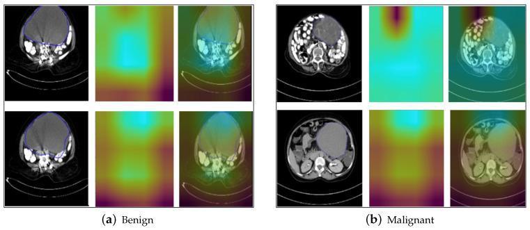
A diagnostic imaging comparison featuring Grad-CAM (Gradient-weighted Class Activation Mapping) results for benign and malignant ovarian tumor classification using axial CT scans. The figure is divided into two sections: (a) Benign and (b) Malignant samples. Each section displays three columns: the original cropped CT image with manual annotation outlines, the standalone Grad-CAM heatmap, and the heatmap overlaid on the CT image. The benign samples demonstrate tumors with relatively smooth, well-defined boundaries and more homogeneous internal textures; the corresponding Grad-CAM heatmaps show concentrated, localized activation areas. In contrast, the malignant samples display tumors with irregular shapes and less distinct margins. The Grad-CAM heatmaps for the malignant class show broader, more dispersed activation regions that extend into the surrounding pelvic tissue, suggesting the model utilizes features of potential local spread or invasion for classification. This comparison illustrates how deep learning interpretability tools identify morphological and peri-tumoral characteristics used to differentiate tumor types in gynecological oncology.
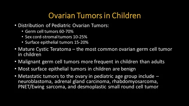
Comprehensive description: Pediatric ovarian tumors distribution and tumor types summarized from Rosai and Ackerman's Surgical Pathology (2011). This educational slide highlights that in children, germ cell tumors account for the largest share of ovarian neoplasms, comprising approximately 60–70%, with sex cord-stromal tumors 10–25% and surface epithelial tumors about 15–20%. Mature cystic teratoma is noted as the most common ovarian germ cell tumor in pediatric patients, often presenting as a benign adnexal mass. The slide emphasizes that malignant germ cell tumors are more frequent in children than in adults, underlining the need for age-appropriate diagnostic and therapeutic strategies. It also states that most surface epithelial tumors in children are benign, influencing surgical planning and prognosis. Metastatic involvement of the ovary in the pediatric population is listed to include neuroblastoma, adrenal cortical carcinoma, rhabdomyosarcoma, primitive neuroectodermal tumor (PNET)/Ewing sarcoma, and desmoplastic small round cell tumor, informing differential diagnosis in advanced disease. The content supports clinical correlation with pediatric oncology, gynecologic oncology, and surgical pathology, guiding imaging interpretation, tumor marker assessment, and multidisciplinary management. This slide serves as a concise reference for medical students, residents, and researchers studying ovarian neoplasia in childhood and adolescence.
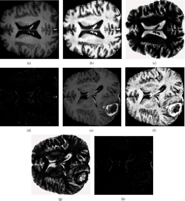
This composite image displays axial MRI brain scans processed through neutrosophic domain transformations for tumor classification. The figure is divided into two rows: the top row (a-d) represents a benign brain tumor case, while the bottom row (e-h) illustrates a malignant tumor case. For each case, four distinct states are shown: (a, e) Original T1-contrast enhanced (T1ce) MRI slices showing the anatomical structure and tumor mass; (b, f) the Truth (T) domain transformation, which enhances contrast and edge definition of the tumor and brain parenchyma; (c, g) the Falsity (F) domain, representing non-membership or background components with inverted intensity; and (d, h) the Indeterminacy (I) domain, which isolates regions of high uncertainty, specifically highlighting the edges and boundaries of the pathological mass and ventricles. The malignant case (e) demonstrates a more heterogeneous mass with clearer contrast enhancement compared to the benign case. This visualization facilitates the extraction of statistical texture features used in neural network-based diagnostic systems to differentiate between low-grade and high-grade gliomas.
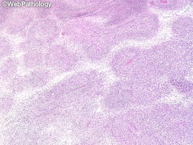
This Histopathology image depicts a malignant peripheral nerve sheath tumor (MPNST) in a soft tissue/nerve-associated mass. The tumor is highly cellular, with spindle-shaped neoplastic cells arranged in intersecting fascicles and occasional palisading and whorled patterns characteristic of nerve sheath neoplasia. Individual cells are predominantly monomorphic with slender, elongated morphology; nuclei are comma-shaped, wavy or buckled and hyperchromatic, and cytoplasm is indistinct and pale. Some tumor areas show plump oval to rounded nuclei, reflecting variable cellular differentiation. The growth demonstrates alternating hypercellular regions and hypocellular zones embedded in a myxoid stroma, indicating mucopolysaccharide-rich extracellular matrix. Focal nuclear palisading and regionally organized whorls resemble tactoid-like differentiation, a feature described in MPNST. Hyalinized cords and nodules may be present; there may be scant mitotic activity in some fields. Vessel wall involvement by tumor cells is noted in places, illustrating aggressive infiltration. Overall, the lesion lacks uniform whorling of benign schwannomas and exhibits malignant cytologic features with heterogeneous architecture. Immunophenotypic correlation (e.g., SOX10/S100) is helpful for confirmation in practice, but not visible here. This histologic profile supports a diagnosis of MPNST and carries important prognostic and therapeutic implications, including surgical excision with clear margins and consideration of adjuvant therapy for patient-specific management and follow-up.
Classification of Neoplasia
1. Definition
- Parenchyma - the neoplastic cells themselves (determines behavior and classification)
- Stroma - reactive connective tissue, blood vessels, and immune cells (supports growth)
2. Primary Classification: Benign vs. Malignant
| Feature | Benign | Malignant |
|---|---|---|
| Differentiation | Well differentiated; resembles tissue of origin | Ranges from well-differentiated to anaplastic (undifferentiated) |
| Rate of growth | Usually slow; mitoses rare and normal | Often rapid; mitoses frequent and may be atypical |
| Local invasion | Non-invasive; remains localized; often encapsulated | Invasive; infiltrates surrounding tissue; no true capsule |
| Metastasis | Does not metastasize | Frequently metastasizes (defining feature of malignancy) |
| Outcome | Usually curable by local excision | Can be fatal; prognosis depends on grade and stage |
Note: "Benign" is not synonymous with "harmless." Benign tumors in critical locations (e.g., intracranial meningioma) can still cause fatal complications.
3. Classification by Tissue of Origin (Nomenclature)
Table: Nomenclature of Selected Tumors
| Tissue of Origin | Benign | Malignant |
|---|---|---|
| CONNECTIVE TISSUE | ||
| Fibrous tissue | Fibroma | Fibrosarcoma |
| Adipose tissue | Lipoma | Liposarcoma |
| Cartilage | Chondroma | Chondrosarcoma |
| Bone | Osteoma | Osteosarcoma |
| ENDOTHELIUM / RELATED | ||
| Blood vessels | Hemangioma | Angiosarcoma |
| Lymph vessels | Lymphangioma | Lymphangiosarcoma |
| Mesothelium | - | Mesothelioma |
| Brain coverings | Meningioma | Invasive meningioma |
| BLOOD CELLS | ||
| Hematopoietic cells | - | Leukemias |
| Lymphoid tissue | - | Lymphomas |
| MUSCLE | ||
| Smooth muscle | Leiomyoma | Leiomyosarcoma |
| Striated muscle | Rhabdomyoma | Rhabdomyosarcoma |
| SKIN / EPITHELIUM | ||
| Stratified squamous | Squamous cell papilloma | Squamous cell / epidermoid carcinoma |
| Basal cells | - | Basal cell carcinoma |
| Melanocytes | Nevus | Melanoma |
| GLANDULAR EPITHELIUM | Adenoma / Cystadenoma / Papilloma | Adenocarcinoma / Cystadenocarcinoma |
| ORGAN-SPECIFIC | ||
| Lung | Bronchial adenoma | Bronchogenic carcinoma |
| Kidney | Renal tubular adenoma | Renal cell carcinoma |
| Liver | Hepatic adenoma | Hepatocellular carcinoma |
| Bladder | Urothelial papilloma | Urothelial carcinoma |
| Placenta | Hydatidiform mole | Choriocarcinoma |
| Testis | - | Seminoma |
| Ovary | Serous/mucinous cystadenoma | Embryonal carcinoma; serous/mucinous cystadenocarcinoma |
| MIXED CELL TUMORS | ||
| Salivary gland | Pleomorphic adenoma (mixed tumor) | Malignant mixed tumor |
| Kidney (embryonal) | - | Wilms tumor (nephroblastoma) |
| GERM CELL TUMORS | Mature teratoma (dermoid cyst) | Immature teratoma |
4. Major Categories Expanded
A. Epithelial Tumors
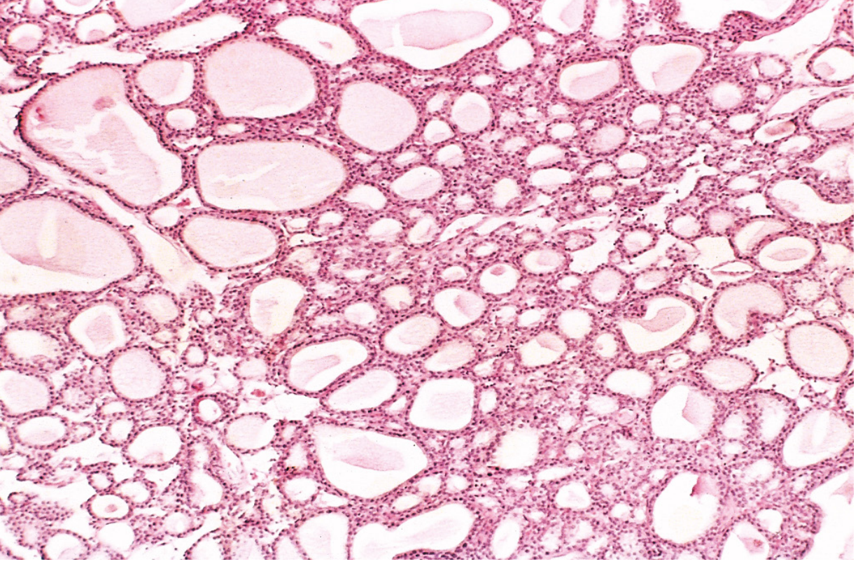
- Adenoma - benign epithelial tumor from glandular tissue (e.g., colonic adenoma, thyroid adenoma)
- Papilloma - fingerlike projections from surface epithelium
- Cystadenoma - large cystic masses (e.g., ovarian cystadenoma)
- Polyp - any tumor projecting from a mucosal surface into a hollow structure (may be benign or malignant)
- Carcinoma - malignant epithelial tumor; most common human cancer
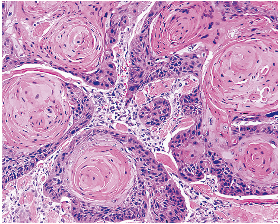
- Squamous cell carcinoma - from squamous/stratified epithelium (skin, oral cavity, cervix, esophagus, lung)
- Adenocarcinoma - glandular differentiation (breast, colon, lung, prostate, endometrium)
- Transitional (urothelial) cell carcinoma - from urothelium
- Basal cell carcinoma - from basal cells of skin
B. Mesenchymal (Connective Tissue) Tumors
- Osteosarcoma (bone), Chondrosarcoma (cartilage)
- Leiomyosarcoma (smooth muscle), Rhabdomyosarcoma (striated muscle)
- Liposarcoma (fat), Fibrosarcoma (fibrous tissue), Angiosarcoma (vessels)
C. Hematopoietic and Lymphoid Tumors
- Leukemias - malignant proliferation of hematopoietic cells in bone marrow and blood
- Lymphomas - solid tumors of lymphoid origin (Hodgkin and Non-Hodgkin)
- Multiple myeloma - malignant plasma cell tumor
D. Neural Tumors
- Benign: Neurofibroma, Schwannoma, Meningioma
- Malignant: Glioblastoma (GBM), Neuroblastoma, Medulloblastoma, Malignant peripheral nerve sheath tumor (MPNST)
E. Melanocytic Tumors
- Benign: Nevus (mole)
- Malignant: Melanoma - aggressive; arises from melanocytes of skin, eye, mucous membranes
F. Germ Cell Tumors
- Teratoma - contains elements from >1 germ layer
- Mature teratoma (dermoid cyst) - benign; well-differentiated tissues (skin, hair, teeth)
- Immature teratoma - malignant; embryonal tissue
- Seminoma / Dysgerminoma - most common testicular malignancy
- Choriocarcinoma - highly malignant; from trophoblastic tissue
- Yolk sac (endodermal sinus) tumor - most common germ cell tumor in children
G. Mixed Tumors
- Pleomorphic adenoma (parotid salivary gland) - benign mixed epithelial/mesenchymal tumor
- Fibroadenoma of the breast - benign mixed glandular and stromal proliferation
- Wilms tumor (nephroblastoma) - malignant; contains blastema, stroma, and epithelial elements
H. Teratomas (Totipotential Cells)
- Mature (benign) vs. Immature (malignant)
5. Eponyms and Exceptions to Nomenclature Rules
| Name | Origin | Behavior |
|---|---|---|
| Melanoma | Melanocytes | Malignant |
| Lymphoma | Lymphoid cells | Malignant |
| Leukemia | Hematopoietic cells | Malignant |
| Seminoma | Testicular germ cells | Malignant |
| Mesothelioma | Mesothelium | Malignant |
| Hepatoblastoma | Liver embryonal cells | Malignant |
| Hamartoma | Disorganized native tissue | Benign (not a true neoplasm) |
| Choristoma | Normal tissue in wrong location | Benign (heterotopic rest) |
6. Differentiation and Grading
- Pleomorphism - variation in cell and nuclear size/shape
- Abnormal nuclear morphology - hyperchromatic nuclei, coarse chromatin, prominent nucleoli
- High nuclear:cytoplasmic (N:C) ratio
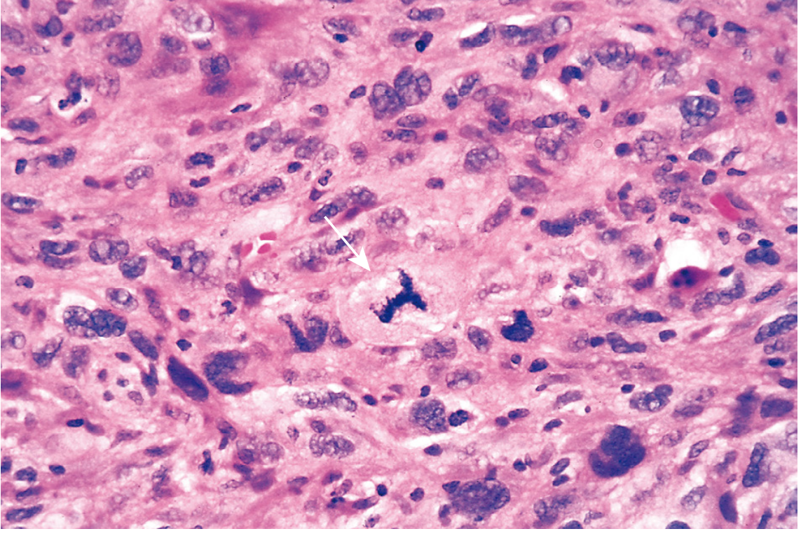
- Atypical mitoses - tripolar or quadripolar spindles
- Loss of polarity - loss of normal cellular orientation
- Grade I (well differentiated) - closely resembles normal tissue
- Grade II (moderately differentiated)
- Grade III (poorly differentiated) - little resemblance to normal
- Grade IV (undifferentiated/anaplastic) - no resemblance
7. Staging (Clinical Extent)
- T (Tumor) = size and local extent (T1-T4)
- N (Nodes) = regional lymph node involvement (N0-N3)
- M (Metastasis) = distant metastasis (M0 or M1)
8. Routes of Metastatic Spread
- Lymphatic spread - most common initial route for carcinomas; follows natural lymphatic drainage (e.g., breast cancer to axillary nodes)
- Hematogenous spread - typical of sarcomas; tumor cells enter veins; liver and lung are most common sites
- Seeding of body cavities - especially peritoneum (e.g., ovarian carcinoma causing pseudomyxoma peritonei), pleura, pericardium, CSF
Histology Gallery
Summary Mnemonic: Benign vs. Malignant
| Benign | Malignant | |
|---|---|---|
| Suffix | -oma | -carcinoma / -sarcoma |
| Differentiation | Good | Poor → anaplastic |
| Capsule | Usually present | Absent |
| Growth rate | Slow | Fast |
| Invasion | No | Yes |
| Metastasis | No | Yes |
| Recurrence | Rare | Common |
| Prognosis | Good | Guarded to poor |