
Perthes disease case presentation
Perthes disease Legg-Calve-Perthes avascular necrosis femoral head X-ray
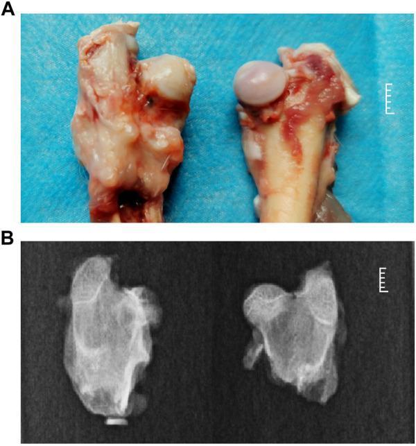
This composite educational image illustrates the phenotypic hallmarks of Legg-Calvé-Perthes disease (Perthes disease) in a rabbit model compared to a healthy control. Panel A presents a gross clinical photograph of excised proximal femurs. The Perthes model (left) shows a misshapen, flattened femoral head with a pale, opaque surface and loss of normal articular gloss. In contrast, the healthy control (right) displays a smooth, spherical, and glistening femoral head. Panel B shows corresponding X-ray diagnostic images. The Perthes disease specimen (left) exhibits a loss of spherical structure, heterogeneous bone density (sclerosis and lucency), and focal collapse of the femoral head epiphysis. The healthy control (right) demonstrates uniform bone density and a well-preserved, rounded epiphysis. This visual comparison highlights the osteonecrotic changes, structural deformity, and cartilage thickening characteristic of ischemic necrosis of the femoral head, modeling the clinical progression observed in pediatric patients. The images serve as a comparative reference for orthopedic pathology and experimental models of avascular necrosis.
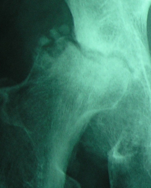
**Modality:** Plain radiography (X-ray) of the hip. **Anatomical Region:** Proximal femur and acetabulum. **Observed Pathology:** Advanced stage of Legg-Calvé-Perthes disease or severe avascular necrosis (AVN) of the femoral head. **Visual Features:** * **Morphology:** Significant collapse and severe flattening of the femoral head epiphysis, resulting in a characteristic "hatchet-like" deformity. * **Articular Surface:** There is a pronounced eccentric depression of the lateral articular surface. * **Alignment:** Evidence of superolateral subluxation of the femoral head relative to the acetabulum. * **Bone Density:** Marked sclerosis within the femoral head, interspersed with subchondral lucencies and defects, indicating ongoing bone remodeling and necrosis. * **Joint Space:** Notable joint space narrowing and incongruity between the femoral head and the acetabular roof. **Diagnostic Features:** The combination of severe epiphyseal flattening, subchondral collapse, and lateral subluxation are hallmark signs of end-stage ischemic necrosis and joint secondary degeneration. This appearance is crucial for classifying disease severity using the Waldenström or Herring systems.
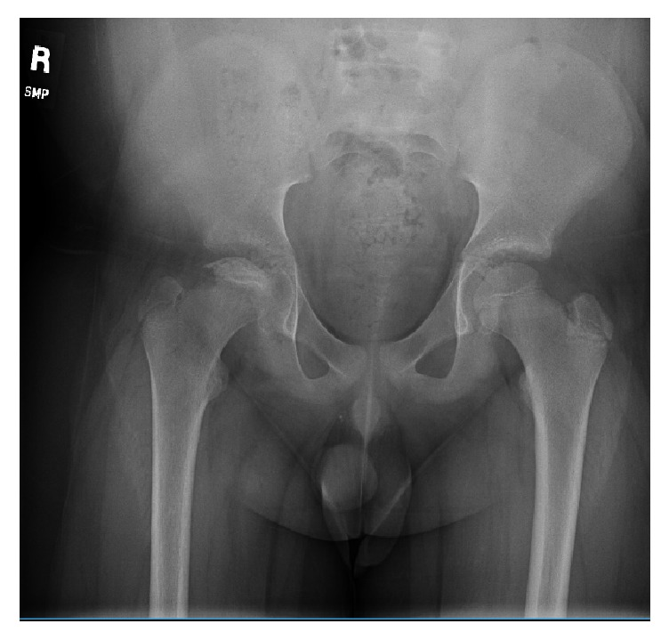
**Modality:** Anterior-posterior (AP) pelvic radiograph. **Anatomy:** Pediatric pelvis including the bilateral proximal femurs, hips, and pelvic ring. **Pathology:** Bilateral Legg-Calvé-Perthes disease (LCPD) or avascular necrosis of the femoral heads. **Visual Features:** - **Right Hip:** Significant fragmentation and flattening of the femoral capital epiphysis. Increased radiodensity (sclerosis) of the epiphysis is visible, indicating the necrotic phase. There is visible widening of the joint space and metaphyseal radiolucency. - **Left Hip:** Subtle subchondral lucency (crescent sign) and mild flattening of the superior femoral head surface, consistent with early articular collapse. - **General:** The triradiate cartilage is open, confirming a pediatric skeleton. The acetabular morphology appears preserved at this stage. **Diagnostic Features:** Asymmetrical involvement of the bilateral femoral heads characterized by collapse, sclerosis, and fragmentation. These findings are diagnostic of osteochondrosis of the capital femoral epiphysis. **Retrieval Keywords:** AP Pelvis X-ray, Legg-Calvé-Perthes disease, femoral head collapse, avascular necrosis, pediatric orthopedics, fragmentation phase, subchondral lucency.
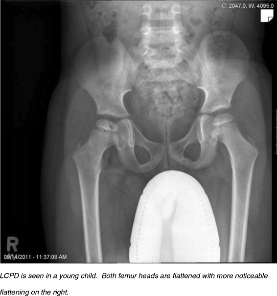
**Imaging Modality:** Anteroposterior (AP) pelvic radiograph (X-ray). **Anatomical Region:** Pediatric pelvis and bilateral hip joints. **Observed Pathology:** Bilateral Legg-Calvé-Perthes Disease (LCPD), characterized by idiopathic avascular necrosis of the proximal femoral epiphyses. **Characteristic Visual Features:** * **Femoral Heads:** Significant bilateral flattening (coxa plana), fragmentation, and increased radiopacity (sclerosis) of the capital femoral epiphyses. * **Symmetry:** The findings are asymmetrical, with more pronounced collapse and fragmentation observed on the right femoral head compared to the left. * **Joint Space:** Preservation of the joint space is noted, though the right femoral head shows lateral subluxation/extrusion relative to the acetabulum. * **Femoral Necks:** Relative widening and shortening of the bilateral femoral necks (coxa magna) are visible, consistent with remodeling secondary to epiphyseal ischemia. **Diagnostic Differentiating Features:** The combination of epiphyseal flattening, fragmentation, and sclerosis in a skeletally immature patient is pathognomonic for the fragmentation or re-ossification stages of Legg-Calvé-Perthes disease. The absence of destructive articular changes helps differentiate this from septic arthritis or inflammatory arthropathies.
Legg-Calve-Perthes disease management
Perthes Disease (Legg-Calve-Perthes Disease) - Case Presentation
Case Vignette
- Insidious onset of intermittent right hip pain, worse with activity, relieved by rest
- Referred pain noticed in the right thigh and knee (no knee pathology found on examination)
- Mild limp noticed by parents; worsening over the last 2 weeks
- No history of trauma, fever, or systemic illness
- No history of steroid use, sickle cell disease, or prior hip surgery
Examination Findings
| Finding | Detail |
|---|---|
| Gait | Antalgic limp (right side) |
| Trendelenburg sign | Positive right |
| Hip movements | Reduced internal rotation and abduction (right hip); other movements relatively preserved early in disease |
| Muscle wasting | Buttock, thigh, and calf atrophy may develop |
| Limb length | Up to 2.5 cm discrepancy in advanced disease |
| General | Afebrile; no groin lymphadenopathy |
Key clinical pearl: Pain is often referred to the knee - always examine the hip when a child presents with knee pain. - Rosen's Emergency Medicine, p. 2685
Epidemiology & Aetiology
- Incidence: More common in boys (3:1 male predominance); peak age 4-7 years (range 3-11 years)
- Bilateral disease: 10-15% of cases; bilateral disease raises suspicion for an underlying systemic cause
- Aetiology: Unknown/idiopathic. Proposed mechanisms include coagulation disorder with thrombotic occlusion of femoral head vasculature. Socioeconomic deprivation and passive smoking are implicated risk factors.
- Important: Later onset (>8 years) carries a poor prognosis - Rosen's Emergency Medicine, p. 2685
- Steroid use
- Infection/post-surgical
- Sickle cell disease
- Hypothyroidism
- Multiple epiphyseal dysplasia
Pathology & Disease Stages
| Stage (Waldenström) | Pathology | Radiographic Features |
|---|---|---|
| 1. Initial (Ischaemic) | Femoral head loses blood supply; subchondral collapse begins | Medial joint space widening; small femoral head; subchondral fracture = Crescent sign |
| 2. Fragmentation | Resorption of dead bone; femoral head begins fragmenting; new bone forms | Fragmentation of epiphysis; sclerosis + lucency; head deformity develops |
| 3. Reossification | Revascularisation; bone density progressively restored | Progressive re-ossification; head reshaping |
| 4. Healed | Bony healing complete; residual deformity permanent | Residual coxa plana, coxa magna, or normal femoral head |
The cartilage "follows" the shape of the reossifying epiphysis - deformity that occurs during fragmentation is irreversible. - Bailey and Love's, p. 640
Investigations
Imaging
- May be normal in first 6 weeks of symptoms
- Follow-up radiograph at 6 weeks if still symptomatic with normal initial X-ray
- Waldenström stages can be identified
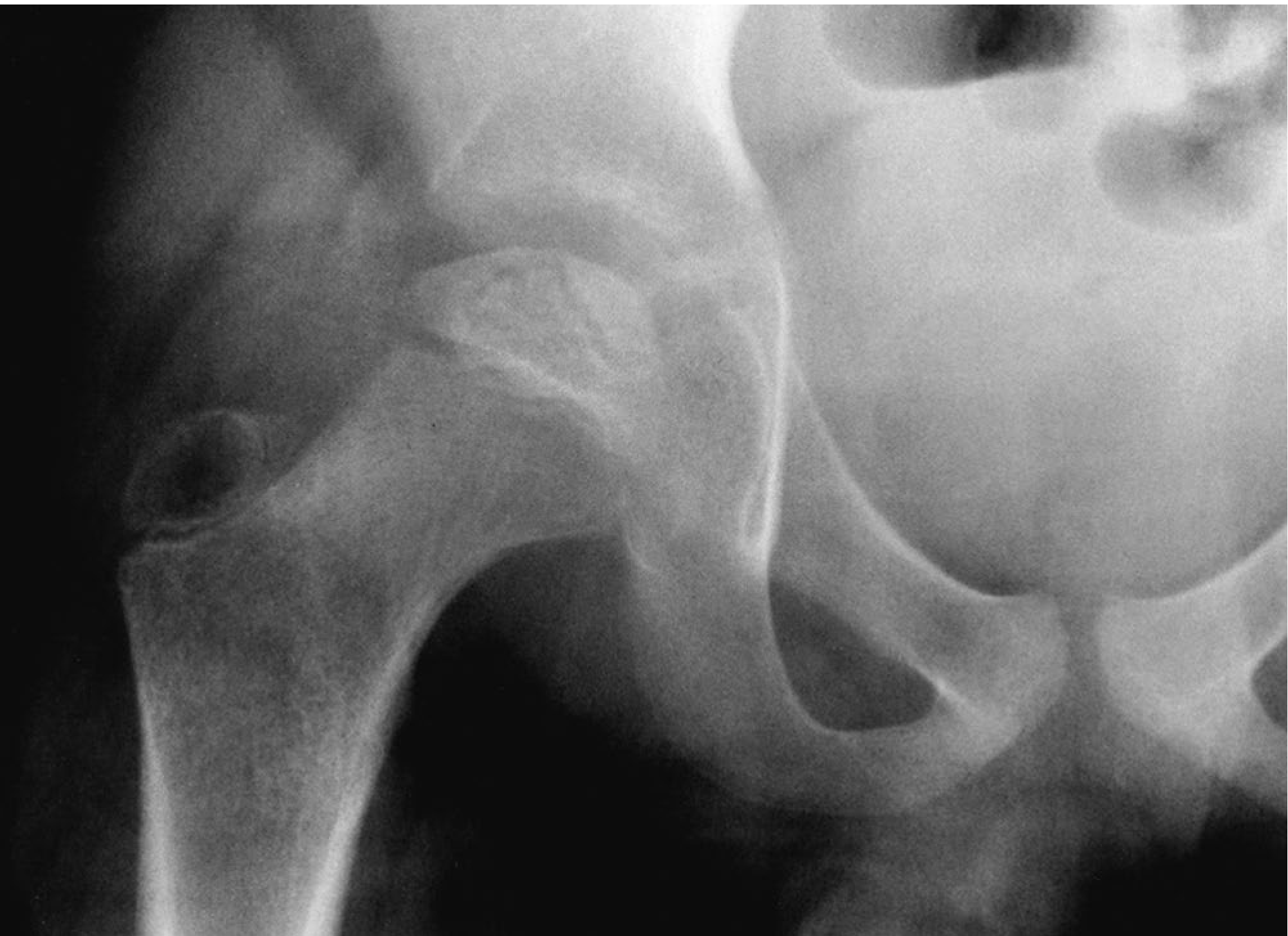
- Most sensitive for early AVN changes before X-ray changes appear
- Recommended when X-rays are normal but symptoms persist >6 weeks
- A 2026 systematic review (PMID: 41308557) confirms diffusion-weighted MRI has high diagnostic performance for LCPD
Classification Systems
1. Herring Lateral Pillar Classification (most widely used - best interobserver reliability)
| Grade | Lateral Pillar Height | Prognosis |
|---|---|---|
| A | Full height maintained; no density change | Uniformly good |
| B | >50% height maintained | Poor if bone age >9 years |
| B/C Border | Narrowed (~50% height) | Intermediate |
| C | <50% height maintained | Poor in all patients |
- Applied at the fragmentation stage (usually ~6 months after symptom onset)
- Based on AP pelvis radiograph - Campbell's Operative Orthopaedics 15th Ed, p. 1476
2. Catterall Classification (extent of epiphyseal involvement)
| Group | Description |
|---|---|
| I | Anterior epiphysis only |
| II | Anterior epiphysis + clear sequestrum |
| III | Only a small portion of epiphysis uninvolved |
| IV | Total head involvement |
- Gage sign - V-shaped radiolucency in lateral epiphysis/metaphysis
- Calcification lateral to the epiphysis
- Lateral subluxation of the femoral head
- Horizontal proximal femoral physis
3. Salter-Thompson Classification
- Type A: Subchondral fracture <50% of dome - good prognosis
- Type B: Subchondral fracture >50% of dome - surgery recommended
4. Stulberg Classification (outcome at skeletal maturity)
- Class I-II: Round femoral head (good long-term outcome)
- Class III-IV: Aspherical head (moderate/poor outcome)
- Class V: Flat femoral head in dysplastic acetabulum (poor outcome)
Management
General Principles
- Younger age at onset = better prognosis (more remodelling potential)
- Herring A: conservative management
- Herring B in patients <8 years: either conservative or surgical (comparable outcomes)
- Herring B/C or C: surgery generally recommended
- Age >8 years with Herring B or C: surgery
Conservative (Non-Surgical)
| Treatment | Detail |
|---|---|
| Analgesia + physiotherapy | Maintain range of joint movement; core treatment |
| Activity restriction | Limitation of weight-bearing in active phase |
| Orthopaedic follow-up | Mandatory for all suspected cases |
| Crutches/wheelchairs | Discouraged - promote flexion/adduction posture |
| Abduction bracing | Does not alter natural history per Bailey and Love's |
Surgical Options
- Proximal femoral varus osteotomy - redirects femoral head into acetabulum
- Innominate (Salter) osteotomy - redirects acetabulum to contain the head
- Combined femoral + pelvic osteotomy
- Shelf acetabuloplasty
- Chiari osteotomy
- Surgical hip dislocation with relative femoral neck lengthening
- Arthrodiastasis (distraction arthroplasty) for late-onset disease
- Hip arthroplasty in adults with sequelae (13% of LCPD patients ultimately require THA) - Campbell's Operative Orthopaedics, p. 1533
Prognosis
- Better prognosis: Age <6 years, male sex, Herring A, Catterall I-II, unilateral disease
- Poor prognosis: Age >8 years, Herring B/C-C, Catterall III-IV, bilateral involvement, head-at-risk signs
- Bilateral disease: 48% rate as Stulberg III/IV at maturity
- Secondary osteoarthritis risk in adulthood; 13% eventually require hip arthroplasty
Key Points Summary
- Most common in boys aged 4-7 years
- AVN leads to femoral head collapse; return of blood supply heralds resorption, reossification ("healing")
- Prognosis better in younger children who have more remodelling potential before skeletal maturity
- Management aims to maintain femoral head sphericity
- Treatment is non-surgical (maximise ROM) or surgical (early for containment, late for salvage)
- Campbell's Operative Orthopaedics 15th Ed (2026), Chapter 34
- Bailey and Love's Short Practice of Surgery 28th Ed, Chapter 44
- Rosen's Emergency Medicine: Concepts and Clinical Practice, Chapter 170
- Mohebbi et al. (2026). Diagnostic performance of DWI in LCPD: Systematic Review. Radiography (Lond).