
Soft tissue sarcoma
Reading File
Now I'll search the medical library for soft tissue sarcoma content in parallel.
Finding Sources
Searching PubMed
"soft tissue sarcoma"[MeSH Terms] AND treatment
Excellent - several strong sources found. Let me read the key sections in parallel.
Reading File
Reading File
Searching PubMed
"sarcoma"[MeSH Terms] AND "treatment outcome"[MeSH Terms]
Good content. Let me read more from the Sabiston chapter (full epidemiology, staging, treatment) and the Miller's clinical pearls table in parallel.
Reading File
Reading File
Good content from Sabiston. Let me now read the clinical pearls table and more Sabiston on staging/treatment.
Reading File
Reading File
Excellent - I now have very comprehensive content. Let me fetch the taxonomy figure and check the Robbins pathology section for histopathology details.
Loading Image
Reading File
Excellent - I have very comprehensive content now. I have enough to write a thorough, well-structured answer covering epidemiology, classification, risk factors, diagnosis, staging, treatment, and key clinical pearls. Let me also check the Sabiston staging section which was truncated.
Reading File
I now have comprehensive content from multiple authoritative sources. Let me compile the full answer.
Soft Tissue Sarcoma (STS)
Overview
Soft tissue sarcomas are a diverse group of approximately 70 distinct malignant neoplasms arising from mesenchymal tissues - skeletal muscle, adipose cells, blood and lymphatic vessels, and other connective tissue - as well as peripheral nerves (neuroectodermal origin). They are relatively rare, accounting for ~1% of cancer incidence and ~2% of cancer-related deaths in the United States (~13,400 new cases estimated annually).
The taxonomy of STS is extensive. The phylogeny below illustrates the major lineages:
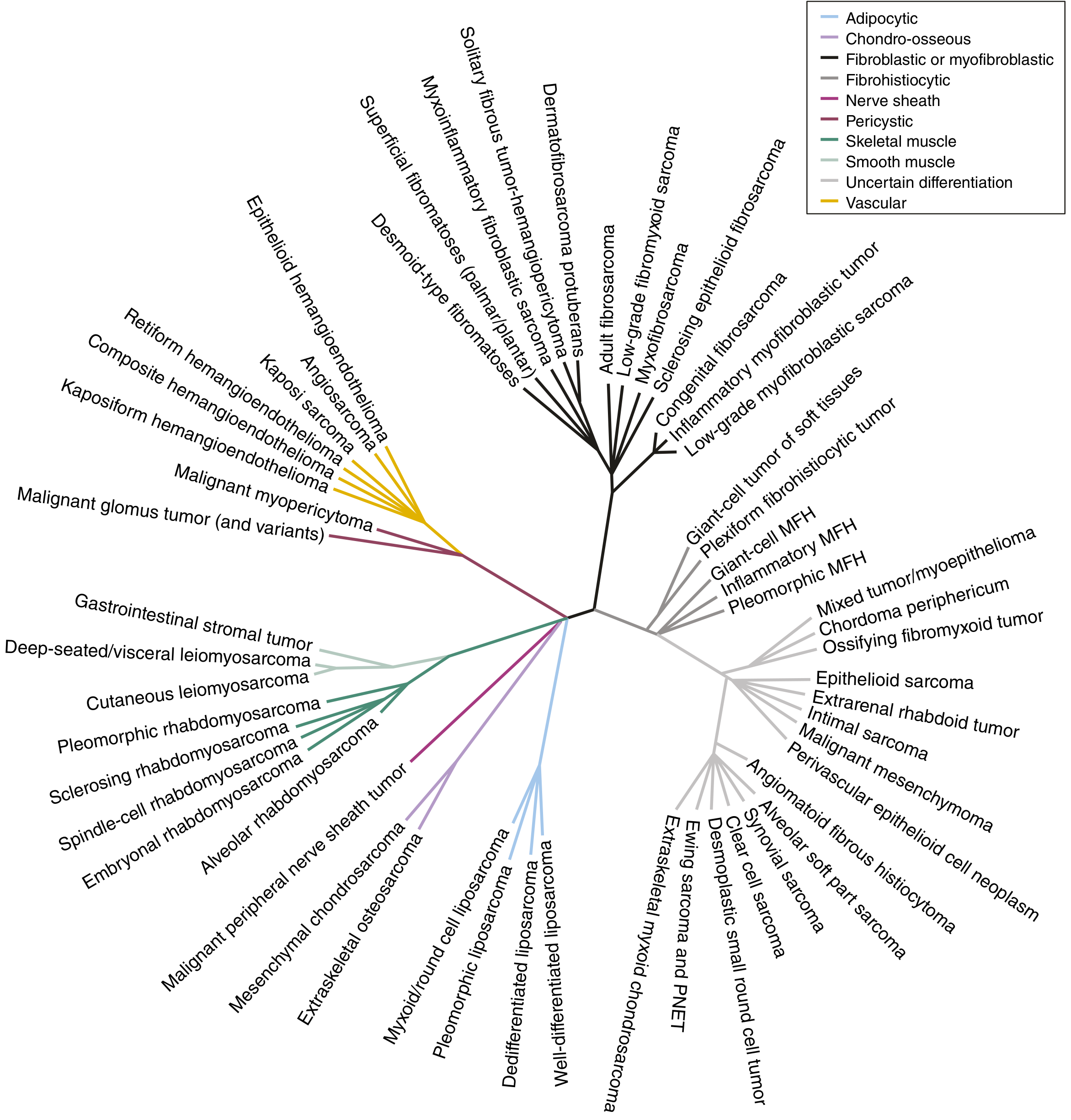
FIGURE 64.1 from Sabiston Textbook of Surgery: Unrooted phylogeny of ~60 STS subtypes, color-coded by lineage (adipocytic, smooth muscle, skeletal muscle, nerve sheath, vascular, etc.)
Epidemiology & Distribution
- Most common site: Thigh (44% of extremity STS), followed by other proximal limb locations
- Extremity > retroperitoneum > intraperitoneal in frequency
- Age linked to histologic subtype: Rhabdomyosarcoma, alveolar soft part sarcoma, and neurofibroma affect children/young adults; most adult STS is sporadic in middle-aged to older patients
- Wide clinical spectrum: from indolent low-grade neoplasms (e.g., well-differentiated liposarcoma) to highly aggressive tumors (e.g., angiosarcoma, desmoplastic small round cell tumor)
Risk Factors
Germline / Hereditary Syndromes
| Syndrome | Gene | Associated STS |
|---|---|---|
| Li-Fraumeni | TP53 (17p13.1) | Rhabdomyosarcoma, UPS, pleomorphic sarcoma |
| Neurofibromatosis type 1 (NF-1) | NF1 | Malignant peripheral nerve sheath tumor (MPNST) |
| FAP / Gardner syndrome | APC (5q21) | Desmoid tumors (esp. post-colectomy) |
| Hereditary retinoblastoma | RB1 (13q14) | Osteosarcoma, STS |
| Carney-Stratakis syndrome | SDHB/C/D | GIST + paraganglioma |
Environmental / Acquired
- Radiation exposure - post-radiation angiosarcoma; Stewart-Treves syndrome (lymphangiosarcoma in chronic lymphedema post-mastectomy)
- Chemical carcinogens (vinyl chloride, arsenic, dioxin) - hepatic angiosarcoma
Classification: Major Subtypes
1. Undifferentiated Pleomorphic Sarcoma (UPS)
- Previously called malignant fibrous histiocytoma (MFH)
- Location: Deep soft tissues of extremities, especially the thigh; middle-aged/older adults
- Histology: Sheets of large anaplastic polygonal cells with hyperchromatic bizarre nuclei; storiform (cartwheel) pattern of spindle and histiocytic cells around slit-like vessels; abundant atypical mitotic figures
- Complex karyotype from genomic instability
- Metastases in 17-50% of cases; generally poor prognosis
2. Liposarcoma
- Rarely arise in the subcutaneous tissues
- Hallmark: lipoblasts (signet ring-shaped cells); IHC: MDM2-positive
- Subtypes:
- Well-differentiated (low-grade, t(12;16) or 12q13-15 amplification)
- Dedifferentiated
- Myxoid/round cell - translocation t(12;16)(q13;p11) - FUS-DDIT3 fusion
- Pleomorphic (high-grade)
3. Rhabdomyosarcoma (RMS)
- Most common pediatric STS
- Alveolar subtype: translocation t(2;13) - PAX3-FOXO1 fusion
- Treatment: chemotherapy ± limb salvage surgery (unlike most other STS)
4. Synovial Sarcoma
- Common in young adults; foot and lower extremity
- Translocation t(X;18) - SYT-SSX fusion products
- One of the ESARC tumors (lymph node metastasis risk)
5. MPNST (Malignant Peripheral Nerve Sheath Tumor)
- NF-1 associated or de novo
- Histology: spindle cells in sweeping fascicles, whorled/nodular areas; S-100 positive on IHC
6. Leiomyosarcoma
- Histology: fascicular growth with spindle cells intersecting at right angles; IHC: SMA and desmin positive
7. Angiosarcoma
- Cells resemble vascular endothelium
- Highly malignant; lymph node and cutaneous metastases common
- Associated with Stewart-Treves syndrome
- May require amputation for local control
8. Epithelioid Sarcoma
- Most common STS of the hand; occurs in young adults
- Part of ESARC group (lymph node metastasis)
9. Clear Cell Sarcoma
- Common tumor of lower extremity/foot; young adults
- Lymph node metastasis risk (ESARC)
ESARC tumors (those with higher risk of lymph node metastases):
Epithelioid sarcoma, Synovial sarcoma, Angiosarcoma, Rhabdomyosarcoma, Clear cell sarcoma
Staging
Key prognostic factors:
| Factor | Adverse |
|---|---|
| Size | ≥5 cm |
| Grade | High grade |
| Depth | Deep to superficial fascia |
| Location | Retroperitoneal worse than extremity |
AJCC staging uses TNM + histologic grade (G1-3). High-grade, deep, >5 cm tumors are Stage III regardless of nodal status.
Diagnosis
Presentation
- Most common presentation: painless mass without prior evaluation
- Mimics include hypertrophic scar, myositis ossificans, hematoma, abscess, lipoma, cyst
- Red flags warranting workup: mass >5 cm, deep to fascia, fixed, growing
Imaging
- MRI - imaging modality of choice for trunk/extremity STS
- T1: decreased signal
- T2: increased signal
- Characterizes tumor relationship to neurovascular structures
- Chest CT - most frequent site of metastasis is the lung
- Additional imaging per subtype: abdomen/pelvis CT for myxoid liposarcoma, epithelioid sarcoma, leiomyosarcoma; paraspinal MRI for myxoid liposarcoma; brain imaging for alveolar soft part sarcoma, clear cell, angiosarcoma
Biopsy
- Core-needle biopsy is the standard approach (percutaneous, image-guided)
- Open incisional biopsy if core needle is non-diagnostic
- Biopsy tract must be oriented so it can be resected en bloc with the specimen
- Small (≤2 cm), superficial, mobile masses may be excised upfront with wide margins in selective cases
- Molecular/NGS testing is increasingly routine - changed diagnosis in 13% of patients in one series; FISH/RT-PCR for specific translocations
Treatment
Trunk and Extremity STS
Surgical principles:
- Wide local resection with negative margins is the cornerstone of treatment
- Limb salvage surgery (LSS) has replaced amputation as the standard; equivalent survival outcomes with better functional results
Radiation:
- Lesions >5 cm: LSS + radiation (XRT) - pre- or postoperative
- Lesions <5 cm, subcutaneous: LSS alone may suffice
- Preoperative radiation: lower total dose, smaller field, higher rate of wound complications
- Postoperative radiation: lower wound complication rate, but larger field and higher total dose
Chemotherapy:
- Standard first-line: doxorubicin ± ifosfamide
- Rhabdomyosarcoma: chemotherapy is a primary treatment modality (VAC regimen)
- Subtype-specific agents: trabectedin for leiomyosarcoma and myxoid liposarcoma; pazopanib for non-adipocytic STS; imatinib for GIST (KIT/PDGFRA mutation)
Retroperitoneal STS
- Surgery remains the cornerstone but complete resection is more challenging
- Higher local recurrence rates than extremity STS
- Role of preoperative radiation is being actively studied
Molecular Features Summary
| STS Subtype | Translocation / Mutation | Fusion Product |
|---|---|---|
| Synovial sarcoma | t(X;18) | SYT-SSX |
| Myxoid liposarcoma | t(12;16)(q13;p11) | FUS-DDIT3 |
| Alveolar rhabdomyosarcoma | t(2;13) | PAX3-FOXO1 |
| MPNST | NF1 loss | - |
| DFSP | t(17;22) | COL1A1-PDGFB |
| GIST | KIT / PDGFRA mutations | - |
| Well-diff. liposarcoma | 12q13-15 amplification | MDM2/CDK4 |
Recent Evidence (2024-2025)
Three notable recent RCTs are worth highlighting:
-
Pembrolizumab + radiation + surgery vs. radiation + surgery for Stage III extremity STS (SU2C-SARC032, Lancet 2024, PMID 39547252) - evaluating immunotherapy integration in localized disease
-
Doxorubicin-trabectedin + trabectedin maintenance vs. doxorubicin alone in leiomyosarcoma (NEJM 2024, PMID 39231341) - showed improved progression-free survival with combination therapy
Key Clinical Pearls (Miller's Orthopaedics)
- All STS subtypes share the same presentation, imaging, treatment, and differential - it is histology (and molecular signature) that distinguishes them
- ESARC = the 5 STS subtypes prone to lymph node metastasis
- NF-1 → MPNST; Stewart-Treves → angiosarcoma; FAP → desmoid
- Characteristic locations: Hand = epithelioid sarcoma; Foot = clear cell / synovial sarcoma
- Biopsy tract orientation is critical - must be excisable en bloc with the tumor
Sources:
- Sabiston Textbook of Surgery, 21st ed., Ch. 64 (Soft Tissue and Retroperitoneal Sarcoma)
- Miller's Review of Orthopaedics, 9th ed., Ch. 9 (Table 9.19, ESARC)
- Robbins, Cotran & Kumar Pathologic Basis of Disease (Undifferentiated Soft Tissue Sarcoma)
This is a shared conversation. Sign in to Orris to start your own chat.