
Atrial septum defect
atrial septal defect types anatomy diagram
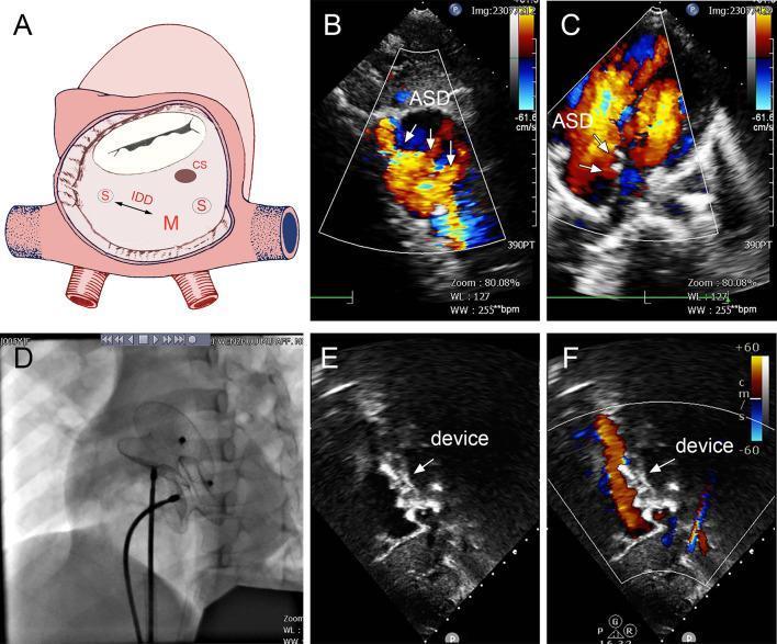
This composite figure illustrates the diagnosis and transcatheter management of multiple atrial septal defects (ASDs). (A) Anatomical diagram showing the interatrial septum with multiple defects: a moderate defect (M) and two small defects (S) labeled with interdefect distance (IDD). (B, C) Transthoracic echocardiography (TTE) with Color Doppler in different planes demonstrating bidirectional, mosaic-colored shunting (red and blue) through the atrial septal defects, confirming turbulent flow. (D) Fluoroscopic radiographic image showing the deployment of two catheter-based occluder devices (MemoPart ASDO) positioned within the heart. (E) Post-operative grayscale TTE showing the echogenic occluder devices securely positioned across the septum. (F) Post-operative Color Doppler echocardiography demonstrating the absence of residual shunting across the atrial septum, indicating successful procedural closure of the multiple defects. This content is designed for cardiology and pediatric educational purposes, highlighting the use of multiple occluders for complex septal anatomy.
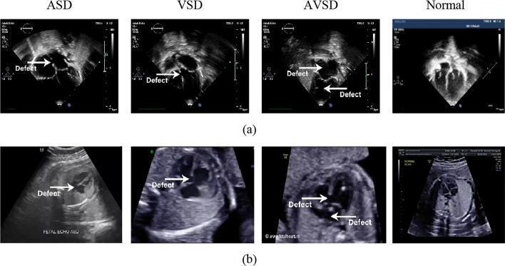
This diagnostic comparison chart features two rows of grayscale ultrasound images demonstrating congenital cardiac septal defects. Row (a) presents postnatal echocardiograms, and row (b) displays prenatal (fetal) echocardiograms. The columns categorize the findings into four types: Atrial Septal Defect (ASD), Ventricular Septal Defect (VSD), Atrioventricular Septal Defect (AVSD), and Normal anatomy. Across both rows, white arrows labeled 'Defect' indicate specific anatomical interruptions. The ASD images highlight a gap in the interatrial septum, while the VSD images show an opening in the interventricular septum. The AVSD panel demonstrates a larger communication involving both septa, resulting in a common atrioventricular junction. The 'Normal' column provides a reference for intact septal integrity and clear chamber separation in the apical four-chamber view. The prenatal scans (row b) demonstrate typical fetal imaging characteristics, including surrounding uterine and fetal structures, compared to the more focused cardiac imaging of the postnatal scans. This visual comparison is designed for pediatric cardiology education, focusing on the identification of septal morphology across developmental stages.
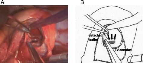
This composite image consists of an intraoperative clinical photograph (A) and a corresponding anatomical line diagram (B) demonstrating the surgical repair of a ventricular septal rupture (VSR). The view is from a right atrial approach after a right atrial oblique incision. In Figure A, surgical forceps and retractors are shown manipulating cardiac tissue to expose the ventricular septum. In Figure B, the anatomical landmarks are clearly labeled, including the tricuspid valve (TV) annulus and a detached portion of the tricuspid valve leaflet. Three black arrows in the diagram point to the site of the ventricular septal rupture, which is located in the basal posterior part of the ventricular septum, immediately posterior to the tricuspid valve annulus. The detachment of the tricuspid valve leaflet is a key surgical maneuver used here to improve visualization and access to the septal defect. This material illustrates complex cardiac surgical anatomy and the transatrial approach for post-infarct VSR repair, a critical procedure in cardiothoracic surgery.
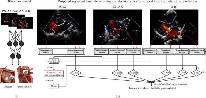
Educational diagram illustrating two artificial intelligence frameworks for Atrial Septal Defect (ASD) treatment planning. (a) A 'Black-box model' uses a neural network to map multiview Doppler transthoracic echocardiogram (TTE) images (PSSAX, SXLAX, A4C) directly to binary treatment options: surgical or transcatheter closure. (b) The 'Deep Keypoint Stadiometry' (DKS) model employs an interpretable algorithmic approach. It extracts clinical measurements from three echocardiographic views: Parasternal Short Axis (PSSAX) for defect diameter and atrial posterior wall distance; Subxiphoid Long Axis (SXLAX) for defect diameter and superior/inferior vena cava distances; and Apical Four-Chamber (A4C) for defect diameter, atrial roof, atrial annulus, and septum length. These anatomical measurements feed into a Boolean decision logic using clinical thresholds (e.g., >5mm, >7mm). If all criteria are satisfied, the model recommends transcatheter closure and calculates the proposed ASD occluder size based on the maximum defect diameter plus a 4mm margin. The diagram highlights the transition from opaque deep learning to interpretable, expert-knowledge-based clinical decision support.
"atrial septal defect" AND management
Atrial Septal Defect (ASD)
Definition & Epidemiology
Embryology
- The septum primum grows down from the roof of the common atrium, leaving an anterior gap called the ostium primum (allows right-to-left shunting in the fetus)
- Before the septum primum obliterates the ostium primum, a posterior opening - the ostium secundum - forms within it
- The septum secundum then grows to the right of the septum primum, covering the ostium secundum but leaving a channel - the foramen ovale
- In fetal life, right atrial pressure exceeds left atrial pressure (lungs non-functional), keeping the foramen ovale open
- At birth, lung expansion drops pulmonary vascular resistance, left atrial pressure rises above right, and the valve of the foramen ovale closes - permanently sealed in ~80% by age 2
- Robbins Pathologic Basis of Disease, p. 504
Types of ASD
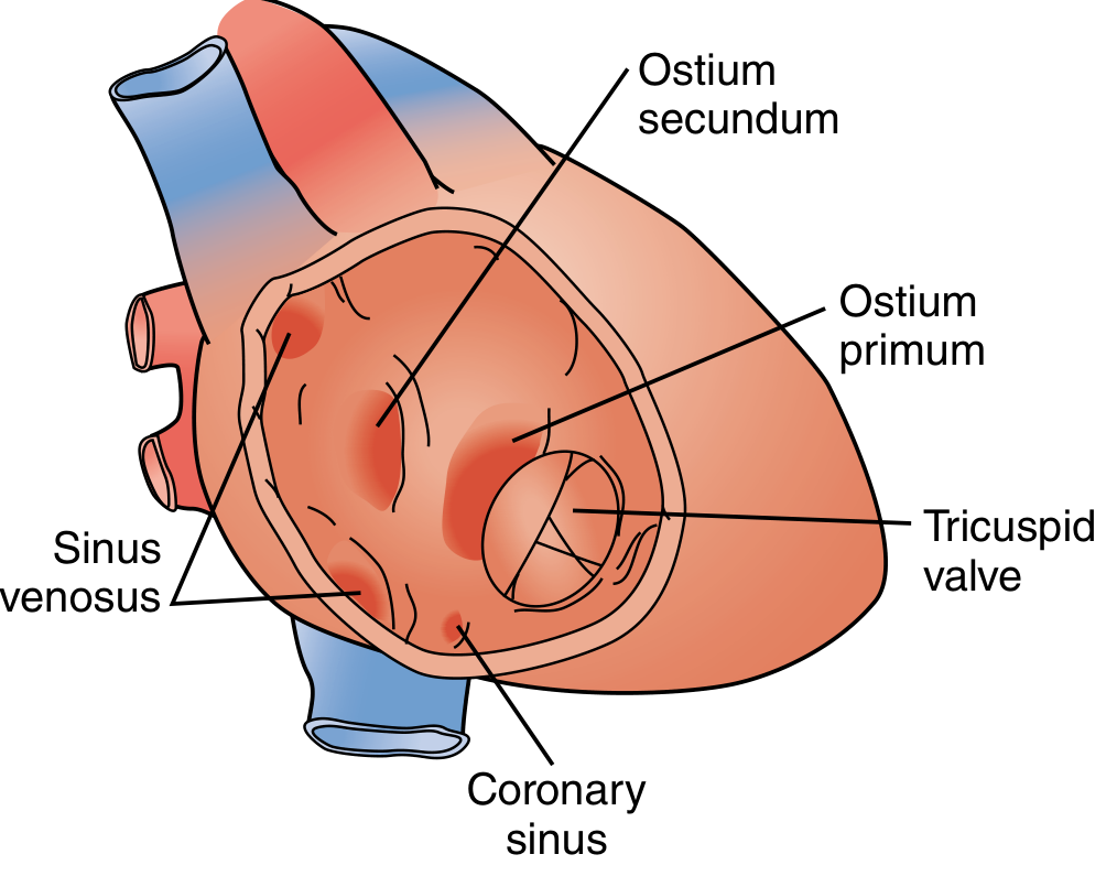
| Type | % of ASDs | Location | Associations |
|---|---|---|---|
| Secundum ASD | 90% | Center of septum, fossa ovalis region - deficient septum secundum | Usually isolated; can be single, multiple, or fenestrated |
| Primum ASD | 5% | Adjacent to AV valves | AV valve abnormalities, VSD (part of endocardial cushion defect / AV canal) |
| Sinus venosus | 5% | Near SVC (superior) or IVC (inferior) entry | Partial anomalous pulmonary venous return (PAPVR) |
| Coronary sinus defect | Rare | Unroofed coronary sinus | Persistent left SVC |
- Robbins Pathologic Basis of Disease; Sabiston Textbook of Surgery
Pathophysiology
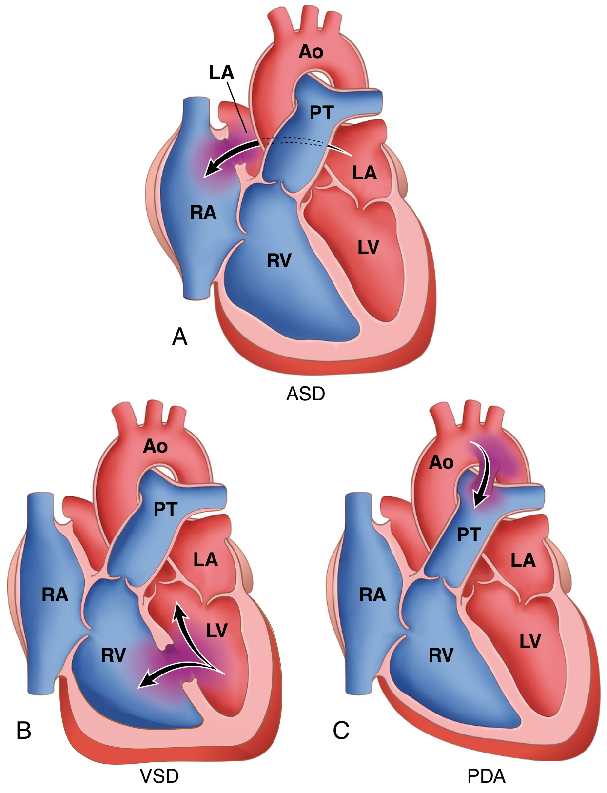
- Lower pulmonary vascular resistance (PVR) vs. systemic resistance
- Greater compliance of the right ventricle vs. left ventricle
-
Right heart volume overload and enlargement (RA + RV dilation)
-
Pulmonary plethora (increased pulmonary blood flow, 2-8x normal)
-
Transient right-to-left shunting occurs during increased intrathoracic pressure (Valsalva, coughing), creating risk for paradoxical embolism and stroke
-
Pulmonary hypertension is unusual in secundum ASD but can develop late
-
Irreversible pulmonary vascular disease is rare compared to VSD
-
Sabiston Textbook of Surgery; Goldman-Cecil Medicine
Clinical Features
Symptoms
- Most children are asymptomatic or have mild exercise intolerance / recurrent respiratory infections
-
70% become symptomatic by the fifth decade
- Adults: dyspnea on exertion, palpitations, fatigue, atrial arrhythmias (AF, flutter)
- Right heart failure signs with advanced disease
- Stroke or TIA from paradoxical embolism
Physical Examination
-
Wide, fixed splitting of S2 - hallmark sign (fixed because increased venous return in inspiration raises RA pressure and reduces shunt, offsetting respiratory S2 variation; P2 delayed due to RV overload)
-
Soft midsystolic ejection murmur at 2nd left intercostal space (increased flow across pulmonary valve)
-
Mid-diastolic murmur at lower left sternal border (increased tricuspid flow in large shunts)
-
Right ventricular heave if significant shunt
-
Dilated pulmonary artery palpable in 2nd left ICS
-
Goldman-Cecil Medicine, p. 984
Investigations
ECG
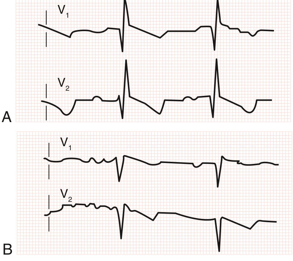
- Right axis deviation
- Prolonged PR interval
- Atrial fibrillation or flutter in adults
- Left axis deviation in primum ASD (due to AV node displacement)
Chest X-Ray
- Pulmonary vascular plethora (increased markings bilaterally)
- Dilated main pulmonary artery and branches
- Right atrial and right ventricular enlargement
Echocardiography (Diagnostic)
- Transthoracic echo: identifies ostium primum and secundum ASDs directly
- Sinus venosus ASD can be missed on TTE - must be specifically sought
- Doppler: quantifies pulmonary artery pressures and estimates shunt ratio (Qp/Qs)
- Transesophageal echo (TEE): better for sinus venosus defects and pre-procedure planning
Cardiac Catheterization
-
Demonstrates step-up in oxygen saturation at the RA level
-
Quantifies Qp/Qs and pulmonary vascular resistance
-
Goldman-Cecil Medicine, p. 985-986
Management
Indications for Closure
- Evidence of right heart enlargement (with or without symptoms)
- Symptomatic patients
- Qp/Qs > 1.5:1
- History of paradoxical embolism
1. Transcatheter (Percutaneous) Closure
- Now used in >60% of ASD interventions worldwide
- Suitable for: centrally located (secundum) ASDs up to 3.5 cm; requires adequate septal rims
- Device: Amplatzer Septal Occluder (most widely used)
- Avoids sternotomy and cardiopulmonary bypass
- Not suitable for primum ASD or sinus venosus ASD
2. Surgical Closure
-
Open repair via cardiopulmonary bypass - standard since the late 1950s
-
Techniques: direct suture closure, autologous pericardial patch, or prosthetic patch
-
Indicated for:
- Large/complex defects not amenable to catheter closure
- Primum ASD (with AV valve repair)
- Sinus venosus ASD (+ redirection of anomalous pulmonary veins)
- Associated congenital anomalies requiring concomitant repair
-
Virtually no residual or recurrent defects
-
Minimally invasive surgical approaches (robotic, mini-thoracotomy) now common with good outcomes - 2025 systematic review (PMID 40517112) confirms equivalence to open sternotomy
-
Sabiston Textbook of Surgery; Goldman-Cecil Medicine
Prognosis
- Surgical/transcatheter mortality is low; postoperative survival is comparable to unaffected population when corrected before pulmonary hypertension develops
-
70% develop impairment by the 5th decade if left untreated
- Risk of atrial arrhythmias persists even after closure, especially if repaired late
- Small ASDs (<5 mm) may close spontaneously in children
Patent Foramen Ovale (PFO) - Distinction
-
No resting shunt detectable on physical exam
-
No ECG abnormality
-
Diagnosis: TEE with agitated saline (bubble study) during Valsalva maneuver
-
Clinical relevance: paradoxical embolism, cryptogenic stroke
-
Robbins Pathologic Basis of Disease, p. 914; Goldman-Cecil Medicine, p. 989