
Bacteria classified 7 groups with images and difference between gram postive and negative bacteria with images
Reading File
Now let me search the medical library and web simultaneously for bacteria classification and gram staining content with images.
Finding Sources
Finding Sources
Searching the Web
bacteria 7 classification groups with images microbiology
Reading File
Finding Sources
Searching the Web
gram positive vs gram negative bacteria difference cell wall structure diagram
Reading File
Reading File
Loading Image
Loading Image
Now let me fetch the textbook gram stain diagram and a few more key images:
Loading Image
Finding Sources
Searching the Web
7 groups classification of bacteria cocci bacilli spirochetes mycoplasma rickettsiae actinomycetes chlamydiae
I now have rich textbook content, diagrams, and images. Let me also fetch a bacteria classification chart image:
Loading Image
Reading File
I now have all the content needed for a comprehensive, image-rich answer. Here is the full response:
Classification of Bacteria into 7 Groups
Bacteria are extraordinarily diverse. In medical microbiology, they are most practically classified by morphology (shape and arrangement), which forms 7 major groups. - Jawetz, Melnick & Adelberg's Medical Microbiology, 28th Ed.
Shapes of Bacteria
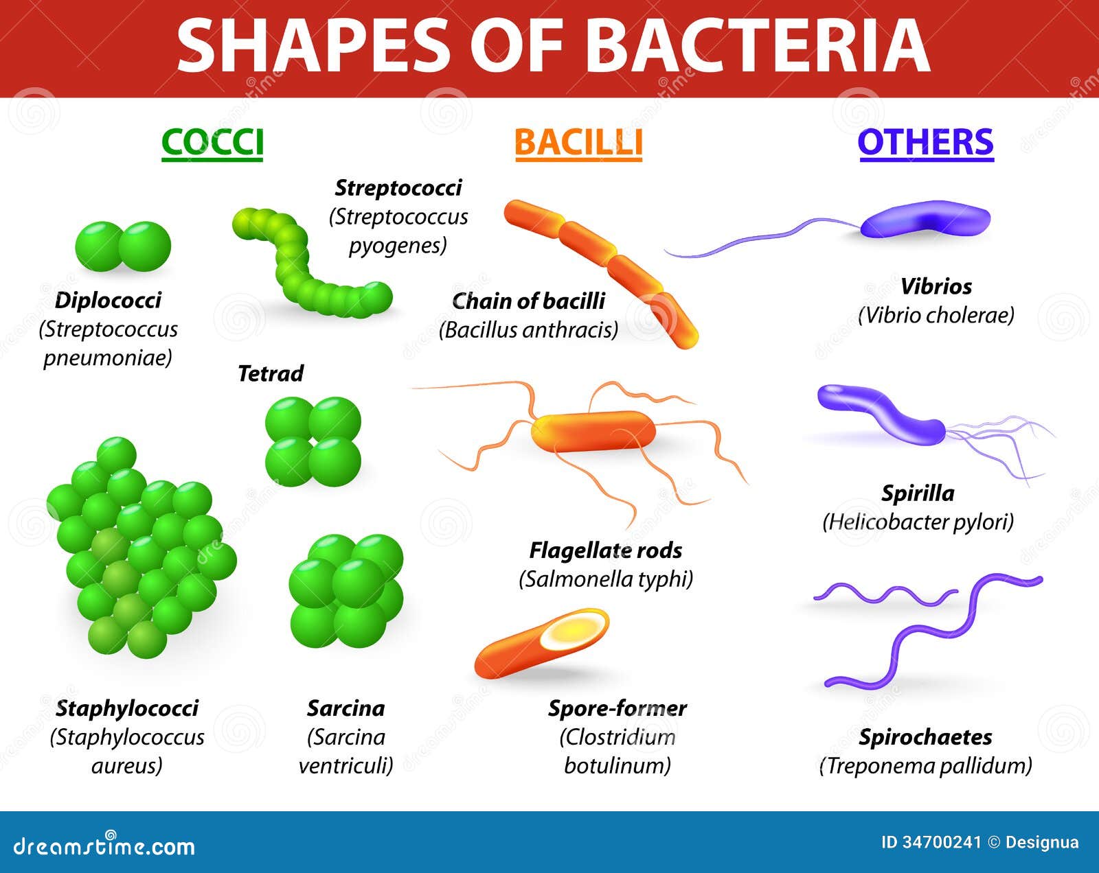
Group 1 - Cocci (Spherical Bacteria)
Round or oval-shaped bacteria. They appear in characteristic arrangements:
- Diplococci - pairs (e.g., Streptococcus pneumoniae, Neisseria meningitidis)
- Streptococci - chains (e.g., Streptococcus pyogenes)
- Staphylococci - grape-like clusters (e.g., Staphylococcus aureus)
- Tetrads - groups of 4 (e.g., Micrococcus)
- Sarcinae - cuboidal packets of 8 (e.g., Sarcina ventriculi)
Key diseases: Pneumonia, meningitis, skin infections, strep throat, food poisoning.
Group 2 - Bacilli (Rod-Shaped Bacteria)
Cylindrical or rod-shaped bacteria. The most common shape in nature.
- Single rods: Escherichia coli, Klebsiella pneumoniae
- Chains of rods: Bacillus anthracis (anthrax)
- Flagellate rods: Salmonella typhi
- Spore-forming rods: Clostridium botulinum, Bacillus anthracis
- Coccobacilli (very short rods): Haemophilus influenzae, Brucella
Key diseases: Typhoid fever, anthrax, food poisoning, urinary tract infections.
Group 3 - Spirilla (Rigid Spiral Bacteria)
Rigid, helical bacteria with external flagella. They are spiral-shaped but cannot flex.
- Example: Helicobacter pylori (peptic ulcer disease), Campylobacter jejuni (gastroenteritis)
- Move by flagella
Key diseases: Peptic ulcers, gastroenteritis, food poisoning.
Group 4 - Spirochetes (Flexible Spiral Bacteria)
Flexible, corkscrew-shaped bacteria. Unlike spirilla, they move by axial filaments (endoflagella) that run inside the outer sheath.
- Treponema pallidum - syphilis
- Borrelia burgdorferi - Lyme disease
- Leptospira interrogans - leptospirosis
Key feature: Too thin to be seen on Gram stain; require darkfield microscopy or silver staining.
Group 5 - Actinomycetes (Filamentous Branching Bacteria)
Filamentous bacteria that form branching hyphae resembling fungi. They produce spores.
- Actinomyces israelii - actinomycosis (lumpy jaw)
- Nocardia asteroides - nocardiosis (lung infections)
- Gram-positive; some are acid-fast (Nocardia)
Key feature: Form sulfur granules in tissue; important source of antibiotics (Streptomycin, Tetracycline from Streptomyces).
Group 6 - Mycoplasmas (Wall-less Bacteria)
The smallest free-living bacteria. They have no cell wall at all, making them naturally resistant to penicillin and beta-lactam antibiotics.
- Mycoplasma pneumoniae - atypical ("walking") pneumonia
- Mycoplasma genitalium - urogenital infections
- Ureaplasma urealyticum - urethritis, neonatal infections
Key feature: Pleomorphic (no fixed shape); require cholesterol for membrane stability; stain poorly on Gram stain.
Group 7 - Rickettsiae & Chlamydiae (Obligate Intracellular Bacteria)
These bacteria cannot survive outside a host cell - they lack metabolic machinery to generate their own ATP.
Rickettsiae:
- Transmitted by arthropod vectors (ticks, lice, fleas)
- Rickettsia rickettsii - Rocky Mountain spotted fever
- Rickettsia prowazekii - epidemic typhus
Chlamydiae:
- Transmitted via respiratory droplets or sexual contact
- Chlamydia trachomatis - STI, trachoma (blindness)
- Chlamydophila pneumoniae - atypical pneumonia
Key feature: Both are Gram-negative but stain poorly; require special staining (Giemsa, immunofluorescence).
Bacterial Classification Chart

Gram-Positive vs. Gram-Negative Bacteria
The Gram stain, developed in 1884 by Hans Christian Gram, is the single most important differential stain in microbiology. The result reflects fundamental differences in the bacterial cell envelope. - Jawetz, Melnick & Adelberg's Medical Microbiology, 28th Ed., p. 29
The Gram Staining Procedure (Step by Step)
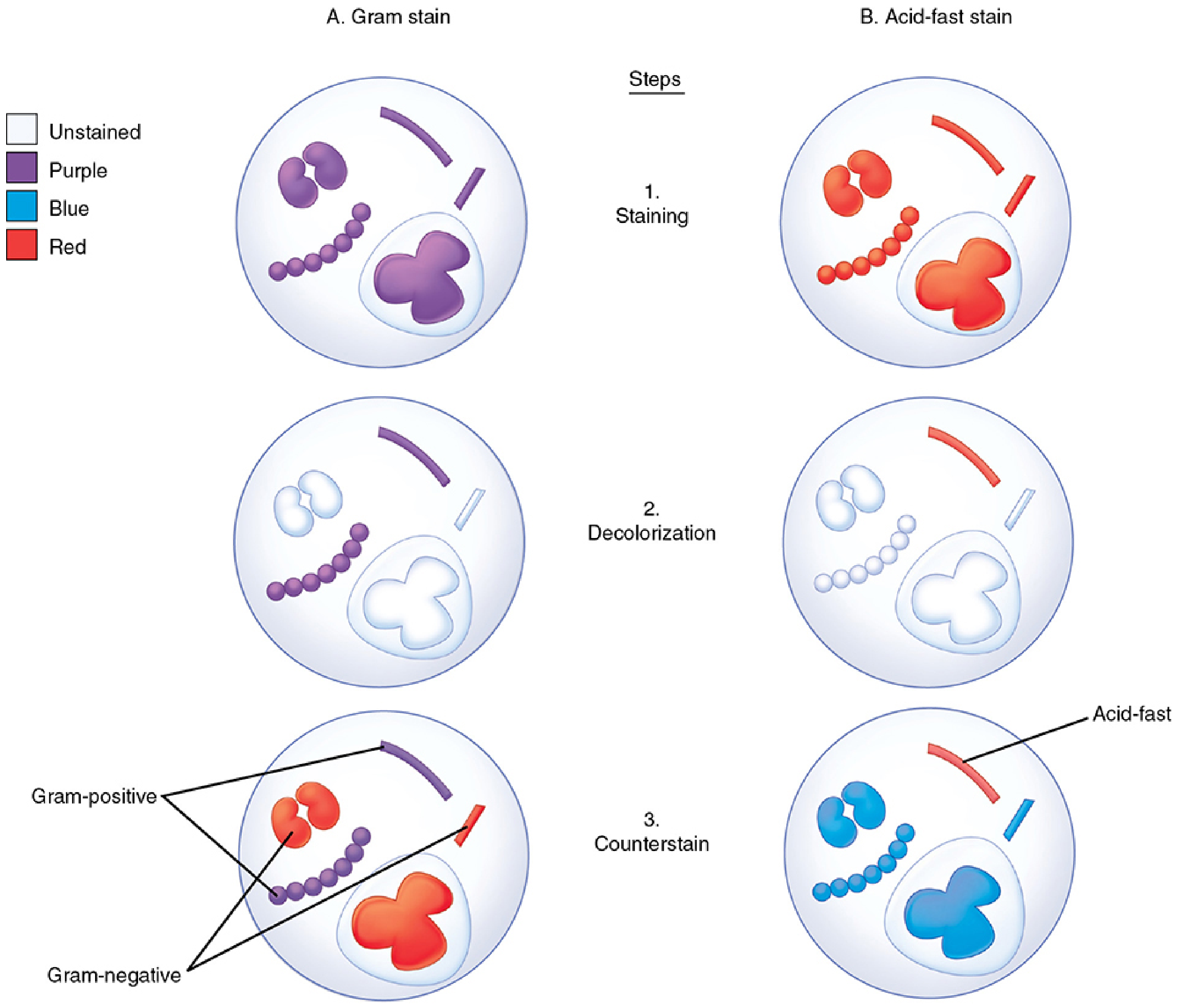
Steps: (1) Apply crystal violet - all bacteria stain purple. (2) Add iodine mordant - forms dye complex. (3) Decolorize with acetone/alcohol - Gram-negative cells lose dye. (4) Counterstain with safranin - Gram-negative cells turn pink/red.
Cell Wall Structure Comparison

Detailed Differences: Gram-Positive vs. Gram-Negative
| Feature | Gram-Positive | Gram-Negative |
|---|---|---|
| Color after Gram stain | Purple / Violet | Pink / Red |
| Peptidoglycan layer | Thick (20-80 nm, many layers) | Thin (10 nm, 1-2 layers) |
| Outer membrane | Absent | Present |
| Teichoic acids | Present (wall & lipoteichoic) | Absent |
| Lipopolysaccharide (LPS / Endotoxin) | Absent | Present |
| Porin proteins | Absent (no outer membrane) | Present (allow molecular passage) |
| Periplasmic space | Absent or minimal | Present (between membranes) |
| Susceptibility to penicillin | More susceptible | Less susceptible (outer membrane barrier) |
| Resistance to physical disruption | More resistant | Less resistant |
| Endotoxin (sepsis risk) | Lower | Higher (LPS triggers fever & sepsis) |
| Exotoxins | Commonly produced | Some produce (e.g., E. coli LT toxin) |
| Examples | Staphylococcus, Streptococcus, Bacillus, Clostridium | E. coli, Salmonella, Pseudomonas, Neisseria |
Source: Jawetz, Melnick & Adelberg's Medical Microbiology 28th Ed., Table 2-1
Why Does Gram Staining Work This Way?
- Gram-positive bacteria have a thick peptidoglycan meshwork that traps the crystal violet-iodine complex after decolorization. They stay purple.
- Gram-negative bacteria have a thin peptidoglycan layer plus an outer lipid membrane. The acetone/alcohol decolorizer dissolves this outer lipid layer, washing out the dye. They become colorless, then pick up the safranin counterstain (red/pink).
- An intact cell wall is required for a positive result - old, dead, or antibiotic-damaged Gram-positive bacteria may fail to retain the stain. - Sherris & Ryan's Medical Microbiology, 8th Ed.
Clinical Importance of Gram Staining
The Gram stain result directly guides antibiotic therapy before culture results are available:
- Gram-positive cocci in clusters → Think Staphylococcus → Use anti-staphylococcal agents
- Gram-positive cocci in chains → Think Streptococcus/Enterococcus → Use penicillin/ampicillin
- Gram-negative rods → Think Enterobacteriaceae (E. coli, Klebsiella) or Pseudomonas → Use broader-spectrum coverage
- Gram-negative diplococci → Think Neisseria → Use ceftriaxone
- Gram-positive rods → Think Bacillus or Clostridium → Adjust accordingly
The Gram-stained smear can achieve 70-80% sensitivity and 90-95% specificity for pneumococcal and H. influenzae pneumonia when performed correctly. - Sherris & Ryan's Medical Microbiology, 8th Ed.
This is a shared conversation. Sign in to Orris to start your own chat.