
Everything about anatomy of diaphragm in brief
Reading File
Now let me search the medical library for diaphragm anatomy content, in parallel with a web search.
Finding Sources
Searching the Web
anatomy of diaphragm origin insertion nerve supply openings
Gray's Anatomy for Students and Murray & Nadel's are the most relevant sources. Let me read them in parallel.
Reading File
Finding Sources
Let me get the detailed anatomy from the THIEME atlas which has a dedicated diaphragm table, and also search for nerve supply and openings.
Finding Sources
Finding Sources
Excellent - I now have strong data from Fischer's Mastery of Surgery and Current Surgical Therapy. Let me read the detailed anatomy section from Fischer's and the THIEME atlas.
Reading File
Reading File
I now have comprehensive content. Let me also fetch the diaphragm diagram image from Fischer's for visual clarity.
Reading File
Reading File
I now have very comprehensive information. Let me also fetch the diagram image.
Loading Image
Loading Image
I now have everything needed for a comprehensive, well-illustrated answer. Here is the full summary:
Anatomy of the Diaphragm
Overview
The diaphragm is a dome-shaped musculotendinous partition that separates the thoracic and abdominal cavities by sealing the inferior thoracic aperture. It is the primary muscle of inspiration - when it contracts, its domes flatten and thoracic volume increases, drawing air into the lungs. - Fischer's Mastery of Surgery, 8th ed.
Structure
The diaphragm has two components:
1. Muscular Part - Peripheral fibers arise radially from the margins of the inferior thoracic aperture and converge centrally.
2. Central Tendon - The aponeurotic center into which all muscle fibers insert. It is trifoliate (three-leafed) in shape, extends further anteriorly than posteriorly, and blends with the fibrous pericardium above. During respiration, the central tendon moves while the peripheral attachments remain fixed. - Fischer's Mastery of Surgery
The right dome rises higher (reaching rib V) than the left dome, partly because the liver supports it from below. - Gray's Anatomy for Students
Origin (Peripheral Attachments)
The diaphragm has three parts of origin:
| Part | Origin |
|---|---|
| Sternal part | Two muscular slips from the posterior surface of the xiphoid process |
| Costal part | Inner aspect of costal cartilages and ribs 7-12 |
| Lumbar part | Right crus (L1-L3/4), Left crus (L1-L2/3), via anterior longitudinal ligament |
Arcuate ligaments of the lumbar part:
- Median arcuate ligament - joins the two crura in the midline, arches over the aorta
- Medial arcuate ligament (medial lumbocostal arch) - from the body of L2 to its transverse process, over psoas major
- Lateral arcuate ligament (lateral lumbocostal arch) - from the transverse process of L2 to the tip of the 12th rib, over quadratus lumborum
A muscular weak spot called the lumbocostal trigone (Bochdalek triangle) exists between the costal and lumbar parts - a common site of congenital diaphragmatic hernia. - Fischer's Mastery of Surgery
Openings (Hiatuses)
There are three major openings, memorized by the mnemonic "8, 10, 12":
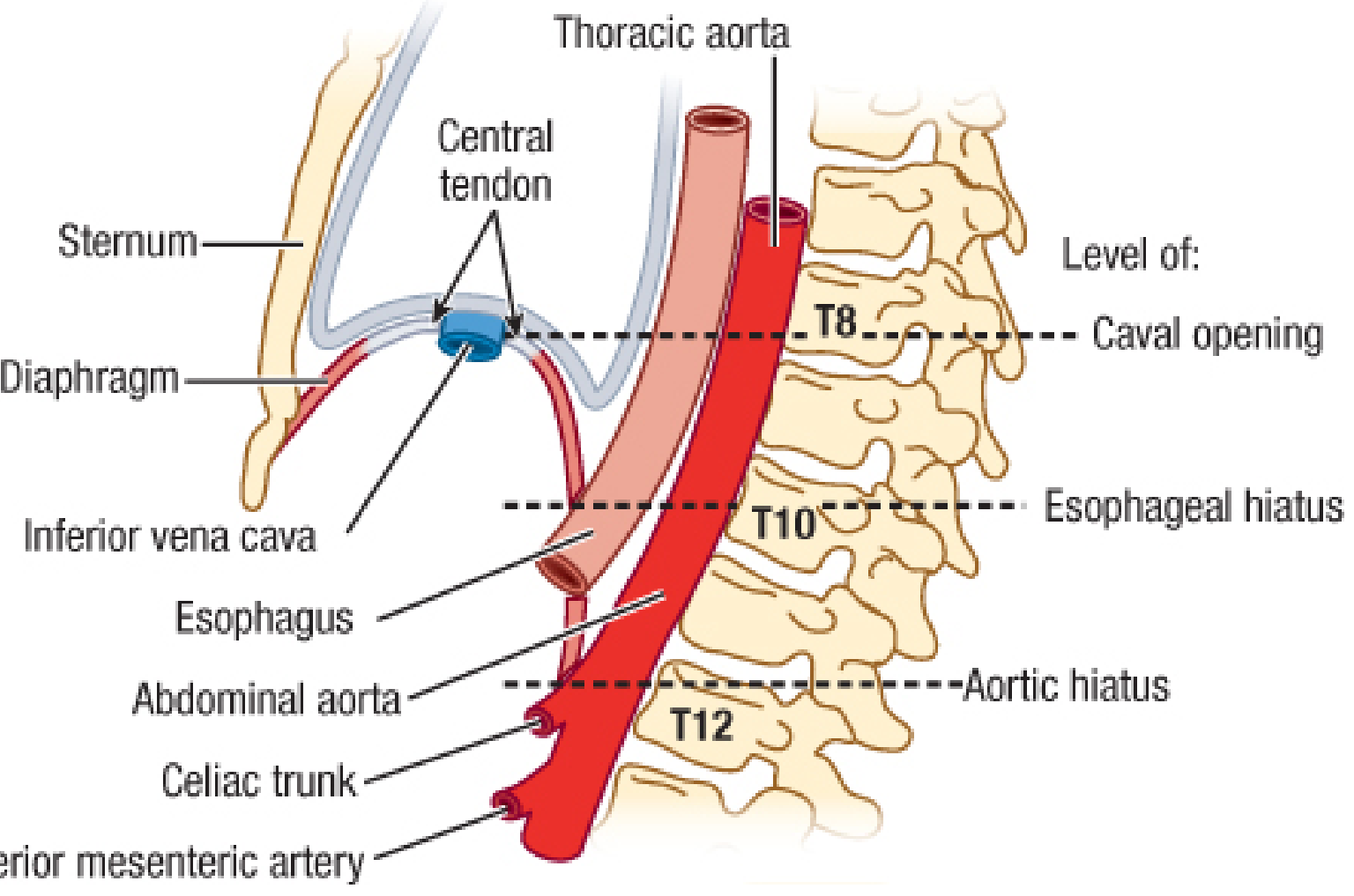
| Opening | Level | Structures Passing Through |
|---|---|---|
| Caval opening | T8 | IVC, right phrenic nerve, lymphatics |
| Esophageal hiatus | T10 | Esophagus, anterior + posterior vagal trunks, left gastric artery and vein |
| Aortic hiatus | T12 | Aorta (posterior to median arcuate ligament), thoracic duct, azygos vein |
Additional smaller openings:
- Sternocostal hiatus (foramen of Morgagni / space of Larrey) - between sternal and costal parts; transmits the superior epigastric artery (branch of internal thoracic artery) and lymphatics from the liver. A retrosternal/parasternal hernia can occur here.
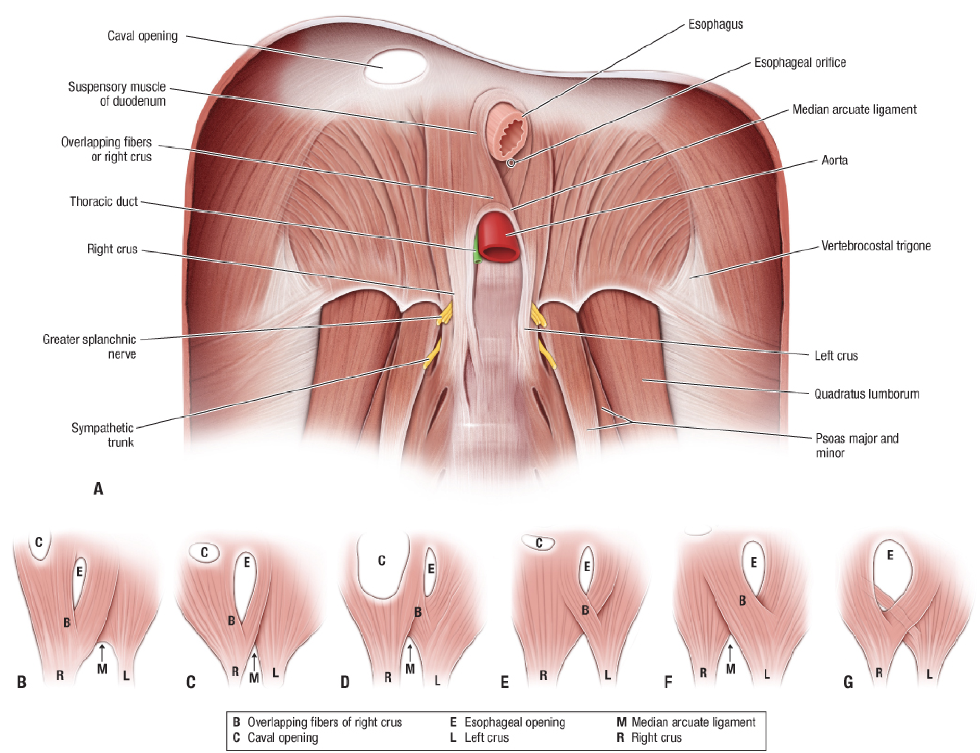
The esophageal hiatus is formed mainly by fibers of the right crus, which split to surround the esophagus like a sphincter. There is considerable variation (shown in the diagram below): - Fischer's Mastery of Surgery
Nerve Supply
Motor + Sensory (central):
- Phrenic nerves (C3, C4, C5 - "C3, 4, 5 keeps the diaphragm alive")
- They descend on the lateral surface of the fibrous pericardium, anterior to the lung root, and enter the diaphragm medially
- The left phrenic nerve enters the muscular part of the left hemidiaphragm just lateral to the cardiac surface
- The right phrenic nerve passes through the caval opening or central tendon
- Each phrenic nerve branches into: sternal (anteromedial), anterolateral, posterolateral, and crural branches
Sensory (peripheral):
- Intercostal nerves 9-11 supply the periphery of the diaphragm
Sympathetic fibers also reach the diaphragm via the inferior phrenic plexus (from the celiac plexus). - Fischer's Mastery of Surgery
Blood Supply
| Surface | Arterial Supply | Venous Drainage |
|---|---|---|
| Superior (thoracic) | Superior phrenic aa. (from thoracic aorta), musculophrenic + pericardiophrenic aa. (from internal thoracic) | Superior phrenic veins → azygos/hemiazygos |
| Inferior (abdominal) | Inferior phrenic aa. (branches of abdominal aorta or celiac trunk) | Inferior phrenic veins → IVC and left suprarenal vein |
The inferior phrenic arteries are the main supply to the abdominal surface. - Fischer's Mastery of Surgery
Lymphatic Drainage
- Superior surface - drains to anterior, middle, and posterior phrenic lymph nodes
- Inferior surface - drains to phrenic nodes (lymphatics pierce the diaphragm) and to aortic (para-aortic) nodes (following the inferior phrenic vessels)
Relations
| Side | Relations |
|---|---|
| Superior (thoracic) | Heart + pericardium (centrally), pleura + lungs (peripherally) |
| Inferior (abdominal) | Liver (right), stomach + spleen (left), kidneys and adrenals (posteriorly) |
The costodiaphragmatic recess - the pleural recess between the thoracic wall and diaphragm - is the most clinically important recess; it accommodates lung expansion during deep inspiration. - Gray's Anatomy for Students
Structures That Pass Posterior to the Diaphragm (Not Through It)
The sympathetic trunk, splanchnic nerves, and the azygos/hemiazygos veins pass posterior to the diaphragm under the medial and lateral arcuate ligaments rather than through a formal hiatus.
Clinical Correlates
- Hiatus hernia - herniation through an enlarged esophageal hiatus (most common diaphragmatic hernia in adults)
- Bochdalek hernia - congenital defect through the lumbocostal trigone, usually on the left; presents in neonates with respiratory distress
- Morgagni hernia - through the sternocostal hiatus; usually right-sided, often asymptomatic in adults
- Referred pain - phrenic nerve irritation (e.g., subphrenic abscess, ruptured spleen) causes referred pain to the shoulder tip (C4 dermatome)
Verterbral origin
The vertebral (lumbar) origin of the diaphragm is via the crura and arcuate ligaments:
- Right crus: L1 - L3 (sometimes L4)
- Left crus: L1 - L2 (sometimes L3)
- Median arcuate ligament: joins the two crura, arches over the aorta
- Medial arcuate ligament: L2 body → L2 transverse process (over psoas major)
- Lateral arcuate ligament: L2 transverse process → 12th rib tip (over quadratus lumborum)
From the previous textbook content already in context (Fischer's Mastery of Surgery), here is the detailed breakdown:
Vertebral Origin of the Diaphragm (Lumbar Part)
Crura
The crura are tendinous at their origin, blending with the anterior longitudinal ligament of the vertebral column:
| Crus | Vertebral Levels |
|---|---|
| Right crus | L1 to L3/L4 vertebral bodies, intervertebral discs, and anterior longitudinal ligament |
| Left crus | L1 to L2/L3 vertebral bodies, intervertebral discs, and anterior longitudinal ligament |
- The right crus is longer and larger than the left, because the aorta pushes the origin further to the right.
- The two crura are joined anteriorly by the median arcuate ligament, which arches over the front of the aorta at the T12/L1 level to form the aortic hiatus.
- Fibers of the right crus diverge and reconverge around the esophagus to form the esophageal hiatus (in most people).
Arcuate Ligaments
These are thickenings of the fascia covering posterior abdominal muscles - they serve as additional "ligamentous" vertebral origins:
| Ligament | From | To | Structure it arches over |
|---|---|---|---|
| Median arcuate ligament | Left crus | Right crus (midline junction) | Aorta (+ thoracic duct) |
| Medial arcuate ligament | Body of L2 | Transverse process of L2 | Psoas major muscle |
| Lateral arcuate ligament | Transverse process of L2 | Tip of 12th rib | Quadratus lumborum muscle |
Weak Points at the Vertebral Origin
-
Lumbocostal trigone (Bochdalek triangle) - a small triangular gap between the costal and lumbar parts of the diaphragm, just medial to the lateral arcuate ligament. It is covered only by connective tissue (no muscle). This is the commonest site of congenital diaphragmatic hernia (Bochdalek hernia), which is typically left-sided (because the right side is protected by the liver).
-
Structures that sneak under the arcuate ligaments (do not pierce the diaphragm):
- Under the medial arcuate ligament: sympathetic trunk, greater/lesser/least splanchnic nerves
- Under the lateral arcuate ligament: subcostal nerve and vessels
-
Fischer's Mastery of Surgery, 8th ed.
This is a shared conversation. Sign in to Orris to start your own chat.