reflex testing in neuro demonstration step by step [proper
Reading File
Finding Sources
Finding Sources
Searching Images
deep tendon reflex testing neurological examination technique
Finding Sources
Finding Sources
Reading File
Loading Image
Loading Image
Loading Image
Loading Image
Loading Image
Here is a comprehensive, step-by-step guide to reflex testing in a neurological demonstration, based on clinical anatomy and neurological examination standards.
Reflex Testing — Neurological Examination (Step-by-Step)
Basics First: What You're Testing
When you strike a tendon, the muscle briefly stretches → muscle spindle fires → afferent Ia fibers → spinal cord → efferent alpha motor neuron → muscle contracts. This monosynaptic reflex arc tests both the sensory (afferent) and motor (efferent) limbs, plus the spinal cord segment in between.
Key principle: Always compare left vs. right — asymmetry is more significant than absolute grade. — General Anatomy and Musculoskeletal System, THIEME Atlas
Equipment
- Reflex hammer (Taylor/tomahawk, Queen Square, or Babinski/Tromner type)
- Patient on examination table or seated
Grading Scale (NINDS Standard)
| Grade | Meaning |
|---|---|
| 0 | Absent — no response |
| 1+ | Diminished / trace |
| 2+ | Normal |
| 3+ | Brisk (without clonus) |
| 4+ | Very brisk + clonus |
General Technique (All Reflexes)
- Explain the procedure to the patient.
- Position the joint at a neutral angle — the muscle must be slightly stretched but relaxed.
- Identify the exact tendon/trigger point by palpation.
- Hold the hammer loosely between thumb and index finger; use a quick, wrist-flick swing — not arm movement.
- Strike the tendon directly (or your own finger placed over it).
- Observe and feel the muscle contraction and limb movement.
- If absent, use the Jendrassik maneuver (reinforcement) — see below.
- Compare bilaterally before moving on.
Step-by-Step: Individual Reflexes
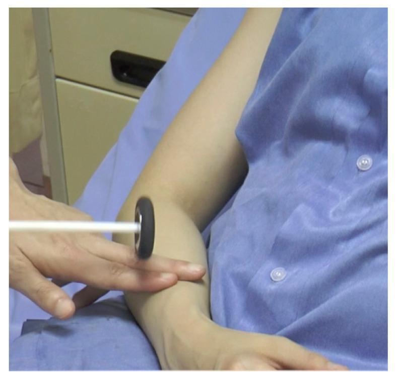
1. Biceps Reflex (C5–C6, Musculocutaneous nerve)
| Step | Action |
|---|---|
| Position | Patient seated; elbow flexed ~90°, forearm supinated, resting on your arm |
| Your hand | Place your thumb over the biceps tendon in the antecubital fossa |
| Strike | Strike your thumb with the hammer |
| Normal response | Elbow flexion and forearm supination |
| ↓ or absent | C5/C6 root lesion or musculocutaneous nerve lesion |
| ↑ (very brisk) | Upper motor neuron lesion |
2. Brachioradialis Reflex (C5–C6, Radial nerve)
| Step | Action |
|---|---|
| Position | Elbow flexed ~90°, forearm in semi-pronation resting on patient's thigh |
| Strike | Tap the brachioradialis tendon directly, ~2 cm above radial styloid |
| Normal response | Elbow flexion + forearm supination |
| Inverted supinator sign | Finger flexion instead of forearm supination → C5/C6 lesion with UMN involvement |
3. Triceps Reflex (C6–C8, Radial nerve)
| Step | Action |
|---|---|
| Position | Hold patient's arm at ~90° abduction; elbow hangs flexed; OR patient's arm crosses chest |
| Strike | Strike the triceps tendon directly ~2–3 cm above the olecranon |
| Normal response | Elbow extension |
| ↓ or absent | C7/C8 root lesion |
4. Patellar (Knee Jerk) Reflex (L3–L4, Femoral nerve)
| Step | Action |
|---|---|
| Position | Patient seated with legs dangling freely, feet off ground — OR supine with knee supported in slight flexion |
| Strike | Strike the patellar tendon just below the patella |
| Normal response | Knee extension (quadriceps contraction) |
| ↓ or absent | L3/L4 root lesion, femoral neuropathy, or peripheral neuropathy |
| ↑ | UMN lesion (corticospinal tract lesion above L3) |
Jendrassik maneuver: If the reflex is absent, ask the patient to interlock their fingers and pull their hands apart just before you strike. This enhances the reflex by reducing cortical inhibition.
5. Achilles (Ankle Jerk) Reflex (S1–S2, Tibial nerve)
| Step | Action |
|---|---|
| Position | Seated: foot hangs freely, or cross one leg over the other; Supine: slightly dorsiflex the foot by hand |
| Strike | Strike the Achilles tendon directly, ~2–3 cm above calcaneal insertion |
| Normal response | Plantar flexion of the foot |
| ↓ or absent | S1 radiculopathy (e.g., disc herniation), diabetic neuropathy, hypothyroidism |
| ↑ or clonus | UMN lesion |
6. Plantar Reflex — Babinski Sign (L5–S1)
| Step | Action |
|---|---|
| Position | Supine, hip extended, knee extended |
| Stimulus | Use a key or blunt object; stroke the lateral sole from heel → ball of foot, then curve medially across the ball |
| Normal (flexor) response | Downward (plantarflexion) of big toe + toe curling |
| Abnormal (Babinski +) | Upward (dorsiflexion) of big toe + fanning of other toes |
| Significance of Babinski + | UMN lesion (corticospinal tract damage) |
7. Hoffman's Reflex (Upper limb UMN sign — cervical cord equivalent of Babinski)
| Step | Action |
|---|---|
| Position | Patient's hand relaxed, middle finger extended |
| Technique | Hold the middle finger and flick the fingernail downward with a quick snap |
| Positive sign | Thumb and index finger flex/adduct reflexively |
| Significance | Suggests UMN lesion above C5 (cervical myelopathy) |
Reinforcement Technique (Jendrassik Maneuver)
When reflexes are absent or diminished:
- Upper limb reflexes: clench teeth, or squeeze thighs together
- Lower limb reflexes: ask patient to interlock fingers and pull apart just as you strike
This increases gamma motor neuron firing, pre-stretching the muscle spindle and making the reflex arc more excitable.
Interpreting Findings
| Finding | Localization |
|---|---|
| Absent/diminished reflex | LMN lesion (nerve root, peripheral nerve, anterior horn cell) |
| Hyperreflexia (3–4+) | UMN lesion (corticospinal tract above that segment) |
| Asymmetric reflex | Localized nerve or root pathology on the diminished side |
| Clonus | Severe UMN lesion |
| Babinski positive | UMN lesion (except normal in infants <1 year) |
| All reflexes absent | Areflexia — consider GBS, severe polyneuropathy, cerebellar shock |
Quick Summary — Reflex → Root → Nerve
| Reflex | Spinal Level | Nerve |
|---|---|---|
| Biceps | C5–C6 | Musculocutaneous |
| Brachioradialis | C5–C6 | Radial |
| Triceps | C6–C8 (mainly C7) | Radial |
| Patellar | L3–L4 | Femoral |
| Achilles | S1–S2 | Tibial |
— General Anatomy and Musculoskeletal System, THIEME Atlas of Anatomy
— Ganong's Review of Medical Physiology, 26th Ed.
— Bradley and Daroff's Neurology in Clinical Practice
Key teaching points for demonstration:
- Always start with the patient relaxed — tense muscles abolish reflexes.
- Use a brisk wrist snap, not a forceful arm blow.
- Bilateral comparison is essential — one side tells you nothing in isolation.
- An absent reflex with a positive Babinski is a classic combination of mixed UMN + LMN disease (e.g., ALS, conus medullaris lesion).
This is a shared conversation. Sign in to Orris to start your own chat.