Cavernous Sinus - Tributaries and Communications
Location
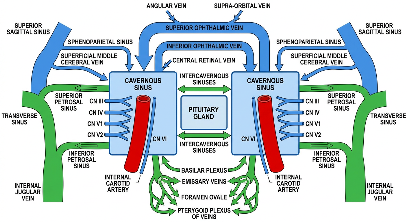
The cavernous sinus lies on the lateral aspect of the sphenoid body, flanking the sella turcica on each side. It extends from the superior orbital fissure anteriorly to the apex of the petrous temporal bone posteriorly.
TRIBUTARIES (Inflow into the Cavernous Sinus)
From the Orbit
| Vein | Route |
|---|
| Superior ophthalmic vein | Formed by angular vein + supra-orbital vein; travels through the orbit and enters via the superior orbital fissure - the main orbital tributary |
| Inferior ophthalmic vein | Smaller; enters directly via the superior orbital fissure OR joins the superior ophthalmic vein first; can also drain into the pterygoid plexus via the inferior orbital fissure |
| Central retinal vein | Small contribution |
From the Brain / Dura
| Vein | Route |
|---|
| Sphenoparietal sinus | Runs along the lesser wing of the sphenoid; receives diploic and meningeal veins; drains into the anterior end of the cavernous sinus |
| Superficial middle cerebral vein | Drains the lateral cerebral cortex into the sphenoparietal sinus and then cavernous sinus |
| Hypophyseal veins | Lateral hypophyseal veins drain the pituitary gland |
From the Face (indirect, via communications)
Facial and nasal infections can reach the cavernous sinus via the angular vein → superior ophthalmic vein route, which is valveless.
COMMUNICATIONS AND DRAINAGE (Outflow)
| Channel | Where it Goes |
|---|
| Superior petrosal sinus | Begins at the posterior end of the cavernous sinus; runs along the superior border of the petrous temporal bone → transverse sinus |
| Inferior petrosal sinus | Also begins at the posterior end; runs in the petro-occipital suture → internal jugular vein; also receives labyrinthine veins from the inner ear |
| Intercavernous sinuses | Connect the right and left cavernous sinuses around the pituitary stalk (anterior and posterior intercavernous sinuses form a venous ring) |
| Basilar plexus | Connects the two inferior petrosal sinuses across the clivus; communicates with the vertebral venous plexus |
| Emissary vein via foramen ovale | Connects cavernous sinus to the pterygoid plexus of veins in the infratemporal fossa |
STRUCTURES INSIDE / RELATED TO THE CAVERNOUS SINUS
Passing through the sinus:
- Internal carotid artery (ICA) - with its sympathetic plexus
- CN VI (abducens) - inferolateral to the ICA
Embedded in the lateral wall (superior to inferior):
- CN III (oculomotor)
- CN IV (trochlear)
- CN V1 (ophthalmic division of trigeminal)
- CN V2 (maxillary division of trigeminal)
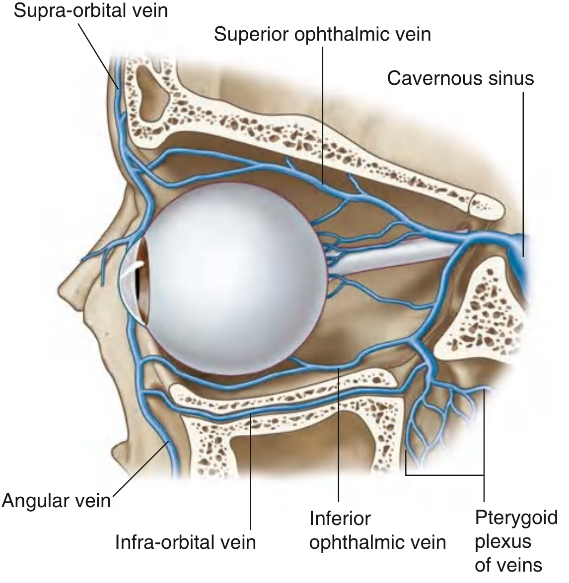
Textbook Image - Orbital Venous Drainage into Cavernous Sinus
This image from Gray's Anatomy for Students shows the orbital tributaries:
Fig. 8.103 - Venous Drainage of the Orbit and Eyeball (Gray's Anatomy for Students)
Clinical Relevance
Because the ophthalmic veins are valveless and communicate freely with the cavernous sinus, infections of the face, nose, sinuses, and orbit can spread intracranially to cause cavernous sinus thrombosis. The pterygoid plexus also connects indirectly via emissary veins through the foramen ovale, providing a second route for spread from the infratemporal fossa.
Sources: Gray's Anatomy for Students (pp. 1082, 1026-27); Color Atlas of Human Anatomy Vol. 2 (pp. 132-34); Scott-Brown's Otorhinolaryngology Head & Neck Surgery Vol. 1 (p. 664); Shambaugh Surgery of the Ear